Comparative Study
. 2005 Nov 1;568(Pt 3):725-35.
doi: 10.1113/jphysiol.2005.087734. Epub 2005 Jul 7.

Proton sensitivity of ASIC1 appeared with the rise of fishes by changes of residues in the region that follows TM1 in the ectodomain of the channel

Affiliations
Comparative Study

Proton sensitivity of ASIC1 appeared with the rise of fishes by changes of residues in the region that follows TM1 in the ectodomain of the channel

Tatjana Coric et al. J Physiol. .

Abstract

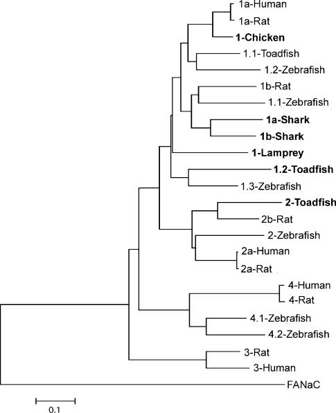
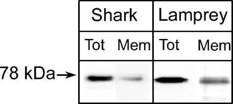
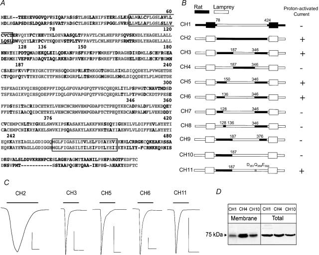
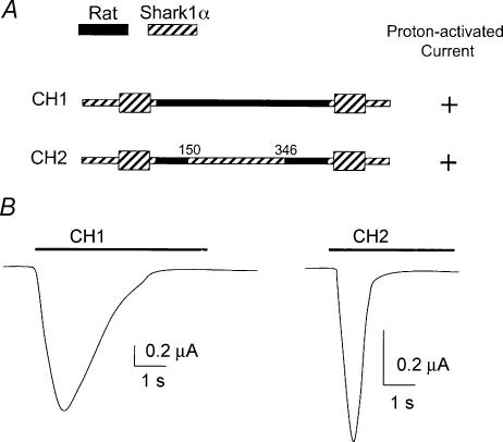
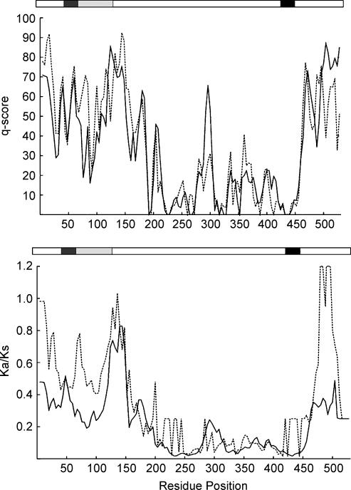
The acid-sensitive ion channel 1 (ASIC1) is a neuronal Na+ channel insensitive to changes in membrane potential but is gated by external protons. Proton sensitivity is believed to be essential for the role of ASIC1 in modulating synaptic transmission and nociception in the mammalian nervous system. To examine the structural determinants that confer proton sensitivity, we cloned and functionally characterized ASIC1 from different species of the chordate lineage: lamprey, shark, toadfish and chicken. We observed that ASIC1s from early vertebrates (lamprey and shark) were proton insensitive in spite of a high degree of amino acid conservation (66-67%) with their mammalian counterparts. Sequence analysis showed that proton-sensitive ASIC1s could not be distinguished from proton-insensitive channels by any signature in the protein sequence. Chimeras made with rat ASIC1 (rASIC1) and lamprey or shark indicated that most of the ectodomain of rASIC1 was required to confer proton sensitivity and the distinct kinetics of activation and desensitization of the rat channel. Proton-sensitive chimeras contained the segment D78-E136, together with residues D351, Q358 and E359 of the rat sequence. However, none of the functional chimeras containing only part of the rat extracellular domain retained the kinetics of activation and desensitization of rASIC1, suggesting that residues distributed in several regions of the ectodomain contribute to allosteric changes underlying activation and desensitization. The results also demonstrate that gating by protons is not a feature common to all ASIC1 channels. Proton sensitivity arose recently in evolution, implying that agonists different from protons activate ASIC1 in lower vertebrates.


Figures

Figure 1
Figure 1. Phylogenetic analysis of ASIC gene family generated using mega version 2.1 (Kumar et al. 2001)
The tree for the phylogram was established by Neighbour-Joining, with the alignment form clustal_x. The tree was rooted with FaNaC as an outgroup. The bar represents genetic distance in substitutions per amino acid. New sequences reported in this work are in bold. The corresponding nucleotide sequences have been submitted to GenBank: the accession numbers are AY278028, AY275841, AY275840, AY956390, AY956391, AY956392 and AY956393 for ASIC 1-toadfish, 2-toadfish, 1.2-toadfish, 1-lamprey, 1a-shark, 1b-shark and 1-chicken, respectively.
Figure 2
Figure 2. Proton-gated currents of fish ASICs
Whole-cell currents elicited by a change in pHo from 7.4 to 5.0 (bars above the current traces) were recorded with two-electrode voltage clamp (TEVC) from injected oocytes. The bath solution contained 150 mm NaCl and the membrane potential was held at –60 mV. A, representative examples of current trances from oocytes injected with single cRNAs corresponding to each of the toadfish ASICs, and oocytes injected with the indicated combinations of cRNAs. B, relative currents induced by pHo 5.0. The data represent the summary of five independent experiments where the peak currents were normalized to the values of fASIC1. Error bars are the standard deviation, n = 8 oocytes for each column. All conditions were statistically different from fASIC1 (P < 0.001).
Figure 3
Figure 3. Expression of fish ASICs Xenopus oocytes
A, identification of fASIC1-HA, fASIC2-FLAG and fASIC1.2-FLAG from injected oocytes by Western blotting using HA and FLAG monoclonal antibodies. fASIC2G431F and fASIC1.2G458F mutants are also shown. B, Western blotting of surface biotinylated proteins of cells expressing toadfish ASIC1.2 or ASIC2, or rat ASIC1, revealed with anti-FLAG monoclonal. C, co-immunoprecipitation of fish ASICs from injected Xenopus oocytes. Oocytes were injected with equal amounts of cRNAs from fASIC1 + fASIC2 or fASIC1 + fASIC1.2. Two days after injection, oocytes were examined for proton-gated currents and saved for co-immunoprecipitation with anti-HA rabbit polyclonal antibody under non-denaturing conditions. Immunocomplexes were resolved on SDS-PAGE and then transferred to membranes for Western blotting. Signals corresponding to fASIC2 and fASIC1.2 were detected with anti-FLAG monoclonal antibody. Arrows on the left of the gels indicate molecular mass markers.
Figure 4
Figure 4. Functional expression of homomeric shark or lamprey channels and heteromeric rat–shark and rat–lamprey ASIC1 channels in Xenopus oocytes
A, 3 days after injection of oocytes with cRNA of rat and/or sASIC1α in ratios indicated below the columns, the oocytes were examined for expression of proton-induced currents with the TEVC. Oocytes were exposed to a preconditioning solution of pHo 7.4 and activated with pHo 4.0 for 5 s. The mean of the values ±s.d. of whole-cell currents (μA oocyte−1) are shown in the ordinate (n = 8). B, similar experiments conducted with oocytes injected with rat and/or lamprey shark cRNAs.
Figure 5
Figure 5. Expression of shark and lamprey ASIC1 in the plasma membrane of oocytes
Western blot of oocytes injected with shark or lamprey ASIC1 cRNA. Oocytes were first treated with sulfo-NHS-SS-Biotin and the biotinylated proteins were isolated with streptavidin beads. Total cell lysate (Tot) and biotinylated proteins (Mem) were resolved in 10% SDS-PAGE, transferred to membranes and processed for Western blotting with V5 (shark) or FLAG (lamprey) monoclonal antibodies, respectively.
Figure 6
Figure 6. Rat–lamprey chimeras
A, alignment of rat and lamprey ASIC1. Identical residues are shown in red and different ones in black. Numbers above the sequence correspond to the amino acid positions in the lamprey sequence. Boxes enclose TM1 and TM2. B, schematic representation of rat–lamprey CHs. Sequences corresponding to rat and lamprey are indicated in black and white, respectively. The names of the chimeras are on the left. The numbers over each chimera are the same as in A. On the right is indicated the response to a pulse of pHo 4.0. C, representative current traces of oocytes expressing proton-sensitive CHs activated by pHo 4.0 indicated by the bars over the current traces. The time and current scale bars under each trace correspond to 1 s and 1 μA. D, Western blots of surface biotinylated CHs (Membrane) and a fraction of cell lysate (Total) revealed by anti-FLAG monoclonal antibody.
Figure 7
Figure 7. Rat–shark1α chimeras
A, schematic of rat–shark1α chimeras. Black and hatched represent sequences from rat and shark, respectively. Numbers are amino acid positions in the shark sequence. B, representative examples of whole-cell currents elicited by changes in pHo from 7.4 to 4.0 (bar above traces) obtained with TEVC. Bars indicate current and time scales.
Figure 8
Figure 8. Sequence analysis of proton-sensitive and -insensitive ASIC1s
A, conservation of amino acid composition within proton-sensitive (continuous line) and -insensitive (dashed line) ASIC1s assessed by conservation score (q-scores). A low score indicates that a region is conserved. B, sliding-window analysis of Ka/Ks ratios performed on proton-sensitive (continuous line) and proton-insensitive (dashed line) ASIC1s. Window of 90 nt with 12 nt step was used for the study. The x-axis indicates the residue numbered according to the rat ASIC1α sequence. Protein domains are drawn schematically above each graph, with transmembrane segments shown in black. Regions that were found important for proton sensitivity in functional studies are shaded in grey.

References

    1. Babini E, Paukert M, Geisler HS, Gründer S. Alternative splicing and interaction with di- and polyvalent cations control the dynamic range of acid-sensing ion channel 1 (ASIC1) J Biol Chem. 2002;277:41597–41603. - PubMed
    1. Champigny G, Voilley N, Waldmann R, Lazdunski M. Mutations causing neurodegeneration in Caenorhabditis elegans drastically alter the pH sensitivity and inactivation of the mammalian H+-gated Na+ channel MDEG1. J Biol Chem. 1998;273:15418–15422. - PubMed
    1. Chen CC, England S, Akopian AN, Wood JN. A sensory neuron-specific, proton-gated ion channel. Proc Natl Acad Sci U S A. 1998;95:10240–10245. - PMC - PubMed
    1. Coric T, Zhang P, Todorovic N, Canessa CM. The extracellular domain determines the kinetics of desensitization in acid-sensitive ion channel 1. J Biol Chem. 2003;278:45240–45247. - PubMed
    1. Huang M, Chalfie M. Gene interactions affecting mechanosensory transduction in Caenorhabditis elegans. Nature. 1994;367:467–470. - PubMed

Publication types

MeSH terms

LinkOut - more resources