Comparative Study
. 2005 Jul 19;102(29):10327-32.
doi: 10.1073/pnas.0408529102. Epub 2005 Jul 7.

Replication and packaging of Norwalk virus RNA in cultured mammalian cells

Affiliations
Comparative Study

Replication and packaging of Norwalk virus RNA in cultured mammalian cells

Miyuki Asanaka et al. Proc Natl Acad Sci U S A. .

Abstract

Human noroviruses, the most common cause of nonbacterial gastroenteritis, are characterized by high infectivity rate, low infectious dose, and unusually high stability outside the host. However, human norovirus research is hindered by the lack of a cell culture system and a small animal model of infection. Norwalk virus (NV) is the prototype strain of human noroviruses. We report here replication of NV viral RNA and its packaging into virus particles in mammalian cells by intracellular expression of native forms of NV viral RNA devoid of extraneous nucleotide sequences derived from the expression vector by the use of replication-deficient vaccinia virus MVA encoding the bacteriophage T7 RNA polymerase (MVA/T7). Expressed genomic RNA was found to replicate; NV subgenomic RNA was transcribed from genomic RNA by use of NV nonstructural proteins expressed from genomic RNA and was subsequently translated into NV capsid protein VP1. Viral genomic RNA was packaged into virus particles generated in mammalian cells. The cesium chloride (CsCl) density gradient profile of virus particles containing genomic RNA was similar to that of NV purified from stool. These observations indicate that the NV cDNA constructed here is a biologically infectious clone, and that mammalian cells have the ability to replicate NV genomic RNA. This work establishes a mammalian cell-based system for analysis of human norovirus replication and, thus, makes it feasible to investigate antiviral agents in mammalian cells.


Figures

Fig. 1.
Fig. 1.
Plasmid constructs for the expression of native forms of Norwalk viral genomic and subgenomic RNA in mammalian cells. (A) Schematic of the complete NV genome, indicating the location of the nonstructural protein cleavage products within the ORF1 polyprotein, the viral structural proteins VP1 and VP2 encoded by ORF2 and ORF3, respectively, and the 5′ and 3′ untranslated regions (solid lines). (B) Diagram of the plasmids used for viral RNA expression (7). T7Ψ, HδR, and T7ϕ refer to the position of the T7 promoter, hepatitis δ virus antigenomic ribozyme, and T7 terminator sequence, respectively. Arrows indicate the transcription initiation site and the self-cleavage site. Numbers refer to the nucleotide positions of the NV genome (GenBank accession no. NC001959).
Fig. 2.
Fig. 2.
Expression and replication of genomic NV RNA in mammalian cells. 293T cells were infected with MVA/T7 and then transfected with plasmids encoding NV cDNAs. (A) Detection of nonstructural and structural NV proteins in cells expressing NV RNA by radioimmunoprecipitation (Left) and immunofluorescence (Right) with antisera to the proteins. Radiolabeled NV proteins synthesized by in vitro translation were processed in parallel. Molecular size markers (kilodaltons) are on the right. Arrows highlight the NV proteins. (B) Detection of subgenomic RNA synthesized in cells expressing genomic RNA. Poly(A)+ RNA, isolated at 4–12 h pt, and was subjected to Northern blotting. In vitro transcripts of subgenomic RNA were analyzed in parallel. Arrow indicates subgenomic RNA. RNA size markers (nucleotides) are on the left.
Fig. 3.
Fig. 3.
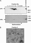
Generation of virus particles from cells expressing subgenomic RNA. Virus particles were isolated from the culture supernatants by isopycnic CsCl gradient centrifugation, and particles in individual gradient fractionations were sedimented. (A) The presence of virus particles in each fraction was examined by Western blotting with anti-rNV hyperimmune serum that reacts with VP1 (18) (Upper) and anti-VP2 antibody (24) (Lower). As a positive control, virus particles produced by the baculovirus expression system (24) were analyzed in parallel. Arrows indicate detected viral proteins. Size markers (kilodaltons) are on the right. (B) Electron micrograph of negatively stained purified virus particles from fraction 5. (Scale bar: 100 nm.)
Fig. 4.
Fig. 4.
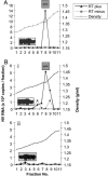
Incorporation of viral RNA into virus particles. Virus particles were isolated from the culture supernatants of cells expressing subgenomic RNA or coexpressing both genomic and subgenomic RNA, followed by sedimentation as described in Fig. 3. The sedimented particles from individual fractions were treated with DNase I and RNase A, and nucleic acid was extracted and subjected to quantitative real-time RT-PCR, performed with or without reverse transcriptase. The copy number of viral RNA present in each fraction was determined by comparison with a standard curve, and the results are plotted against the density. Each fraction was also examined by Western blotting for the presence of VP1, and the results are shown in the inserts. (A) Detection of genomic RNA incorporated into virus particles generated by the coexpression of genomic and subgenomic RNA by using primers corresponding to the Pol region. (B) Detection of subgenomic RNA incorporated into virus particles generated by coexpression of genomic and subgenomic RNA (i) or subgenomic RNA alone (ii) by using primers corresponding to the capsid region.

References

    1. Billgren, M., Christenson, B., Hedlund, K. O. & Vinje, J. (2002) J. Infect. 44, 26-32. - PubMed
    1. Fankhauser, R. L., Noel, J. S., Monroe, S. S., Ando, T. & Glass, R. I. (1998) J. Infect. Dis. 178, 1571-1578. - PubMed
    1. Hutson, A. M., Atmar, R. L. & Estes, M. K. (2004) Trends Microbiol. 12, 279-287. - PMC - PubMed
    1. Jiang, X., Wang, M., Wang, K. & Estes, M. K. (1993) Virology 195, 51-61. - PubMed
    1. Green, K. Y., Chanock, R. M. & Kapikian, A. Z. (2001) in Fields Virology, eds. Knipe, D. M. & Howley, P. M. (Lippincott Williams & Wilkins, Philadephia), pp. 841-874.

Publication types

MeSH terms