. 2005 Jul;4(7):1228-38.
doi: 10.1128/EC.4.7.1228-1238.2005.

A CDC42 homologue in Claviceps purpurea is involved in vegetative differentiation and is essential for pathogenicity

Affiliations

A CDC42 homologue in Claviceps purpurea is involved in vegetative differentiation and is essential for pathogenicity

Jan Scheffer et al. Eukaryot Cell. 2005 Jul.

Abstract

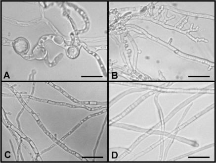
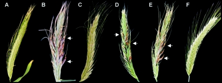
Claviceps purpurea, a biotrophic pathogen of cereals, has developed a unique pathogenic strategy including an extended period of unbranched directed growth in the host's style and ovarian tissue to tap the vascular system. Since the small GTPase Cdc42 has been shown to be involved in cytoskeleton organization and polarity in other fungi, we investigated the role of Cdc42 in the development and pathogenicity of C. purpurea. Expression of heterologous dominant-active (DA) and dominant-negative (DN) alleles of Colletotrichum trifolii in a wild strain of C. purpurea had significant impact on vegetative differentiation: whereas DA Ctcdc42 resulted in loss of conidiation and in aberrant cell shape, expression of DN Ctcdc42 stimulated branching and conidiation. Deletion of the endogenous Cpcdc42 gene was not lethal but led to a phenotype comparable to that of DN Ctcdc42 transformants. DeltaCpcdc42 mutants were nonpathogenic; i.e., they induced no disease symptoms. Cytological analysis (light microscopy and electron microscopy) revealed that the mutants can penetrate and invade the stylar tissue. However, invasive growth was arrested in an early stage, presumably induced by plant defense reactions (necrosis or increased production of reactive oxygen species), which were never observed in wild-type infection. The data show a significant impact of Cpcdc42 on vegetative differentiation and pathogenicity in C. purpurea.


Figures

FIG. 1.
FIG. 1.
Protein sequence alignment of the CDC42 homologs (with accession numbers) from C. purpurea (CpCDC42; AJ879079), C. trifolii (CtCdc42; AAK31624), Gibberella zeae (CDC42; EAA75264), M. grisea (MgCdc42; AAF73431); S. cerevisiae (Cdc42p; NP_013330), and H. sapiens (CDC42, AAH18266). Amino acids mutated in the Ctcdc42 alleles (see text) are indicated by arrows. The known functional domains are indicated as follows: dashed box, GTP binding/hydrolysis domains; bold black box, effector domain; dotted box, Rho insert domain; and thin black box, membrane localization domain.
FIG. 2.
FIG. 2.
Morphology of C. purpurea transformants overexpressing Ctcdc42(G14V) (A), Ctcdc42(T19N) (B), and unmodified Ctcdc42 (C). C. purpurea wild-type strain 20.1 (D). Strains were grown for 5 days on Mantle medium. For details see text. Scale bars, 10 μm.
FIG. 3.
FIG. 3.
Generation of ΔCpcdc42 mutants. (A) Gene replacement approach (Δ) and complementation construct (C) for Cpcdc42. For the replacement approach, the replacement vector pΔcpcdc42 was constructed by cloning 3′ and 5′ parts of Cpcdc42 on each side of a hygromycin resistance cassette (hph) in the pAN7-1UM plasmid (see Material and Methods for details). The resistance cassette, excised using a NotI-KpnI restriction, was used to transform the C. purpurea wild-type strain 20.1. Δcdc42 mutants were generated following the disruption of the wild-type gene (WT) by homologous recombination through a double crossover event. The Cpcdc42 coding sequence and introns are represented with black and white boxes, respectively. The ATG indicates the start of translation of Cpcdc42, and the black arrow indicates the orientation of the hygromycin resistance cassette within the replacement construct. Primers DCDCLF1/DCDCLF2 (a and b) and DCDCRF1/DCDCRF2 (c and d) used for the amplification of the upstream and downstream flanks, respectively, are indicated by black arrows. The positions of primers DCDC-hIL1/DCDC-hIL2 (e and f) and DCDC-hIR1/DCDC-hIR2 (g and h), used for the identification of homologous integrations, and primers DCDC-WT1/DCDC-WT2 (i and j), used for detection of the Cpcdc42 wild-type copy, are indicated with black triangles. The DNA fragment used as a probe in the Southern blot shown in panel B is indicated as a striped box. The full-length clone of Cpcdc42 for complementation is shown (see text for details). To check the reinsertion of Cpcdc42 for complementation, the primers DCDC-hIL1/CCDC42-1 (e and k) were used. Abbreviations for restriction enzymes: CI, ClaI; EV, EcoRV, KI, KpnI; NI, NotI; SI, SacI; XI, XbaI. (B) Southern analysis of deletion mutants ΔCpcdc42-1, ΔCpcdc42-2, complemented strain Ccpcdc42-1, and wild-type 20.1. Genomic DNA of selected strains was digested with ClaI, separated in an agarose gel, blotted, and probed with the right flank of the replacement vector pΔcpcdc42. A successful deletion by gene replacement is demonstrated by the shift of the wild-type band (2.6 kb) to 5.75 kb in the lanes of ΔCpcdc42-1 and ΔCpcdc42-2. The complementation leads to the reappearance of the 2.6-kb band and an additional band (0.64 kb). For restriction sites and location of the probe, see panel A.
FIG. 4.
FIG. 4.
Axenic growth of the ΔCpcdc42-1 mutant (A to C), complemented mutant Ccpcdc42-1 (D and E), ΔCpcdc42 mutant overexpressing DA Ctcdc42(G14V) (ΔCpcdc42-DA Ctcdc42-1) (F to I), and ΔCpcdc42 mutant overexpressing wild-type Ctcdc42 (ΔCpcdc42-WT Ctcdc42-1) (J to L). For ΔCpcdc42-1 young mycelium with phialidic branches are shown in panels A and B in detail. Massive conidiation occurs after 2 days (C). In the complemented strain Ccpcdc42-1, complementation restored hyphal morphology, as shown in panel D (E, detailed view). For ΔCpcdc42-DA Ctcdc42-1, swollen hyphae (F to H) and bulbous hyphal tips are shown (I). In ΔCpcdc42-WT Ctcdc42-1, hyphal morphology is restored (J and K). Occasionally, an unusual branching pattern was observed (L), which, however, did not form conidia as did ΔCpcdc42-1. Compare wild type with Fig. 2D. Strains were grown on Mantle medium. Pictures were taken 5 dpi. Scale bars, 10 μm.
FIG. 5.
FIG. 5.
Pathogenicity assays of C. purpurea transformants on rye. Rye florets were infected with water (A) and conidial suspensions from wild type (B), ΔCpcdc42-1 mutant (C), the complemented strain Ccpcdc42-1 (D), a ΔCpcdc42 mutant overexpressing wild-type Ctcdc42 (E), and a ΔCpcdc42 mutant overexpressing Ctcdc42(G14V) (F). Pictures were taken 4 weeks postinoculation. Arrows indicate sclerotia.
FIG. 6.
FIG. 6.
Effect of the inactivation of Cpcdc42 on pathogenicity of C. purpurea. Ovaries were infected in vitro with conidial suspensions from wild type (A, D, and F) and ΔCpcdc42-1 mutant (B, C, E, and H). A schematic overview of a rye ovary is given in panel G. The solid-lined box represents details shown in pictures panels A to E, while the dotted box represents pictures in panels F and H. Hyphal growth within the stigmas and stigmatic hairs is visible in both the wild-type (A and D) and the ΔCpcdc42-1 mutant (B, C, and E). In contrast to the massive colonization of the transmitting tissue by the wild type (F), no hyphae could be detected in this area after infection with the ΔCpcdc42-1 mutant. Ovaries were collected at 5 days postinoculation, stained with aniline blue, and observed using epifluorescence microscopy. Arrows indicate hyphae.
FIG. 7.
FIG. 7.
Light and electron microscopic analyses of rye stylar tissues infected by wild type and ΔCpcdc42. (A and B) longitudinal section of a style colonized by wild-type C. purpurea showing mostly intercellular growth between the prosenchymatic host tissue. (C) Rye stylar tissue inoculated with ΔCpcdc42. The transition from normal-looking, highly vacuolated host cells to an area of obviously collapsed host cells showing dense staining of the whole cell compartment is visible. (D to F and I to K) In situ detection of H2O2 using the CeCl3 technique. Electron-dense precipitate of ceriumperhydroxide represents the areas where H2O2 was formed. (D) Subcuticular wild-type hypha which shows cell wall-bound and secreted H2O2. Note that the host cell completely lacks any signs of H2O2 generation. (E) Wild-type hyphae, growing both epicuticular (asterisk) and subcuticular, showing intense formation of H2O2. Note also that the host epidermal cells produce H2O2 at this interaction site. (F) Wild-type hyphae among the prosenchymatic transmitting tissue. In this tissue virtually no generation of H2O2 takes place. (G) A ΔCpcdc42 hypha inside the stigmatic trichomes. (H) Sparse colonization of stylar tissue by ΔCpcdc42 hyphae (arrow). (I) H2O2 production in noncollapsed prosenchymatic host tissue (as depicted in the upper part of panel C adjacent to ΔCpcdc42 hyphae). Strong signals can be found in the area of the plasmalemma, vacuoles, and host cell wall (arrow). The host cells show clear signs of breakdown of cell compartments like disintegration of the vacuolar system. (J) H2O2 detection in collapsed host tissue (as depicted in the lower part of panel C). A plasma membrane-bound signal is visible. (K) Collapsed host tissue (CeCl3 treatment was omitted). Granular electron-dense particles are visible in large parts of the cell compartments pointing to the occurrence of phenolic/tannic substances in this area. (L) Noncollapsed host tissue (CeCl3 treatment was omitted). No electron-dense precipitation structures are detectable. Toluidine blue (pH 6.8) staining was used in panels A to C, G, and H. Scale bars: 150 μm (A and C), 10 μm (B, G, and H), 5 μm (D, E, and J to L), and 2 μm (F and I). f, fungus; hc, host cell.

References

    1. Adams, A. E., D. I. Johnson, R. M. Longnecker, B. F. Sloat, and J. R. Pringle. 1990. CDC42 and CDC43, two additional genes involved in budding and the establishment of cell polarity in the yeast Saccharomyces cerevisiae. J. Cell Biol. 111:131-142. - PMC - PubMed
    1. Altschul, S. F., W. Gish, W. Miller, E. W. Myers, and D. J. Lipman. 1990. Basic local alignment search tool. J. Mol. Biol. 215:403-410. - PubMed
    1. Austin, B., R. M. Hall, and B. M. Tyler. 1990. Optimized vectors and selection for transformation of Neurospora crassa and Aspergillus nidulans to bleomycin and phleomycin resistance. Gene 93:157-162. - PubMed
    1. Ausubel, F. M., R. Brent, R. E. Kingston, D. D. Moore, J. G. Seidmann, J. A. Smith, and K. Struhl. 1987. Current protocols in molecular biology. John Wiley and Sons, New York, N.Y.
    1. Bestwick, C. S., I. R. Brown, M. H. R. Bennett, and J. W. Mansfield. 1997. Localization of hydrogen peroxide accumulation during the hypersensitive reaction of lettuce cells to Pseudomonas syringae pv phaseolicola. Plant Cell 9:209-221. - PMC - PubMed

Publication types

Associated data