. 2005 Nov 15;392(Pt 1):103-16.
doi: 10.1042/BJ20050826.

Phylogenetic analysis of the phytochrome superfamily reveals distinct microbial subfamilies of photoreceptors

Affiliations

Phylogenetic analysis of the phytochrome superfamily reveals distinct microbial subfamilies of photoreceptors

Baruch Karniol et al. Biochem J. .

Abstract

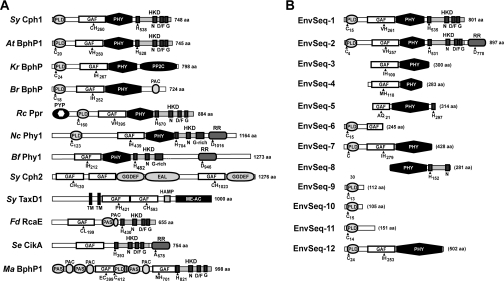
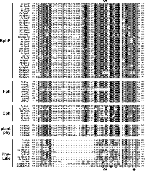
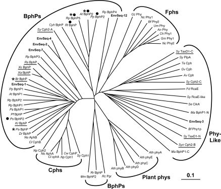
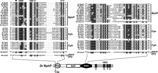
Phys (phytochromes) are a superfamily of photochromic photoreceptors that employ a bilin-type chromophore to sense red and far-red light. Although originally thought to be restricted to plants, accumulating genetic and genomic analyses now indicate that they are also prevalent among micro-organisms. By a combination of phylogenetic and biochemical studies, we have expanded the Phy superfamily and organized its members into distinct functional clades which include the phys (plant Phys), BphPs (bacteriophytochromes), Cphs (cyanobacterial Phys), Fphs (fungal Phys) and a collection of Phy-like sequences. All contain a signature GAF (cGMP phosphodiesterase/adenylate cyclase/FhlA) domain, which houses the bilin lyase activity. A PHY domain (uppercase letters are used to denote the PHY domain specifically), which helps stabilize the Pfr form (far-red-light-absorbing form of Phy), is downstream of the GAF region in all but the Phy-like sequences. The phy, Cph, BphP and Fph families also include a PLD [N-terminal PAS (Per/Arnt/Sim)-like domain] upstream of the GAF domain. Site-directed mutagenesis of conserved residues within the GAF and PLD motifs supports their importance in chromophore binding and/or spectral activity. In agreement with Lamparter, Carrascal, Michael, Martinez, Rottwinkel and Abian [(2004) Biochemistry 43, 3659-3669], a conserved cysteine within the PLD of several BphPs was found to be necessary for binding the chromophore via the C-3 vinyl side chain on the bilin A ring. Phy-type sequences were also discovered in the actinobacterium Kineococcus radiotolerans and collections of microorganisms obtained from marine and extremely acidic environments, thus expanding further the range of these photoreceptors. Based on their organization and distribution, the evolution of the Phy superfamily into distinct photoreceptor types is proposed.


Figures

Figure 1
Figure 1. Domain structures of representative members from the BphP, Fph, Cph and Phy-like clades (A) and predicted members of the Phy superfamily (EnvSeq) present in a random collection of DNA sequences from a marine environment (EnvSeq-1 to -11 [29]) and an acidophilic mine biofilm (EnvSeq-12 [30]) (B)
Species designations can be found in the legend of Figure 3. The amino acid (aa) length of each protein is indicated on the right; parentheses indicate partial sequence. Abbreviations not otherwise defined in the main text: EAL, motif bearing a consensus Glu/Ala/Leu sequence; GGDEF, motif bearing a consensus Gly/Gly/Asp/Glu/Phe sequence; HAMP, HK/adenylate cyclase/methyl-binding protein/phosphatase domain. HKD, HK domain (the H, N, D/F and G boxes are indicated); HKRD, HK-related domain. Me-Ac, methyl-accepting chemotaxis protein domain. PAC, PAS-like domain C-terminal to PAS; PYP, photoactive yellow protein domain; TM, transmembrane. The positions of signature amino acids in the PLD, GAF, HKD and RR domains are indicated.
Figure 2
Figure 2. Amino-acid-sequence alignment of a portion of the GAF domain from members of the Phy superfamily
Alignment of the entire domain can be found in Supplemental Figure 1 (http://www.BiochemJ.org/bj/392/bj3920103add.htm). The sequences are grouped by their proposed inclusion in the BphP, Fph, Cph, plant phy and Phy-like families. The numbers indicate the amino acid positions within each GAF sequence. Black and grey boxes denote identical and similar residues respectively. The Cys and His residues important for bilin binding are identified by the open and closed arrowheads respectively (see Figure 6). The diamond locates the Pro conserved among Phy-like proteins. Species and sequence designations can be found in the legend of Figure 3.
Figure 3
Figure 3. Phylogenetic analysis of the Phy superfamily based on an alignment of the GAF domain
The BphP, Fph, Cph, plant phy and Phy-like families are indicated. The sequences were aligned by CLUSTALX and the unrooted phylogenetic tree was generated by the neighbour-joining method and displayed using Treeview. Underline indicates proteins without an obvious HK domain. Asterisks indicate known members of the bathyBphP subfamily. Closed circles denote HK proteins that belong to the HWE group [40]. Genus/species abbreviations shown in the Figure: Agrobacterium tumefaciens (At), Anabaena variabilis (Av), Arabidopsis thaliana (Ath), Aspergillus nidulans (An), Botryotinia fuckeliana (Bf), Bradyrhizobium ORS278 (Br), Calorthrix PCC7601 (Cl), Cochliobolus heterostrophus (Ch), Crocosphaera watsonii (Cw), Cytophaga hutchinsonii (Cyh), Deinococcus radiodurans (Dr), Fremyella diplosiphon (Fd), Gibberella moniliformis (Gm), Gibberella zeae (Gz), Gleobacter violaceus (Gv), Kineococcus radiotolerans (Kr), Methanosarcina acetivorans (Ma), Neurospora crassa (Nc), Nostoc PCC 7120 (No), Nostoc punctiforme (Np), Pseudomonas aeruginosa (Pa), Pseudomonas putida (Pp), Pseudomonas syringae (Ps), Rhizobium leguminosarium (Rl), Rhodospirillum centenum (Rc), Rhodopseudomonas palustris (Rp), Rhodospirilum rubrum (Rr), Synechocystis PCC6803 (Sy), Thermosynechococcus elongatus (Te), Ustilago maydis (Um), Xanthomonas axonopodis (Xa) and Xanthomonas campestris (Xc). EnvSeq-1, -2, -3, -4 and -7 were identified in a collection of genomic fragments randomly sequenced from a marine environmental sequence database [29]. EnvSeq-12 is a random DNA sequence isolated from an acidophilic mine biofilm [30]. Both the N-terminal (N) and C-terminal (C) GAF domains in Synechocystis TaxD1 and M. acetivorans BphP1, and two of the GAF domains (A and C) in Synechocystis Cph2, were included in the comparison.
Figure 4
Figure 4. Amino acid sequence alignment of portions of PLD (left) and PHY (right) from members of the BphP, Fph, Cph and plant phy clades
Alignments of the entire domains can be found in Supplemental Figures 2 and 3 (http://www.BiochemJ.org/bj/392/bj3920103add.htm). The sequences were grouped by their proposed inclusion into BphP, Fph, Cph and plant phy families. The position of each domain in the linear Phy sequence of D. radiodurans BphP is shown. Black and grey boxes denote identical and similar residues respectively. The numbers identify the amino acid positions for each sequence. The cysteine in the PLD important for bilin binding is identified by the closed arrowhead (Cys-24 in DrBphP). The open arrowheads identify conserved residues in AtBphP2 that were tested for their importance in BV IXα ligation (see Figure 7). The asterisks locate amino acids in the PHY domain identified as being important for the spectral properties of Synechocystis Cph1 [36]. Species and sequence designations can be found in the legend of Figure 3.
Figure 5
Figure 5. Assembly and spectral properties of full-length (FL) and C-terminally truncated forms of Ag. tumefaciens BphP1 and BphP2
The numbers indicate the last amino acid of the truncated polypeptides. (A) Covalent binding of BV IXα. The wild-type (WT) and mutant apoproteins were incubated with BV IXα for 1 h, purified by nickel-chelate affinity chromatography and then subjected to SDS/PAGE and either assayed for the bound bilin by zinc-induced fluorescence (Zn) or stained for protein with Coomassie Blue (Prot). (B) Absorption and R/FR (AtBphP1) or FR/D (AtBphP2) difference spectra of samples from (A). The absorption maxima of the Pr and Pfr forms are indicated. The spectra were adjusted for an equal amount of protein as determined by absorbance at 280 nm.
Figure 6
Figure 6. Requirements of the PLD Cys and the GAF His for bilin binding by D. radiodurans BphP, and BphP1 and BphP2 from Ag. tumefaciens
Codons for the Cys and His residues were converted into those for the indicated amino acids by site-directed mutagenesis of DrBphP(N321), AtBphP1(N320) and AtBphP2(N505). The recombinant wild-type (WT) and mutant polypeptides were incubated with BV IXα for 1 h and purified by nickel-chelate chromatography. (A) Samples were subjected to SDS/PAGE and either assayed for the bound bilin by zinc-induced fluorescence (Zn) or stained for protein with Coomassie Blue (Prot). Apo, apoprotein prior to BV IXα incubation. (B) Absorption spectra of the samples following an extended incubation in darkness. The absorption maxima of the Pr (DrBphP and AtBphP1) and Pfr (AtBphP2) forms are indicated. The absorption spectra were adjusted for an equal amount of protein as determined by absorbance at 280 nm.
Figure 7
Figure 7. Importance of conserved residues within the PLD for BV IXα binding and the spectral properties of Ag. tumefaciens BphP2
(A) Covalent binding of BV IXα to the wild-type and mutant versions of AtBphP2(N505). The recombinant polypeptide was incubated with BV for 1 h, purified by nickel-chelate affinity chromatography, and then subjected to SDS/PAGE and either assayed for the bound bilin by zinc-induced fluorescence (Zn) or stained for protein with Coomassie Blue (Prot). Apo, apoprotein prior to BV IXα incubation. (B) Absorption and FR/D difference spectra of samples from (A) following an overnight incubation in darkness (D) or saturating irradiation with FR. The absorption maxima of the Pfr forms are indicated.
Figure 8
Figure 8. Assembly and spectral properties of D. radiodurans BphP and Ag. tumefaciens BphP1 and BphP2 following incubation with BV IXα, PCB and various BV IXα derivatives
The structure of each bilin is shown in (B). (A) Covalent binding of bilins to the apoproteins. The recombinant polypeptides [DrBphP(N321), AtBphP1(N320) and AtBphP2(N505)] were incubated with the bilin for 1 h, purified by nickel-chelate affinity chromatography, and then subjected to SDS/PAGE and either assayed for the bound bilin by zinc-induced fluorescence (Zn) or stained for protein with Coomassie Blue (Prot). Apo, apoprotein without BV IXα incubation. (B) Absorption spectra of the DrBphP(N321) (long dashed lines), AtBphP1(N320) (solid lines) and AtBphP2(N505) (short dashed lines) from (A) following an extended incubation in darkness. The absorbance maxima are indicated. The bar represents 0.18 A, 0.13 A and 0.05 A for DrBphP(N321), AtBphP1(N320) and AtBphP2(N505) respectively.

References

    1. Briggs W. R., Spudich J. L. Weinheim, Germany: Wiley-VCH Verlag GmbH & Co; 2005. Handbook of Photosensory Systems.
    1. Quail P. H. Phytochrome photosensory signalling networks. Nat. Rev. Mol. Cell Biol. 2002;3:85–93. - PubMed
    1. Smith H. Phytochromes and light signal perception by plants – an emerging synthesis. Nature (London) 2000;407:585–591. - PubMed
    1. Vierstra R. D. Cyanophytochromes, bacteriophytochromes, and plant phytochromes: light-regulated kinases related to bacterial two-component regulators. In: Inouya M., Dutta R., editors. Histidine Kinases in Signal Transduction. New York: Academic Press; 2002. pp. 273–295.
    1. Bhoo S. H., Davis S. J., Walker J., Karniol B., Vierstra R. D. Bacteriophytochromes are photochromic histidine kinases using a biliverdin chromophore. Nature (London) 2001;414:776–779. - PubMed

Publication types