

Modulation of epithelial neoplasia and lymphoid hyperplasia in PTEN+/- mice by the p85 regulatory subunits of phosphoinositide 3-kinase
- PMID: 16006513
- PMCID: PMC1174923
- DOI: 10.1073/pnas.0504378102
Modulation of epithelial neoplasia and lymphoid hyperplasia in PTEN+/- mice by the p85 regulatory subunits of phosphoinositide 3-kinase
Abstract
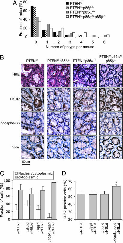
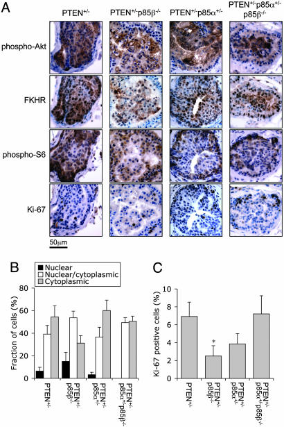
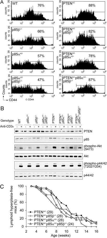
Mice with heterozygous deletion of the PTEN tumor suppressor gene develop a range of epithelial neoplasia as well as lymphoid hyperplasia. Previous studies suggest that PTEN suppresses tumor formation by acting as a phosphoinositide phosphatase to limit signaling by phosphoinositide 3-kinase (PI3K). Here, we examined the effect of deleting various regulatory subunits of PI3K (p85alpha and p85beta) on epithelial neoplasia and lymphoid hyperplasia in PTEN+/- mice. Interestingly, we found the loss of one p85alpha allele with or without the loss of p85beta led to increased incidence of intestinal polyps. Signaling downstream of PI3K was enhanced in the PTEN+/-p85alpha+/-p85beta-/- polyps, as judged by an increased fraction of both cells with cytoplasmic staining of the transcription factor FKHR and cells with positive staining for the proliferation marker Ki-67. In contrast, the incidence of prostate intraepithelial neoplasia was not significantly altered in PTEN+/- mice heterozygous for p85alpha or null for p85beta, whereas the fraction of proliferating cells in prostate intraepithelial neoplasia was reduced in mice lacking p85beta. Finally, there was no significant change in T lymphocyte hyperplasia in the PTEN+/- mice with various p85 deletions, although anti-CD3-stimulated AKT activation was somewhat reduced in the p85alpha+/- background. These results indicate that decreasing the levels of different p85 regulatory subunits can result in enhanced PI3K signaling in some tissues and decreased PI3K signaling in others, supporting the model that, although p85 proteins are essential for class I(A) PI3K signaling, they can function as inhibitors of PI3K signaling in some tissues and thus suppress tumor formation.
Figures
References
-
- Cantley, L. C. (2002) Science 296, 1655-1657. - PubMed
-
- Vivanco, I. & Sawyers, C. L. (2002) Nat. Rev. Cancer 2, 489-501. - PubMed
-
- Sarbassov, D. D., Guertin, D. A., Ali, S. M. & Sabatini, D. M. (2005) Science 307, 1098-1101. - PubMed
-
- Burgering, B. M. & Medema, R. H. (2003) J. Leukoc. Biol. 73, 689-701. - PubMed
-
- Fingar, D. C. & Blenis, J. (2004) Oncogene 23, 3151-3171. - PubMed
Publication types
MeSH terms
Substances
Grants and funding
LinkOut - more resources
Full Text Sources
Medical
Molecular Biology Databases
Research Materials
Miscellaneous
