Comparative Study
. 2005 Aug 9;102(32):11408-13.
doi: 10.1073/pnas.0504197102. Epub 2005 Jul 8.

Identification of mast cell progenitors in adult mice

Affiliations
Comparative Study

Identification of mast cell progenitors in adult mice

Ching-Cheng Chen et al. Proc Natl Acad Sci U S A. .

Abstract

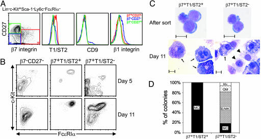
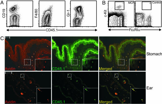
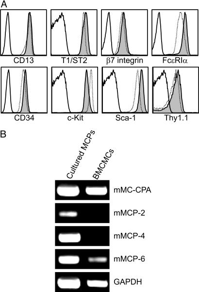
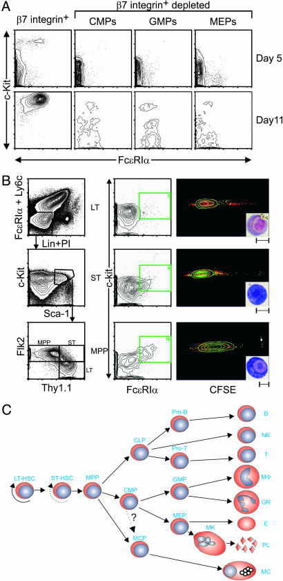
It is well known that mast cells are derived from hematopoietic stem cells. However, in adult hematopoiesis, a committed mast cell progenitor has not yet been identified in any species, nor is it clear at what point during adult hematopoiesis commitment to the mast cell lineage occurs. We identified a cell population in adult mouse bone marrow, characterized as Lin(-)c-Kit(+)Sca-1(-)-Ly6c(-)FcepsilonRIalpha(-)CD27(-)beta7(+)T1/ST2+, that gives rise only to mast cells in culture and that can reconstitute the mast cell compartment when transferred into c-kit mutant mast cell-deficient mice. In addition, our experiments strongly suggest that these adult mast cell progenitors are derived directly from multipotential progenitors instead of, as previously proposed, common myeloid progenitors or granulocyte/macrophage progenitors.


Figures

Fig. 1.
Fig. 1.
Identification of MCPs in adult mouse bone marrow. (A) Lin-c-Kit+Sca-1-Ly6c-FcεRIα- cells were subdivided into β7+CD27-, β7-CD27-, and β7-CD27+ populations (red, blue, and green, respectively) and analyzed for their surface expression of T1/ST2, CD9, and β1 integrin. (B) Double-sorted β7-CD27-, β7+T1/ST2+ (MCPs), and β7+T1/ST2- cells were cultured with SCF, EPO, TPO, Flt-3L, GM-CSF, and IL-1, -3, -6, -7, -9, and -11 and harvested at days 5 and 11 for flow cytometry analysis for mast cells to be detected by expression of c-Kit and FcεRIα.(C) Cytospin of double-sorted β7+T1/ST2+ (MCPs) and β7+T1/ST2- cells at isolation (Upper) and after 11 days in culture (Lower) and May Grunwald-Giemsa staining of mast cells (arrows) and macrophages (arrowheads). (Scale bars, 10 μm.) (D) Day 10 results of colony-forming assay in methylcellulose containing SCF, EPO, TPO, Flt-3L, GM-CSF, and IL-1, -3, -6, -7, -9, and -11. Data shown are representative of those obtained in three (A, C, and D) or four (B) experiments. MC, mast cells; Mix, mixed; GM, granulocyte/macrophage; E/MK, erythrocyte/megakaryocyte.
Fig. 2.
Fig. 2.
MCPs reconstitute mast cell compartments in mast cell-deficient KitW-sh/KitW-sh mice. Lethally irradiated KitW-sh/KitW-sh mice were injected i.v. with double-sorted CD45.1+ MCPs (104 cells per mouse) mixed with rescuing KitW-sh/KitW-sh bone marrow cells (3 × 10⁵ cells per mouse) or with rescuing KitW-sh/KitW-sh bone marrow cells only (negative control). Mice were killed 4 weeks after transplantation. (A) Flow cytometry analysis of the bone marrow from MCP-transplanted KitW-sh/KitW-sh mice. No donor-derived B cells (CD19), macrophages (F4/80), or granulocytes (Gr-1) were found. (B) Peritoneal lavage cells from MCP-transplanted or control KitW-sh/KitW-sh mice were analyzed by flow cytometry. Mast cells (gated population) were identified only in MCP-transplanted animals. (C) Immunofluorescence microscopy analysis of forestomach (Upper) and ear pinna (Lower) of MCP-transplanted KitW-sh/KitW-sh mice. Sections were stained with avidin for mast cells (red) and CD45.1 for donor-derived cells (green). Acquired images were then merged with photoshop (Adobe Systems, San Jose, CA). (Scale bars, 100 μm.) (Insets) Magnified views of the white boxed areas. Data shown are representative of those obtained in three experiments. (Scale bars, 10 μm.)
Fig. 3.
Fig. 3.
MCPs give rise to mast cells in vitro. Double-sorted MCPs were cultured with SCF and IL-3, -6, and -9 for 14 days. (A) Flow cytometry analysis of surface antigen expression of cultured MCPs (shaded), BMCMCs (dotted line), and isotype control (solid line). (B) RT-PCR analysis of mast cell-associated protease expression in cultured MCPs and BMCMCs. Data shown are representative of those obtained in four experiments.
Fig. 4.
Fig. 4.
Evidence that MCPs are derived from MPPs but not from CMPs or GMPs. (A) Lin-c-Kit+Sca-1- cells were sorted into β7+ and β7- fractions. β7- cells were sorted into CMP (FcγRII/IIIloCD34+), GMP (FcγRII/IIIhiCD34+), and MEP (FcγRII/IIIloCD34-) populations. Double-sorted cells were cultured with SCF, EPO, TPO, Flt-3L, GM-CSF, and IL-1, -3, -6, -7, -9, and -11 and harvested at days 5 and 11 for flow cytometry analysis for mast cells detected by c-Kit and FcεRIα. (B) Lin-c-Kit+Sca-1+Ly6c-FcεRIα- cells were double-sorted into LT-HSCs (Thy1.1+Flk2-), ST-HSCs (Thy1.1+Flk2+), and MPPs (Thy1.1-Flk2+). Sorted cells were labeled with CFSE and then cultured with SCF and IL-3, -6, and -9 for 5 days. Harvested cells were stained for c-Kit and FcεRIα to identify mast cells. c-Kit+FcεRIα+ cells (green) were gated, and their CFSE level was analyzed. (Insets) May Grunwald-Giemsa staining of sorted c-Kit+FcεRIα+ cells (Scale bars, 10 μm). (A and B) Data shown are representative of those obtained in three (A) or two (B) experiments. (C) Proposed lineage relationship model of MCPs to other hematopoietic progenitors.

Comment in

  • Mast cell-committed progenitors.
    Kitamura Y, Ito A. Kitamura Y, et al. Proc Natl Acad Sci U S A. 2005 Aug 9;102(32):11129-30. doi: 10.1073/pnas.0505073102. Epub 2005 Aug 1. Proc Natl Acad Sci U S A. 2005. PMID: 16061798 Free PMC article. Review. No abstract available.

References

    1. Kitamura, Y. (1989) Annu. Rev. Immunol. 7, 59-76. - PubMed
    1. Metcalfe, D. D., Baram, D. & Mekori, Y. A. (1997) Physiol. Rev. 77, 1033-1079. - PubMed
    1. Kinet, J. P. (1999) Annu. Rev. Immunol. 17, 931-972. - PubMed
    1. Galli, S. J., Kalesnikoff, J., Grimbaldeston, M. A., Piliponsky, A. M., Williams, C. M. M. & Tsai, M. (2005) Annu. Rev. Immunol. 23, 749-786. - PubMed
    1. Benoist, C. & Mathis, D. (2002) Nature 420, 875-878. - PubMed

Publication types

Substances