. 2005 Aug 11;24(34):5333-43.
doi: 10.1038/sj.onc.1208582.

Adhesion signaling by a novel mitotic substrate of src kinases

Affiliations

Adhesion signaling by a novel mitotic substrate of src kinases

Ami S Bhatt et al. Oncogene. .

Abstract

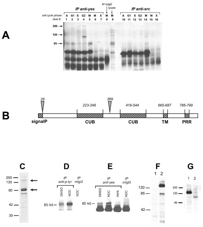
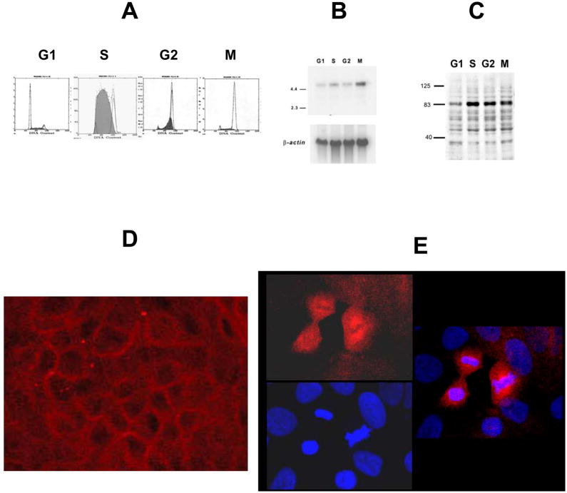
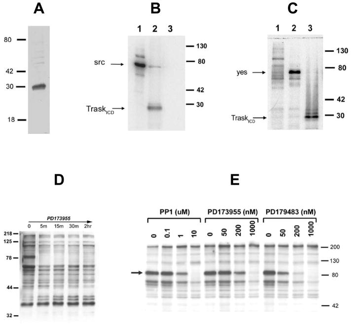
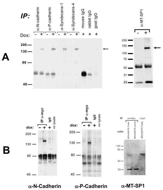
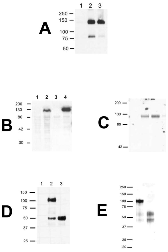
Src kinases are activated and relocalize to the cytoplasm during mitosis, but their mitotic function has remained elusive. We describe here a novel mitotic substrate of src kinases. Trask (transmembrane and associated with src kinases) is a 140 kDa type I transmembrane glycoprotein unrelated to currently known protein families. Src kinases phosphorylate Trask in vitro and mediate its mitotic hyperphosphorylation in vivo. Trask associates with both yes and src, is localized to the cell membrane during interphase, and undergoes cytoplasmic relocalization during mitosis. Overexpression of Trask leads to cell rounding and a loss of adhesion phenotype. Consistent with a function in cell adhesion, Trask interacts with a number of adhesion and matrix proteins including cadherins, syndecans, and the membrane-type serine protease 1 (MT-SP1), and is proteolytically cleaved by MT-SP1. Trask is unique among cell adhesion molecules in that it is under cell cycle regulation and thus links src kinases with the mitotic regulation of cell adhesion. This suggests a potential pathway by which hyperactive src kinases in tumors can deregulate adhesion signaling and mediate the metastatic phenotype.


Figures

Figure 1
Figure 1. Identification and characterization of an 85kd mitotic phosphoprotein associated with yes
A) An 85kd phosphoprotein was identified by anti-phosphotyrosine immunoblotting of anti-yes and anti-src immune complexes. Lanes correspond to asynchronous cells (1&10) or cells synchronized in G1 (2&11), S (3&12), G2(4&13), or M (5&14) phases of the cell cycle using lovastatin, aphidicolin, etoposide, and nocodazole respectively. Cells in lane 6 and 15 were blocked in mitosis by the src-selective tyrosine kinase inhibitor PD173955. Additional lanes include immunoprecipitates lacking cell lysates (7&16), or mitotic mIgG immunoprecipitates (8), or total lysate (9). B)The structural organization of Trask protein. Labels indicate the signal peptide (SignalP), two CUB domains, transmembrane domain (TM), and proline-rich region (PRR). Arrows indicate two known cleavage sites described in the text. C) Immunoblot of MDA-468 cells using anti-Trask 12F3 antibodies. D)Anti-phosphotyrosine immunoprecipitates from asynchronous and mitotic MDA-468 cells were immunoblotted with anti-Trask antibodies. E)Anti-yes immunoprecipitates from MDA-468 cells were immunoblotted with anti-Trask antibodies. Controls include immunoprecipitates from lysis buffer without cell lysate (3rdlane) and mIgG control (4thlane). F) MCF-7 cells were transfected with vector control (1) or pcDNA4-MycHis-Trask (2). An anti-myc immunoblot is shown here. Similar results were obtained by transfecting 293T or MDA-468 cells (not shown).G)MDA-468 cells were transiently transfected with pcDNA4-MycHis-Trask and treated with DMSO (1) or tunicamycin (2) for 24 hours. An anti-myc immunoblot is shown here.
Figure 2
Figure 2. Cellular expression of Trask
A) MDA-468 cells were synchronized in G1, S, G2, or M and cell cycle phase was verified. B)Northern blot analysis of polyA selected RNA from these cells was performed using a 2kb 32P labeled Trask cDNA probe. C) Immunoblot analysis of total lysates from these cells was performed using anti-Trask 12F3 monoclonal antibodies. D) Immunofluorescence microscopy of MCF10A breast epithelial cells using anti-Trask monoclonal antibodies labeled with Rhodamine-red. The majority of the cells show cell membrane staining as shown in this image. Mitotic cells were identified by their characteristic chromatin condensation and show cytoplasmic staining. A representative high power field is shown in image E.
Figure 3
Figure 3. Src kinases phosphorylate Trask in vitro and in vivo
A) The purity of recombinant Trask ICD was verified by SDS-PAGE separation and coomassie blue staining. B) In vitro src kinase reaction. Lanes 1–3 correspond to reactions containing src alone, src and TraskICD, and TraskICD alone. C) In vitro Yes kinase reaction. Lanes 1–3 correspond to reactions containing mIgG immunoprecipitates and TraskICD, α-yes immunoprecipitates alone, and α-yes immunoprecipitates and TraskICD. D)Asynchronous MDA-468 cells were treated with 1uM P173955 for the indicated times and lysates were immunoblotted using anti-phosphotyrosine antibodies. The 85 kd phosphotyrosine band indicated by the arrow is Trask, identified previously by purification. E)MDA-468 cells were treated for 20 hours with nocodazole, and mitotic shake-offs were washed and released into media containing the indicated concentrations of src-selective tyrosine kinase inhibitors for 30 minutes. Negative control lanes were treated with vehicle (DMSO) and are labeled as “0”. An anti-phosphotyrosine immunoblot is shown here. The 85kd band identified by the arrow is phosphorylated Trask, and disappears rapidly with increasing concentrations of src inhibitors.
Figure 4
Figure 4. Trask over-expression in MDA-468 cells
A) MDA-468TR cells were transfected with pcDNA4-TO-MycHis-Trask and stable transfectants (named MDA-468TR-Trask) selected in Zeocin. Inducible expression of the myc-tagged Trask protein is shown here by anti-myc immunoblots of 4 clones. Induction of expression of full length Trask cDNA results in expression of a 140kd full length Trask protein as well as an 85kd cleaved product. B)MDA-468TR-Trask cells were grown in the absence or presence of 100ng/ml doxycycline and inspected under inverted light microscopy. Induction with doxycycline results in cell rounding and continued growth in a loosely adherent semi-suspended state morphology. Clones 4 and 11 are shown here and are typical of all transfected clones. Doxycyline treatment of vector transfected controls has no visible effects on cell morphology (not shown).
Figure 5
Figure 5. Trask interactions with membrane and matrix proteins
A) MDA-468TR-Trask cells were induced with doxycyline to express Trask and cell lysates were immunoprecipitated with the indicated specific antibodies. Immune complexes were separated by SDS-PAGE and immunoblotted using anti-myc antibodies to detect the presence of myc-tagged Trask in the complexes. Controls include immune complexes using non-immune IgGs and immune complexes from un-induced cells that do not express the construct. B)The same experiments were performed in reverse order such that lysates were immunoprecipitated with anti-myc antibodies and immunoblotted with the indicated specific antibodies.
Figure 6
Figure 6. Trask cleavage by MT-SP1
A) MDA-468TR-Trask cells were left un-induced (lane 1) or induced to express Trask with 100ng/ml doxycycline (2–3). The induced cells were treated with no drug (2), or with the serine protease inhibitor 7uM ecotin (3) beginning 2 hours before doxycycline induction. 24 hours after doxycycline induction, cell lysates were harvested and immunoblotted using anti-myc antibodies. The expression of p85Trask is reduced by the protease inhibitor. B)293T cells were transfected with a myc/his-tagged TraskECD construct. Anti-myc immunoblots were performed to confirm the expression of TraskECD in cell lysates from controls (1) and transfectants (2), and the secretion of TraskECD in the media of controls (3) and transfectants (4). C) These transfected 293T cells were grown in serum free media and the soluble recombinant TraskECD was purified from conditioned media over a charged nickel column and dialyzed against PBS yielding a pure 120kd soluble TraskECD product shown here by coomassie staining. D)1ug rTraskECD was added to an in vitro protease reaction using recombinant catalytic domain of MT-SP1 (rMT-SP1CD). The mixture was incubated for 1hr at 37C in PBS, the reaction products were denatured, deglycosylated by PNGaseF treatment, separated on SDS-PAGE and immunoblotted using anti-myc antibodies. Lanes correspond to reactions containing rMT-SP1CD alone (1), rTraskECD alone (2), or rMT-SP1CD and rTraskECD (3). The rTraskECD construct is myc-tagged at the carboxy terminus, therefore these anti-myc immunoblots identify uncleaved ECD as well as the carboxy terminal cleavage product, but not the N-terminal cleavage product. E)Silver staining of rTraskECD before (1) and after (2) cleavage with rMT-SP1CD identifies both cleavage products. Our rTraskECD preparation from 293T cells contains some amount of cleaved Trask as seen in both the silver stain and the myc immunoblots here, however addition of rMT-SP1CD results in complete proteolysis. The precise site of the in vitro cleavage was determined by N-terminal sequencing of the reaction product, as described in the text.

References

    1. Bagrodia S, Chackalaparampil I, Kmiecik TE, Shalloway D. Nature. 1991;349:172–175. - PubMed
    1. Bagrodia S, laudano AP, Shalloway D. J Biol Chem. 1994;269:10247–10251. - PubMed
    1. Bhatt AS, Takeuchi T, Ylstra B, Ginzinger D, Albertson D, Shuman MA, Craik CS. Biological Chemistry. 2003;384:257–266. - PubMed
    1. Boyer B, Bourgeois Y, Poupon MF. Oncogene. 2002;21:2347–2356. - PubMed
    1. Brown TA, Yang TM, Zaitsevskaia T, Xia Y, Dunn CA, Sigle RO, Knudsen B, Carter WG. J Biol Chem. 2004;279:14772–14783. - PubMed

Publication types

Associated data