Review
. 2005 Jul;115(7):1688-98.
doi: 10.1172/JCI25669.

Entry of parainfluenza virus into cells as a target for interrupting childhood respiratory disease

Affiliations
Review

Entry of parainfluenza virus into cells as a target for interrupting childhood respiratory disease

Anne Moscona. J Clin Invest. 2005 Jul.

Abstract

Human parainfluenza viruses cause several serious respiratory diseases in children for which there is no effective prevention or therapy. Parainfluenza viruses initiate infection by binding to cell surface receptors and then, via coordinated action of the 2 viral surface glycoproteins, fuse directly with the cell membrane to release the viral replication machinery into the host cell's cytoplasm. During this process, the receptor-binding molecule must trigger the viral fusion protein to mediate fusion and entry of the virus into a cell. This review explores the binding and entry into cells of parainfluenza virus type 3, focusing on how the receptor-binding molecule triggers the fusion process. There are several steps during the process of binding, triggering, and fusion that are now understood at the molecular level, and each of these steps represents potential targets for interrupting infection.


Figures
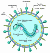
Figure 1
Figure 1
A schematic diagram of the parainfluenza virion. L, large RNA polymerase protein; M, matrix protein; NP, nucleocapsid protein; P, phosphoprotein. Modified with permission from New England Journal of Medicine (91).
Figure 2
Figure 2
A schematic illustration of the parainfluenza viral life cycle. RER, rough endoplasmic reticulum.
Figure 3
Figure 3
Model of class I fusion protein–mediated membrane fusion. (A and B) The trimeric paramyxovirus F protein contains 2 hydrophobic domains: the fusion peptide and the transmembrane-spanning domain. Each is adjacent to 1 of 2 HR regions, HR-N and HR-C. (B) The F protein binds to a receptor on the host cell membrane, causing a conformational change and subsequent insertion of the hydrophobic fusion peptide into the host cell membrane. (C) Multiple F protein trimers are believed to mediate the fusion process. Protein refolding occurs when the host and viral cell membranes bend toward each other (D), and formation of a hemifusion stalk allows the lipids on the outer part of the membranes to interact (E). (F) When protein refolding is completed, the fusion peptide and the transmembrane domain are antiparallel in the same membrane, which creates the most stable form of the fusion protein. Figure modified with permission from Nature (40).
Figure 4
Figure 4
Images of the active site of the HPIV3 HN protein complexed with sialic acid (A) and 4-GU-DANA (zanamivir) (B) (both shown in yellow). Figure modified with permission from the Journal of Molecular Biology (22).
Figure 5
Figure 5
Sites or steps within the viral life cycle that represent potential targets for antiviral molecules. (A) Agents directed at blocking the ability of the HN protein to recruit inflammatory cells to the lung and subsequent cytokine expression may reduce the inflammatory response to the HN protein and ameliorate disease. (B) Molecules that fit into the binding pocket on the HN globular head may inhibit HN-receptor binding and the subsequent F protein triggering action of the HN protein stalk. On the left, the HN protein is shown bound with an inhibitor, precluding the scenario shown on the right, in which the HN protein’s binding to the cell has led to its activation of F protein. (C) F protein peptides may be designed to prevent the refolding event that is essential to fusion during virus entry into the host cell. In addition, the F protein could perhaps be prematurely triggered and become incapacitated before it reaches the target host cell membrane. (D) Finally, the HN protein has NA activity and thus the ability to cleave the sialic acid moieties of the cellular receptors, promoting the release of new virions from the host cell surface. Specific inhibition of this activity may prevent virion entry into additional uninfected cells.

References

    1. Williams JV, et al. Human metapneumovirus and lower respiratory tract disease in otherwise healthy infants and children. N. Engl. J. Med. 2004;350:443–450. - PMC - PubMed
    1. Collins, P., Chanock, R., and McIntosh, K. 1996. Parainfluenza viruses. In Fields virology. B. Fields et al., editors. Lippincott-Raven Publishers. Philadelphia, Pennsylvania, USA. 1205–1241.
    1. Chanock RM. Control of pediatric viral diseases: past successes and future prospects [review] Pediatr. Res. 1990;27(Suppl.):S39–S43. - PubMed
    1. Murphy BR. Current approaches to the development of vaccines effective against parainfluenza viruses [review] Bull. World Health Organ. 1988;66:391–397. - PMC - PubMed
    1. Groothuis J, et al. Prophylactic administration of respiratory syncytial virus immune globulin to high-risk infants and young children. N. Engl. J. Med. 1993;329:1524–1530. - PubMed

Publication types

MeSH terms