. 2005 Jul;115(7):1743-55.
doi: 10.1172/JCI22593. Epub 2005 Jun 2.

Restoration of tubular epithelial cells during repair of the postischemic kidney occurs independently of bone marrow-derived stem cells

Affiliations

Restoration of tubular epithelial cells during repair of the postischemic kidney occurs independently of bone marrow-derived stem cells

Jeremy S Duffield et al. J Clin Invest. 2005 Jul.

Abstract

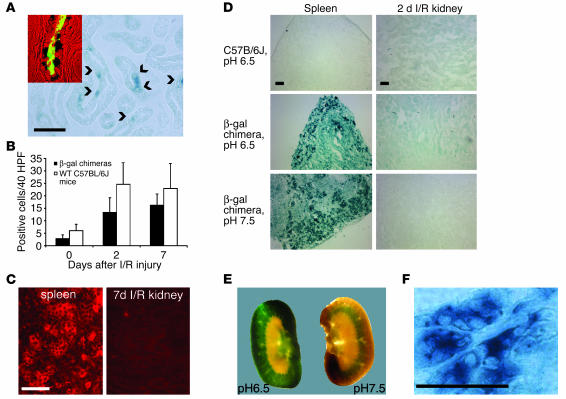
Ischemia causes kidney tubular cell damage and abnormal renal function. The kidney is capable of morphological restoration of tubules and recovery of function. Recently, it has been suggested that cells repopulating the ischemically injured tubule derive from bone marrow stem cells. We studied kidney repair in chimeric mice expressing GFP or bacterial beta-gal or harboring the male Y chromosome exclusively in bone marrow-derived cells. In GFP chimeras, some interstitial cells but not tubular cells expressed GFP after ischemic injury. More than 99% of those GFP interstitial cells were leukocytes. In female mice with male bone marrow, occasional tubular cells (0.06%) appeared to be positive for the Y chromosome, but deconvolution microscopy revealed these to be artifactual. In beta-gal chimeras, some tubular cells also appeared to express beta-gal as assessed by X-gal staining, but following suppression of endogenous (mammalian) beta-gal, no tubular cells could be found that stained with X-gal after ischemic injury. Whereas there was an absence of bone marrow-derived tubular cells, many tubular cells expressed proliferating cell nuclear antigen, which is reflective of a high proliferative rate of endogenous surviving tubular cells. Upon i.v. injection of bone marrow mesenchymal stromal cells, postischemic functional renal impairment was reduced, but there was no evidence of differentiation of these cells into tubular cells of the kidney. Thus, our data indicate that bone marrow-derived cells do not make a significant contribution to the restoration of epithelial integrity after an ischemic insult. It is likely that intrinsic tubular cell proliferation accounts for functionally significant replenishment of the tubular epithelium after ischemia.


Figures

Figure 1
Figure 1
Characterization of I/R injury in chimeric mice. (A) Phalloidin staining of actin cytoskeleton in sham-operated (control) kidneys and at 2, 7, and 21 days following I/R injury. Note the loss of the tubular cell brush border at 2 days and the denuded tubules (arrow) and the progressive restoration with time of both apical actin staining and tubule integrity. (B) PAS-stained sections at 2 and 7 days following I/R injury. Although the necrotic intratubular debris (arrows) is widespread at 2 days, the tubular architecture is significantly restored at 7 days. (C) PCNA staining of tubules in sham-operated (control) and ipsilateral kidney 2 days following I/R injury. (D) Outer medulla intratubular proliferation as assessed by counting mitotic cells in PAS-stained sections and by anti-PCNA immunofluorescence at 0, 2, and 7 days following I/R injury to the kidney. (E) Plasma creatinine levels at 0, 2, and 7 days following bilateral I/R renal injury. Scale bars: 50 μm.
Figure 2
Figure 2
Confirmation of chimerism in mice transplanted with bone marrow from EGFP-chimeric mice. (A) Top panels: Representative FACS analysis for EGFP fluorescence of bone marrow from wild-type mice (left) and a chimeric mouse at 6 weeks after bone marrow transfer (right). Bottom panels: FACS analysis for EGFP fluorescence of blood leukocytes from wild-type mice (left) and chimeric mice at 6 weeks (right). (B) Representative analysis of leukocytes from EGFP chimeras for EGFP fluorescence and expression of the myeloid lineage marker CD11b (left) or lymphoid lineage markers B220, CD4, and CD8 (right). Note that in each case, the majority of circulating leukocyte populations express EGFP at high levels. (C) Tissue sections of spleen and thymus in chimeric mice showing widespread EGFP fluorescence. Scale bar: 50 μm.
Figure 3
Figure 3
Tubular cells in EGFP chimeras do not express EGFP following I/R injury. Photomicrographs of corticomedullary regions of the contralateral kidney (A) and kidney 7 days after I/R in chimeric mice (B). Note bright EGFP in nontubular cells but only dull autofluorescence in tubular cells. (C) Photomicrograph of kidney from an EGFP donor mouse as positive control showing bright EGFP in tubular cells. Scale bar: 50 μm.
Figure 4
Figure 4
Bone marrow EGFP-expressing cells lack leukocyte markers and acquire characteristics of peritubular endothelial cells 7 days following I/R injury. (A) Fluorescence images of 7-day postischemic kidney showing interstitial cells expressing GFP but lacking CD45 (red) (arrowhead). T, tubule. (B) Confocal images of 7-day postischemic kidney from EGFP chimeras labeled with anti-CD31 antibodies (red). Note EGFP-positive cells coexpressing CD31 (arrowheads) in the 2D image. Also note that the endothelial cell nucleus expresses EGFP but not CD31, which is not expressed in nuclei (arrows). (C) Confocal images from 7-day postischemic kidney from EGFP chimeras labeled with antibodies against vWF (red). Note EGFP-positive cell coexpressing vWF in cytoplasmic granules (arrowhead) in the 2D image. Also note that the endothelial cell nucleus expresses EGFP but not vWF, which is not expressed in nuclei (arrows). (D) Quantification of bone marrow–derived cells expressing markers of endothelial cells through determination of the number of peritubular cells coexpressing EGFP and endothelial markers in contralateral (control) and 7-day postischemic kidneys. Data are presented as percent of vWF-positive or CD31-positive cells that express EGFP. Scale bars: 50 μm.
Figure 5
Figure 5
In female mice with male bone marrow, tubular cells are not derived from bone marrow cells following I/R injury to the kidney. (A) Fluorescent image of kidney outer medulla at day 15 following bilateral ischemic injury. The section was hybridized with an FITC-conjugated probe for the Y chromosome and counterstained with lotus lectin (red) highlighting proximal tubular cells and DAPI showing nuclei. Note that many interstitial cells (arrowheads) exhibit the Y chromosome, but none of the regenerated tubular cells stain for the Y chromosome. (B) Section of spleen showing that the majority of nuclei stain positively for the Y chromosome. (C) Detailed view of a proximal tubule, showing a tubular cell nucleus apparently containing a Y chromosome when viewed by epifluorescence. (D) An ultrathin deconvolution image through the same section as shown in C. The fluorescent label can be clearly seen to reside outside of the nucleus. (E and F) Sections hybridized with FITC-conjugated Y chromosome probe were counterstained with anti-vWF antibodies (red). (E) Peritubular capillary showing endothelial cell nucleus with Y chromosome. (F) Section of the image in E obtained by deconvolution microscopy confirming vWF staining and a nucleus containing the Y chromosome all in 1 plane. Scale bars: 50 μm.
Figure 6
Figure 6
Confirmation of chimerism in mice transplanted with β-gal–expressing bone marrow. (A) Fixed bone marrow cells stained blue with X-gal. (B) Percentage of bone marrow cells that stained blue with X-gal in control C57BL/6J mice and chimeric mice (n = 5 per time point) subjected to I/R followed by 2 days and 7 days of recovery. (C) Peritoneal cells as well as cells of thymus, spleen, and lung from chimeric mice all stained positively with X-gal. Scale bars: 50 μm.
Figure 7
Figure 7
X-gal stains tubular cells in the kidney of wild-type mice, reflecting endogenous β-gal activity, but can be distinguished from bacterial β-gal when high-pH X-gal staining or anti–β-gal antibodies are used. (A) Occasional tubular cells stained blue, indicating β-gal, 7 days following I/R injury in chimeric mice (arrowheads). X-gal–stained cells colabeled with the proximal tubular cell marker gp330, seen as green fluorescence (inset). (B) The number of X-gal–positive cortical and outer medullary tubular cells per 40 HPF in contralateral kidneys (0 days) and in kidneys of both chimeric and wild-type C57BL/6J mice following I/R injury. Note that I/R resulted in an increase in the number of β-gal–stained cells in both nonchimeric and chimeric mice. (C) Sections at day 7 after I/R labeled with anti–β-gal antibodies. Note the bright staining in spleen cells (left) but no staining in regenerated tubules (right) (D) Sections (magnification, ×100) of I/R kidneys and spleen from wild-type (n = 3 per time point) and chimeric mice (n = 5 per time point) stained with X-gal solution at pH 6.5 or 7.5. Note faint blue tubules in kidneys of wild-type and chimeric animals (compare top and middle panels). Blue staining in kidney tubules but not spleen is suppressed using X-gal in solution at pH 7.5 in chimeric mice (compare bottom and middle panels). (E) Sagittal sections (0.2 mm) of normal C57BL/6J kidney stained with X-gal solution at pH 6.5 (left) and pH 7.5 (right). Note widespread cortical and outer medullary staining at low pH and milder restricted staining in the outer medulla at higher pH. (F) Kidney section (magnification, ×400) from LacZ donor mouse stained with X-gal, pH 7.5. Note intense blue staining of tubules indicative of bacterial β-gal. Scale bars: 50 μm.
Figure 8
Figure 8
Bone marrow MSCs have multilineage potential and are protective in ischemic injury without differentiating into tubule cells. (A) Flow cytometry of MSCs for cell surface stem cell markers. Compared with isotype control labeling, which was used to define the marker (data not shown), MSCs lacked expression of c-kit and CD31 but expressed CD34 and Sca-1 (values are expressed as percent positive compared with isotype control antibody labeling). (B) Photomicrograph of MSCs differentiated into capillary-like structures (left). The same structure shows strong immunofluorescent labeling with the endothelial marker CD31 (right). (C) Photomicrograph of MSCs differentiated in vitro into adipocytes and labeled with oil red O for lipid. (D) Fluorescence micrograph showing an EGFP-expressing MSC in the cortex of the postischemic kidney 2 hours after intraparenchymal injection of EGFP-labeled MSCs. (E) Plasma creatinine levels 24 hours after 30-minute bilateral I/R renal injury followed by i.v. injection of control PBS or 0.5 × 10⁶ MSCs cultured on plastic (n = 4 per group). (F) Plasma creatinine levels 24 hours after 30-minute bilateral I/R renal injury followed by i.v. injection of control PBS, 0.5 × 10⁶ MSCs cultured on Matrigel, or embryonic fibroblasts (Fibro) cultured on the same matrix. Note that the level of creatinine was significantly higher in PBS-treated mice (n = 7 per group; **P < 0.01, ANOVA). Scale bars: 50 μm.
Figure 9
Figure 9
The proportion of lin blood leukocytes does not increase following I/R injury. Flow cytometry plots (representative of n = 6) of blood leukocytes from healthy mice (left) or mice 2 days following bilateral renal ischemia (right), labeled with PE-fluorescent antibodies against lineage markers (Lin-PE) (CD11b, Gr-1, CD4, CD8, B220, Ter-119) or IgG control antibodies. Note that a minority (<1%) of leukocytes were lin in control mice, and this percentage was not increased in mice 2 days following I/R injury.

Comment in

References

    1. Thadhani R, Pascual M, Bonventre JV. Acute renal failure. N. Engl. J. Med. 1996;334:1448–1460. - PubMed
    1. Zuk A, Bonventre JV, Brown D, Matlin KS. Polarity, integrin, and extracellular matrix dynamics in the postischemic rat kidney. Am. J. Physiol. 1998;275:C711–C731. - PubMed
    1. Witzgall R, Brown D, Schwarz C, Bonventre JV. Localization of proliferating cell nuclear antigen, vimentin, c-Fos, and clusterin in the postischemic kidney. Evidence for a heterogenous genetic response among nephron segments, and a large pool of mitotically active and dedifferentiated cells. J. Clin. Invest. 1994;93:2175–2188. - PMC - PubMed
    1. Witzgall R, et al. Kid-1 expression is high in differentiated renal proximal tubule cells and suppressed in cyst epithelia. Am. J. Physiol. 1998;275:F928–F937. - PubMed
    1. Sutton TA, Fisher CJ, Molitoris BA. Microvascular endothelial injury and dysfunction during ischemic acute renal failure. Kidney Int. 2002;62:1539–1549. - PubMed

Publication types

MeSH terms

Substances