. 2005 Jul;115(7):1756-64.
doi: 10.1172/JCI23015.

Intrarenal cells, not bone marrow-derived cells, are the major source for regeneration in postischemic kidney

Affiliations

Intrarenal cells, not bone marrow-derived cells, are the major source for regeneration in postischemic kidney

Fangming Lin et al. J Clin Invest. 2005 Jul.

Abstract

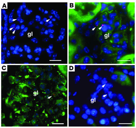
Ischemic injury to the kidney produces acute tubular necrosis and apoptosis followed by tubular regeneration and recovery of renal function. Although mitotic cells are present in the tubules of postischemic kidneys, the origins of the proliferating cells are not known. Bone marrow cells (BMCs) can differentiate across lineages to repair injured organs, including the kidney. However, the relative contribution of intrarenal cells and extrarenal cells to kidney regeneration is not clear. We produced transgenic mice that expressed enhanced GFP (EGFP) specifically and permanently in mature renal tubular epithelial cells. Following ischemia/reperfusion injury (IRI), EGFP-positive cells showed incorporation of BrdU and expression of vimentin, which provides direct evidence that the cells composing regenerating tubules are derived from renal tubular epithelial cells. In BMC-transplanted mice, 89% of proliferating epithelial cells originated from host cells, and 11% originated from donor BMCs. Twenty-eight days after IRI, the kidneys contained 8% donor-derived cells, of which 8.4% were epithelial cells, 10.6% were glomerular cells, and 81% were interstitial cells. No renal functional improvement was observed in mice that were transplanted with exogenous BMCs. These results show that intrarenal cells are the main source of renal repair, and a single injection of BMCs does not make a significant contribution to renal functional or structural recovery.


Figures

Figure 1
Figure 1
Genetic marking of renal tubular epithelial cells with EGFP in creksp;Z/EG mice. (A) Expression of native EGFP fluorescence in frozen section of the kidney. co, cortex; me, medulla. The inset shows that green fluorescence is absent in the kidney of a wild-type littermate photographed under the identical exposure conditions. (BE) Paraffin sections of the kidney stained with anti-EGFP antibody (green) and antibodies or lectins that stain specific nephron segments (red) showing the presence of EGFP in LTA-positive proximal tubules (pt, B); THP-positive thick ascending limbs (tal, C); TSC-positive distal tubules (dt, D); and DBA-positive collecting ducts (cd, E). (F) Absence of EGFP in the glomeruli (gl) and interstitium (int). The nuclei were counterstained with DAPI, and images were merged in (BF) Scale bars: 400 μm in A and 20 μm in BF.
Figure 2
Figure 2
Tubular injury and cell proliferation in EGFP-expressing tubular epithelial cells. PAS staining and immunostaining were performed in the kidney sections of creksp;Z/EG mice with renal IRI. (A) PAS staining of the kidney at 2 days after IRI. Tubular injury is shown by the loss of brush border membrane, cell detachment from the basement membrane, and nuclear condensation in some cells (arrows). (B) Expression of PCNA in tubular cells (red, arrows). (C) Low-power image of BrdU incorporation in renal tubules. Some BrdU-containing cells (red, arrows) colocalized with EGFP-expressing cells (green). The arrowhead indicates BrdU incorporation in an EGFP-negative cell. (D) BrdU incorporation (red, arrow) in the epithelial cells expressing EGFP (green). The nuclei were counterstained with DAPI, and images were merged (BD). Scale bars: 20 μm.
Figure 3
Figure 3
Dedifferentiation of epithelial cells during repair of renal tubules. Immunostaining was performed in kidney sections of creksp;Z/EG mice with renal IRI. (A) Expression of Pax2 (blue, arrows) in LTA-positive (green) proximal tubules. Note the nuclear localization of Pax2 in the injured area lacking brush border staining of LTA. The same tubule also contains cells that incorporated BrdU (red, arrowhead). (B) Downregulation of p21 (red) in cells expressing Pax2 (blue, arrows). (C) Expression of vimentin (red) in LTA-positive (green) proximal tubule. (D) Expression of vimentin (red) in epithelial cells expressing EGFP (green). Cells coexpressing vimentin and EGFP appear yellow. (E) An EGFP-expressing cell (green, arrow) shows incorporation of BrdU (red) and basolateral expression of AQP3 (blue). (F) Cells that incorporated BrdU (green) established intercellular junction and expressed ZO-1 (red, arrows). The nuclei were counterstained with DAPI (C, D, and F), and images were merged. Scale bar: 20 μm.
Figure 4
Figure 4
Effects of BMCs on tubular regeneration (AH) and renal function (I). (A) Abundant Y chromosome signals located at the periphery of the nuclei in the kidneys of male mice (red, arrows), consistent with the typical nuclear localization of the Y chromosome. The sensitivity of FISH analysis was 64% (n = 5). (B) Kidney of a female mouse without male BMC transplant shows complete absence of Y chromosome signal. (C) A tubular cell that incorporated BrdU (green) shows the presence of Y chromosome (red, arrow). (DF) Y chromosome FISH (red) was followed by immunostaining of tubular epithelial cell markers (green). (D) The arrow indicates a Y+ cytokeratin+ tubular epithelial cell. (E) The arrow indicates a Y+ LTA+ proximal tubular cell. (F) The arrow indicates a Y+ AQP3+ collecting duct cell. Note the basolateral staining of the AQP3, which defines the intratubular localization of the Y+ cells. (G) Y chromosome–containing tubular cell (arrows) in the kidney of a wild-type female mouse injected with BMCs from a male creksp;Z/EG donor. Arrowheads indicate interstitial cells. (H) The same cell shown in G is negative for EGFP by immunostaining (arrow). The nuclei were counterstained with DAPI (A, B, and DH), and images were merged. Scale bars: 20 μm. (I) BUN levels in mice with sham operation that did not receive BMC injection (Control; filled triangles), mice with renal IRI that did not receive BMC injection (IRI – BMC; open circles), and mice with renal IRI that received BMC injection (IRI + BMC; filled circles). No statistically significant difference was observed in mice with or without BMC injection. n = 5–13 at each time point; total 84 mice.
Figure 5
Figure 5
Formation of renal cells from bone marrow–derived cells. The total of 8% of bone marrow–derived cells in the kidney consisted of tubular epithelial cell, glomerular cells, and interstitial cells. The relative percentages of different cell types are shown.
Figure 6
Figure 6
Formation of glomerular cells from BMCs. Y chromosome FISH (red) was followed by immunostaining for markers of glomerular cells. (A) Arrows indicate Y+ CD45 cells in the glomerulus. (B) The arrow indicates a Y+ LEL+ endothelial cell. LEL is labeled green. The arrowhead indicates a Y+ LEL cell. (C) The arrow indicates a Y+ vimentin+Wt1 mesangial cell. Wt1 is labeled blue in the nuclei. (D) The arrow indicates Y+ Wt1+ podocyte. Wt1 is labeled green in the nuclei. The arrowhead indicates a Y+ Wt1 cell. The nuclei were counterstained with DAPI (A, B, and D), and images were merged. Scale bars: 20 μm.
Figure 7
Figure 7
Formation of interstitial cells from BMCs. (A) Y chromosome FISH shows abundant Y+ cells (red, arrows) in the interstitium. The inset shows a Y+ CD45+ cell detected in the interstitium. (B) The arrow indicates a Y+ α-SMA+ myofibroblast. (C) Trichrome stain shows prominent collagenous deposit and fibrosis in the kidney of mouse with IRI that received BMC injection. (D) Trichrome staining shows minimal collagenous formation in the kidney of mouse with IRI that did not receive BMC injection. The nuclei were counterstained with DAPI (A and B), and images were merged. Scale bars: 20 μm.

Comment in

  • J Clin Invest. 115:1705.

References

    1. Bonventre JV. Mechanisms of ischemic acute renal failure. Kidney Int. 1993;43:1160–1178. - PubMed
    1. Savill J. Apoptosis and the kidney. J. Am. Soc. Nephrol. 1994;5:12–21. - PubMed
    1. Witzgall R, Brown D, Schwarz C, Bonventre JV. Localization of proliferating cell nuclear antigen, vimentin, c-Fos, and clusterin in the postischemic kidney. Evidence for a heterogenous genetic response among nephron segments, and a large pool of mitotically active and dedifferentiated cells. J. Clin. Invest. 1994;93:2175–2188. - PMC - PubMed
    1. Weissman IL. Stem cells: units of development, units of regeneration, and units in evolution [review] Cell. 2000;100:157–168. - PubMed
    1. Metcalf D. Stem cells, pre-progenitor cells and lineage-committed cells: are our dogmas correct [review]? Ann. N. Y. Acad. Sci. 1999;872:289–303; discussion 303–304. - PubMed

Publication types

MeSH terms

Substances