. 2005 Jul;115(7):1777-84.
doi: 10.1172/JCI24834.

Mutations in lipid transporter ABCA12 in harlequin ichthyosis and functional recovery by corrective gene transfer

Affiliations

Mutations in lipid transporter ABCA12 in harlequin ichthyosis and functional recovery by corrective gene transfer

Masashi Akiyama et al. J Clin Invest. 2005 Jul.

Abstract

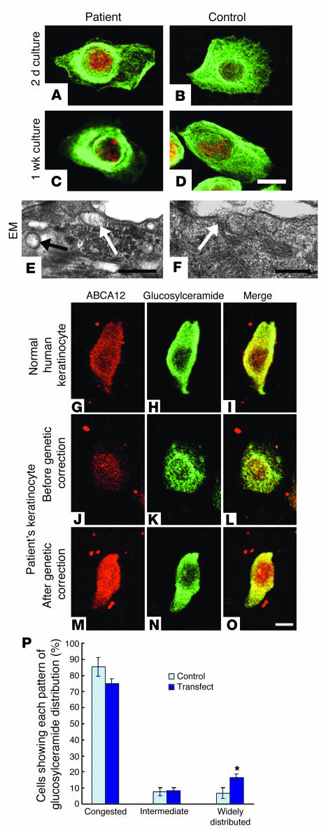
Harlequin ichthyosis (HI) is a devastating skin disorder with an unknown underlying cause. Abnormal keratinocyte lamellar granules (LGs) are a hallmark of HI skin. ABCA12 is a member of the ATP-binding cassette transporter family, and members of the ABCA subfamily are known to have closely related functions as lipid transporters. ABCA3 is involved in lipid secretion via LGs from alveolar type II cells, and missense mutations in ABCA12 have been reported to cause lamellar ichthyosis type 2, a milder form of ichthyosis. Therefore, we hypothesized that HI might be caused by mutations that lead to serious ABCA12 defects. We identify 5 distinct ABCA12 mutations, either in a compound heterozygous or homozygous state, in patients from 4 HI families. All the mutations resulted in truncation or deletion of highly conserved regions of ABCA12. Immunoelectron microscopy revealed that ABCA12 localized to LGs in normal epidermal keratinocytes. We confirmed that ABCA12 defects cause congested lipid secretion in cultured HI keratinocytes and succeeded in obtaining the recovery of LG lipid secretion after corrective gene transfer of ABCA12. We concluded that ABCA12 works as an epidermal keratinocyte lipid transporter and that defective ABCA12 results in a loss of the skin lipid barrier, leading to HI. Our findings not only allow DNA-based early prenatal diagnosis but also suggest the possibility of gene therapy for HI.


Figures

Figure 1
Figure 1
Clinical features of HI patients. (A) Patient 1 from family A harboring a homozygous mutation IVS23-2A→G in ABCA12. (B) Patient 2 from family B with compound heterozygous ABCA12 mutations, IVS23-2A→G and 5848C→T (R1950X). (C) Patient 3 (family C) carrying compound heterozygous ABCA12 mutations, 2021_2022del AA and 4158_4160delTAC (T1387del). (D) An affected fetus from family C aborted at 23 weeks’ gestation showed no serious symptoms, although some abnormal keratinization was observed mainly on the cheeks and the perioral area.
Figure 2
Figure 2
Localization and structure of ABCA12 protein and the sites of HI mutations. (A) ABCA12 protein (green) was localized in the cytoplasm of the upper epidermal keratinocytes (arrows) in healthy skin. (B) No ABCA12 immunolabeling was seen in the epidermis of patient 4. (C) Weak ABCA12 staining (arrows) was observed in the epidermis of patient 1. Asterisks indicate nonspecific staining in the stratum corneum. Red, nuclear counterstain. Scale bar: 10 μm. (DF) By immunoelectron microscopy, ABCA12 protein (5 nm gold particles) was restricted to LGs (arrows) in the upper epidermal cell (D). Lamellar structures were apparent in some ABCA12-positive LGs (E). ABCA12-positive LGs (arrows) were observed close to the keratinocyte-cell membrane (white circles) and they fused with it to secrete their contents into the intercellular space (F). Scale bars: 0.2 μm. (G) Model of ABCA12 function in the skin. ABCA12 transports lipid into the LG, and ABCA12-positive LGs fuse with the cell membrane to secrete lipid into extracellular space to form the intercellular lipid layer. (H) Structure of ABCA12 protein and the 5 mutation sites (red arrows) in HI families. Dark-blue area, cell membrane; bottom of dark-blue area, cytoplasmic surface.
Figure 3
Figure 3
Families with HI and ABCA12 mutations. (A) Patient 1 from family A was a homozygote for the mutation IVS23-2A→G, and both his parents were heterozygous carriers. (B) Patient 2 from family B was a compound heterozygote for the mutations IVS23-2A→G and 5848C→T (R1950X). (C) Patient 3 from family C was a compound heterozygote for the mutations 2021_2022delAA and 4158_4160delTAC (T1387del). (D) Patient 4 from family D was a homozygote for the mutation 1300C→T (R434X), and her 2 parents were heterozygous carriers.
Figure 4
Figure 4
Verification of splice-site mutation IVS23-2A→G and conservation of residues deleted by mutations IVS23-2A→G and 4158_4160delTAC (T1387del). (A) RT-PCR analysis of mRNA fragments around the exon 23–24 boundary indicated that keratinocytes from patient 1 (lane P) showed 2 different mutant transcripts, 674 bp and 513 bp, which were shorter than the control transcript (683 bp) from healthy human keratinocytes (lane C). Lane M, markers. (B) Sequencing of the mutant transcripts and the control transcript revealed that 9 nucleotides and 170 nucleotides were deleted in mutant transcripts. (C) A 9-nucleotide deletion resulted in the loss of 3 amino acids from the N terminal sequence of exon 24 (Y1099_K1101del), and the 170-nucleotide deletion led to a frameshift. (D) ABCA12 amino acid sequence alignment shows the level of conservation in diverse species of the amino acids, Y1099_K1101 and T1387 (red characters), which were deleted by mutations in HI families. Asterisks indicate ABC (abt-4).
Figure 5
Figure 5
Extremely thick stratum corneum and severe disruption of the secretion of LGs in the ABCA12-deficient skin of the present series of HI patients. (A) Strikingly thick stratum corneum (SC; double arrow) in the patient's skin. (B) Control epidermis showing normal, stratum corneum (arrow). (C) By electron microscopy, LG secretion was disturbed, and many abnormal immature (lacking proper lamellar structures) LGs (arrows) were observed in the keratinocytes. (D) In control skin, LGs (arrows) were distributed in a gradually increasing pattern toward the plasma membrane. (E) Abnormal HI LGs (arrows) were localized close to the cell membrane, but not secreted. (F) LGs were secreted into the extracellular space (arrows). (G) Patient's epidermis including stratum corneum (arrows) showed diffuse staining for glucosylceramide (green), a lipid component of LGs. (H) Glucosylceramide staining (green) was restricted and intense in the stratum corneum (arrows) of normal skin. Red, nuclear stain. Scale bars: 50 mm (A, B, G, H); 1 mm (C, D); 0.5 mm (E, F).
Figure 6
Figure 6
Cultured HI keratinocytes carrying ABCA12 mutations showed abnormal congestion of lipid, and this phenotype was recovered by corrective ABCA12 gene transfer. (AD) HI keratinocytes cultured in high Ca2+ conditions showed that glucosylceramide, a major component of LG lipid, was distributed densely around the nuclei (in a congested pattern) (green, FITC). Control normal human keratinocytes showed a widely distributed, diffuse glucosylceramide staining pattern. (E) Electron microscopic (EM) observation revealed, in cultured HI keratinocytes, that apparently amorphous, electron lucent LG-like structures (arrows) formed, but were not secreted. (F) Normal secretion of LG contents (arrow) in a control keratinocyte. (GO) Before genetic correction, an HI patient cell showed weak ABCA12 immunostaining (red, TRITC) and a congested pattern of glucosylceramide staining (green, FITC) (JL). After genetic correction, an HI patient cell demonstrated stronger ABCA12 labeling (red) and a normal distribution pattern of glucosylceramide (green) (MO), similar to those of a normal human keratinocyte (GI). (P) Corrective gene transfer resulted in a statistically significant increase in the number of cells with completely normal, widely distributed glucosylceramide patterns. *P < 0.02, Student's t test. Scale bars: 10 mm (AD, GO); 0.5 mm (E, F).

Comment in

References

    1. Williams ML, Elias PM. Genetically transmitted, generalized disorders of cornification; the ichthyoses. Dermatol. Clin. 1987;5:155–178. - PubMed
    1. Judge, M.R., McLean, W.H.I., and Munro, C.S. 2004. Disorders of keratinization. In Rook’s textbook of dermatology. 7th edition. T. Burns, S. Breathnach, N. Cox, and C. Griffiths, editors. Blackwell Publishers. Oxford, United Kingdom. 34.1–34.111.
    1. Allikmets R, Gerrard B, Hutchinson A, Dean M. Characterization of the human ABC superfamily: isolation and mapping of 21 new genes using the expressed sequence tags database. Hum. Mol. Genet. 1996;5:1649–1655. - PubMed
    1. Dean M, Rzhetsky A, Allikmets R. The human ATP-binding cassette (ABC) transporter superfamily. Genome Res. 2001;11:1156–1166. - PubMed
    1. Borst P, Elferink RO. Mammalian ABC transporters in health and disease. Annu. Rev. Biochem. 2002;71:537–592. - PubMed

Publication types

MeSH terms

Substances