. 2005 Jul;115(7):1942-52.
doi: 10.1172/JCI24354.

Hypertension from targeted ablation of chromogranin A can be rescued by the human ortholog

Affiliations

Hypertension from targeted ablation of chromogranin A can be rescued by the human ortholog

Nitish R Mahapatra et al. J Clin Invest. 2005 Jul.

Abstract

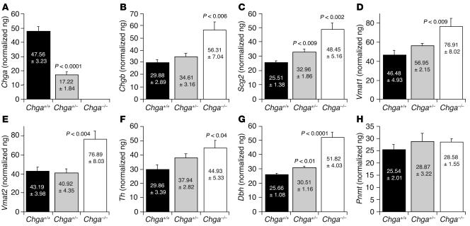
The secretory prohormone chromogranin A (CHGA) is overexpressed in essential hypertension, a complex trait with genetic predisposition, while its catecholamine release-inhibitory fragment catestatin is diminished, and low catestatin predicts augmented adrenergic pressor responses. These findings from studies on humans suggest a mechanism whereby diminished catestatin might increase the risk for hypertension. We generated Chga and humanized mice through transgenic insertion of a human CHGA haplotype in order to probe CHGA and catestatin in vivo. Chga mice displayed extreme phenotypic changes, including: (a) decreased chromaffin granule size and number; (b) elevated BP; (c) loss of diurnal BP variation; (d) increased left ventricular mass and cavity dimensions; (e) decreased adrenal catecholamine, neuropeptide Y (Npy), and ATP contents; (f) increased catecholamine/ATP ratio in the chromaffin granule; and (g) increased plasma catecholamine and Npy levels. Rescue of elevated BP to normalcy was achieved by either exogenous catestatin replacement or humanization of Chga mice. Loss of the physiological "brake" catestatin in Chga mice coupled with dysregulation of transmitter storage and release may act in concert to alter autonomic control of the circulation in vivo, eventuating in hypertension.


Figures

Figure 1
Figure 1
Chga gene targeting. (A) Chga wild-type gene structure. Chga from the isogenic mouse 129/SvJ strain showing the positions of wild-type Chga’s 8 coding exons, as well as restriction sites and the locations of the 3 Chga segments (arms) used to construct the targeting vector. Also shown is an undisrupted, wild-type ˜13-kbp diagnostic BamHI (asterisk) fragment, spanning the promoter, exons 13, and corresponding introns. (B) Structure of the homologously targeted Chga gene. A Southern blot probe (˜1.3 kb BamHI/XhoI fragment) detected a ˜7.3 kbp BamHI (asterisks) fragment after homologous recombination. (C) Southern blot results. While the probe (B; ˜1.3 kbp BamHI/XhoI fragment) detected only a ˜13 kbp fragment in wild-type DNA (A), it detected both ˜13-kbp (wild-type) and ˜7.3-kbp (targeted) BamHI fragments in ES cell genomic DNA after homologous recombination disrupted 1 Chga allele. Lane 1: 1-kbp extension ladder; lane 2: wild-type ES cells (13-kbp band only); lane 3: clone B7 ES cells with homologous recombination in 1 Chga allele; lane 4: clone C5 cells with homologous recombination in 1 Chga allele. (D) Chga gene after homologous integration of the targeting construct into mouse genomic DNA (Chga locus on mouse chromosome 12), followed by Cre-loxP–mediated recombination (type I deletion, between the first and third loxP sites; dashed line). The triangles represent loxP recognition sites. Asterisks indicate BamHI sites flanking the diagnostic 7.3-kbp BamHI fragment.
Figure 2
Figure 2
Verification of Chga, Chgb, and Dbh protein expression in vivo. Immunoblot analysis of Chga, Chgb, and Dbh proteins in adrenal gland homogenates from wild-type (Chga+/+; n = 4) and knockout (Chga–/–; n = 4) mice. Ten micrograms of adrenal protein was loaded in each lane. (A) Chga (goat polyclonal antiserum). (B) Chgb (goat polyclonal antiserum). (C) Dbh (rabbit polyclonal antiserum). As a control, expression of β-actin (goat polyclonal) was measured in each sample.
Figure 3
Figure 3
Expression of other chromaffin cell genes. Quantitative RT-PCR analysis for mRNA expression in adrenal glands from Chga+/+ (n = 6), Chga+/– (n = 6) and Chga–/– (n = 6) mice. Chromogranins/secretogranins (chromaffin granule soluble proteins): (A) Chga (n = 6), (B) Chgb (n = 6), and (C) Scg2 (n = 6). Chromaffin granule membrane proteins: (D) Vmat1 (n = 6) and (E) Vmat2 (n = 6). Catecholamine biosynthetic enzymes: (F) Th (n = 6), (G) Dbh (n = 6), and (H) Pnmt (n = 6). Five nanograms cDNA was used for each probe. Five nanograms adrenal cDNA was used for each amplification. Results were normalized to 18S ribosomal RNA (rRNA) amplified from the same sample.
Figure 4
Figure 4
Microscopic chromaffin cell anatomy. Transmission electron micrograph of the mouse adrenal medulla showing chromaffin granules (arrows) in Chga+/+ and Chga–/– mice. For each group (Chga+/+ and Chga–/–), we evaluated randomly selected chromaffin cells from at least 100 micrographs (>15/mouse). M, mitochondria. Scale bar: 0.5 μm. In the Chga–/– chromaffin cells, note the decreased size, number, and electron density of the granules.
Figure 5
Figure 5
Biochemical consequences in Chga–/– mice. (A). Catecholamine (CA) contents in the adrenal gland of Chga+/+ (n = 6), Chga+/– (n = 6), and Chga–/– (n = 6) mice as measured by a sensitive radioenzymatic assay. (B) Catecholamine concentrations in plasma of wild-type (n = 6), heterozygous (n = 6), and Chga-null (n = 6) mice as measured by sensitive radioenzymatic assay. (C) Adrenal NPY level in wild-type (n = 6) and Chga-null (n = 6) mice, as measured by Npy enzyme immunoassay. (D) Plasma Npy concentration in wild-type (n = 5) and Chga-null (n = 5) mice, as measured by Npy enzyme immunoassay. (E) Adrenal ATP level in wild-type (n = 10) and Chga-null (n = 11) mice, as measured by ATP luminescence assay. (F) Adrenal catecholamine/ATP molar ratios. n = 6 adrenal glands/condition. (G) Adrenal corticosterone level in wild-type (n = 12) and Chga-null (n = 12) mice, as measured by corticosterone enzyme immunoassay.
Figure 6
Figure 6
Autonomic/cardiovascular physiology. (A) SBP measured by the tail-cuff method in Chga+/+ (n = 8), Chga+/– (n = 14), and Chga–/– (n = 8) conscious male mice during restraint at 5–6 months of age. (B) Continuous telemetric data from unrestrained mice on SBP and DBP over a 24-hour period in Chga+/+ (n = 5) and Chga–/– (n = 7) mice. (C) Telemetric data on DBP over a 24-hour period in Chga+/+ (n = 5) and Chga–/– (n = 7) mice. (D) Rescue from elevated SBP by the CHGA catecholamine release–inhibitory fragment catestatin: exaggerated SBP fall in Chga–/– mice. SBP was monitored by telemetry before and after administration of catestatin (20 nmol/25 g body weight, i.p.) at time 0 in Chga–/– (n = 4) and Chga+/+ (n = 4) mice. Results were analyzed by 2-way, repeated-measures ANOVA, evaluating the effects of time (F = 359.9; P < 0.001), mouse strain (Chga+/+ vs. Chga–/–; F = 14.8; P = 0.009), or strain/peptide interaction (F = 79.4; P < 0.001). (E) Transthoracic echocardiography in Chga+/+ (n = 8) and Chga–/– (n = 8) mice. LVIDd, LV dimension at end diastole; LVIDs, LV dimension at end systole; IVSd, interventricular septal thickness at end diastole; IVSs, interventricular septal thickness at end systole; LVPWd, LV posterior wall thickness at end diastole; LVPWs, LV posterior wall thickness at end systole.
Figure 7
Figure 7
Characterization of the humanized CHGA mice. (A) Schematic representation of the BAC clone and founder line showing complete integration of the transgene. The BAC clone RP11-862G15, a component of the human 14 contig NT_026437 spanning the locus between 87,162,128 and 87,372,996, has the complete CHGA (12,150 bp) gene flanked by native human sequences, 44,068 bp upstream and 154,651 bp downstream. Founder line no. 31 has the complete 210-kb insert fragment of RP11-862G15 stably integrated in its genome. (B) RT-PCR analysis of the cDNA prepared from mouse derived from founder no. 31. The expression of both the mouse Chga and human CHGA genes was analyzed in various tissues. The tissues analyzed include adrenal gland, pancreas, pituitary, ovary, olfactory bulb, frontal cortex, striatum, hippocampus, thalamus, hypothalamus, brainstem, cerebellum, liver, and spleen. The experiment was repeated twice, and consistent results were obtained. (C) Rescue of SBP in mice humanized at the CHGA locus. SBP measured by the tail-cuff method in wild-type (Chga+/+, n = 8), null (Chga–/–, n = 8), and humanized (CHGA+/+Chga–/–, n = 6) conscious mice during restraint at 5–6 months of age.

Comment in

References

    1. Winkler H, Fischer-Colbrie R. The chromogranins A and B: the first 25 years and future perspectives. Neuroscience. 1992;49:497–528. - PMC - PubMed
    1. Taupenot L, Harper KL, O’Connor DT. Mechanisms of disease: the chromogranin-secretogranin family. N. Engl. J. Med. 2003;348:1134–1149. - PubMed
    1. Takiyyuddin MA, et al. Is physiologic sympathoadrenal catecholamine release exocytotic in humans? Circulation. 1990;81:185–195. - PubMed
    1. Winkler H, Apps DK, Fischer-Colbrie R. The molecular function of adrenal chromaffin granules: established facts and unresolved topics. Neuroscience. 1986;18:261–290. - PubMed
    1. Videen JS, Mezger MS, Chang YM, O’Connor DT. Calcium and catecholamine interactions with adrenal chromogranins. Comparison of driving forces in binding and aggregation. J. Biol. Chem. 1992;267:3066–3073. - PubMed

Publication types

MeSH terms