. 2005 Jul 18;202(2):249-59.
doi: 10.1084/jem.20041685. Epub 2005 Jul 11.

Role of membrane sphingomyelin and ceramide in platform formation for Fas-mediated apoptosis

Affiliations

Role of membrane sphingomyelin and ceramide in platform formation for Fas-mediated apoptosis

Michihiko Miyaji et al. J Exp Med. .

Abstract

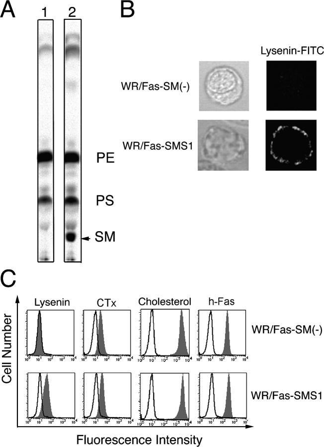
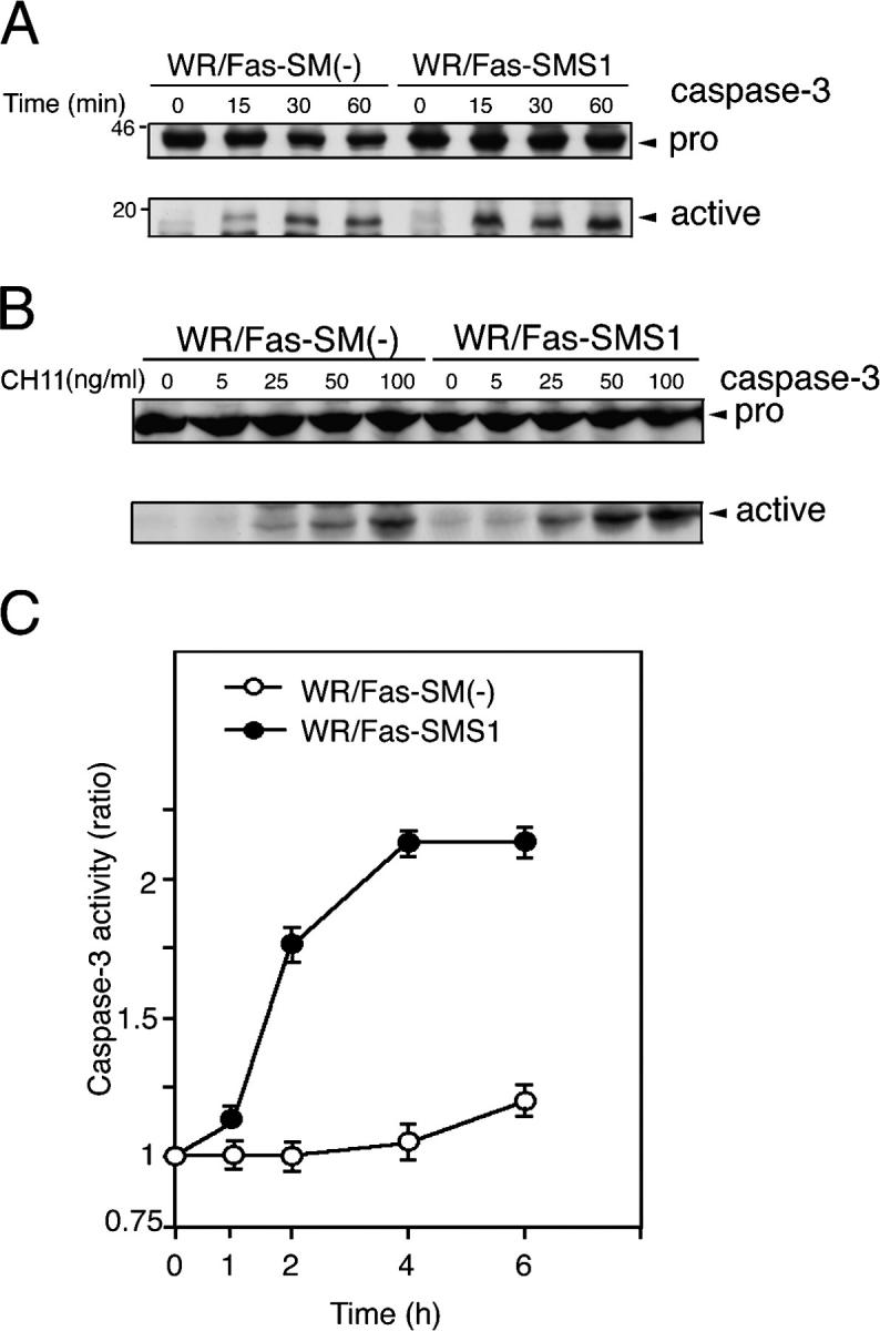
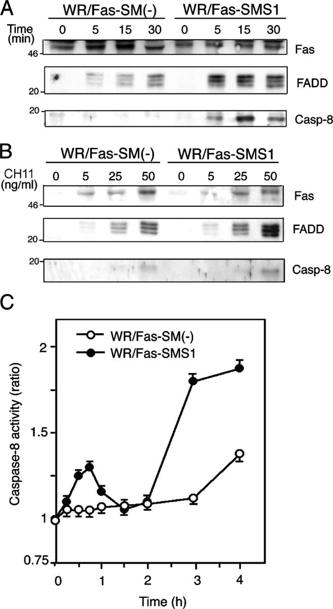
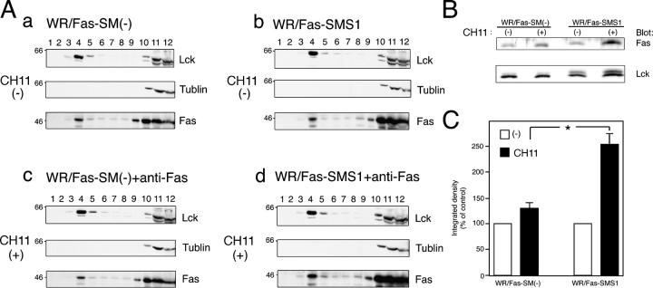
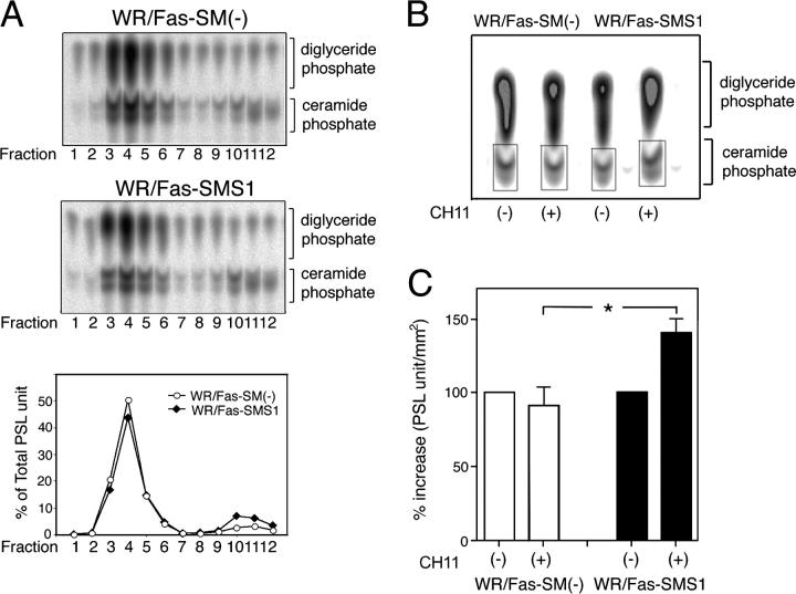
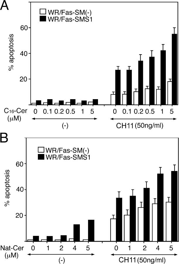
Engagement of the Fas receptor (CD95) initiates multiple signaling pathways that lead to apoptosis, such as the formation of death-inducing signaling complex (DISC), activation of caspase cascades, and the generation of the lipid messenger, ceramide. Sphingomyelin (SM) is a major component of lipid rafts, which are specialized structures that enhance the efficiency of membrane receptor signaling and are a main source of ceramide. However, the functions of SM in Fas-mediated apoptosis have yet to be clearly defined, as the responsible genes have not been identified. After cloning a gene responsible for SM synthesis, SMS1, we established SM synthase-defective WR19L cells transfected with the human Fas gene (WR/Fas-SM(-)), and cells that have been functionally restored by transfection with SMS1 (WR/Fas-SMS1). We show that expression of membrane SM enhances Fas-mediated apoptosis through increasing DISC formation, activation of caspases, efficient translocation of Fas into lipid rafts, and subsequent Fas clustering. Furthermore, WR/Fas-SMS1 cells, but not WR/Fas-SM(-) cells, showed a considerable increase in ceramide generation within lipid rafts upon Fas stimulation. These data suggest that a membrane SM is important for Fas clustering through aggregation of lipid rafts, leading to Fas-mediated apoptosis.


Figures

Figure 1.
Figure 1.
Characterization of WR/Fas-SM() and WR/Fas-SMS1 cells. (A) SM synthase activity of WR/Fas-SM(−) (lane 1) and WR/Fas-SMS1 (lane 2) cells. The cellular lipids were labeled with [14C]serine, extracted by the Bligh and Dyer method (reference 53), and assessed by TLC. PE, phosphatidylethanolamine; PS, phosphatidylserine. (B) Analysis of membrane SM expression by confocal microscopy. Cells were stained with lysenin-MBP, and FITC-conjugated anti–mouse IgG mAb, then examined by laser scan confocal microscopy. (C) FACS analysis of membrane sphingolipids. To detect membrane SM, cells were stained with lysenin-MBP (Lysenin), CH11, and FITC-conjugated anti–mouse IgG mAb. Surface expression of ganglioside GM1, cholesterol, and human Fas were analyzed using FITC-conjugated CTx, cholesterol-PEG (cholesterol), or anti-Fas mAB (h-Fas), respectively.
Figure 2.
Figure 2.
Fas-mediated apoptosis in WR/Fas-SM() and WR/Fas-SMS1 cells. (A and B) Dose dependency of Fas-mediated apoptosis. Cells were incubated for 3 h with the indicated concentration of CH11. After incubation, cells were harvested and analyzed by flow cytometry for DNA fragmentation using nuclear staining with PI. The numbers in each box represent the percentages of apoptotic cells. (C) Time kinetics of Fas-mediated apoptosis. Cells were incubated with 50 ng/ml of CH11 for the indicated time, and apoptosis was analyzed by flow cytometry using PI. Error bars represent SEM. (D) Time–kinetics of loss of ΔΨm in apoptotic cells. Cells were incubated with 50 ng/ml of CH11 for the indicated time. ΔΨm was determined by intracellular staining with DiOC6(3) and flow cytometry. (E) Inhibition of Fas-mediated apoptosis by caspase inhibitors. Cells were stimulated with 50 ng/ml of CH11 for 6 h in the presence of the indicated amounts of Ac-DEVD-CHO or Ac-IETD-CHO. After incubation, apoptosis was analyzed by flow cytometry using PI.
Figure 3.
Figure 3.
Fas-mediated caspase-3 activation in WR/Fas-SM() and WR/Fas-SMS1 cells. Time kinetics (A) and dose dependency (B) of caspase-3 activation by Western blot analysis. Cells were stimulated with 50 ng/ml CH11 for the indicated time (A), or stimulated with the indicated concentration of CH11 for 15 min (B). Total cell lysates were analyzed by Western blot using mouse mAb to caspase-3. The arrows indicate the bands corresponding to 32 kD for procaspase-3 (pro) and 17 kD for the active caspase (active). (C) Cells were stimulated with 50 ng/ml CH11 for the indicated time, and caspase-3 activities were measured in extracts of cell lysates using colorimetric assay kits. Each experiment was done in triplicate. Data are representative of five independent experiments. Error bars represent SEM.
Figure 4.
Figure 4.
Fas-mediated DISC formation and caspase-8 activation in WR/Fas-SM() and WR/Fas-SMS1 cells. Time kinetics (A) and dose dependency (B) of Fas-mediated DISC formation. 2 × 10⁷ cells were stimulated with 50 ng/ml CH11 for the indicated time (A) or stimulated for 15 min with the indicated concentration of CH11 (B). After stimulation, Fas was immunoprecipitated with anti–mouse IgM antibody from Brij 97 lysates. Immunoprecipitates were subjected to 12% SDS-PAGE and immunoblotted with anti-Fas death domain (3D5), anti-FADD, and anti–caspase-8 mAb. Data are representative of five independent experiments. (C) Fas-mediated activation of caspase-8. Cells were stimulated with 50 ng/ml CH11 for the indicated time, and caspase-8 activities were measured in extracts of cell lysates using colorimetric assay kits. Each experiment was done in triplicate. Data are representative of five independent experiments. Error bars represent SEM.
Figure 5.
Figure 5.
Fas clustering and capping in WR/Fas-SM() and WR/Fas-SMS1 cells. Time kinetics (A) and dose dependency (B) of Fas multimer formation. 4 × 10⁷ cells were stimulated with 50 ng/ml CH11 for the indicated time (A), or stimulated for 10 min with the indicated concentration of CH11 (B). After stimulation, cell pellets were snap frozen and treated at 45°C for 1 h in 300 μl of a 1% NP-40–treating solution. DNA in the samples was sheared with a 25-gauge needle, and solubilized samples were loaded on 12% SDS-PAGE. Fas multimers were detected with antibody to the intracellular death domain of human Fas (3D5). Data are representative of more than three independent experiments. (C) Activation-induced capping of Fas. Cells were stained for 20 min with 50 ng/ml CH11 at 4°C. Afterwards, capping was induced by warming cells to 37°C for 30 min in a water bath with mild agitation. Cells were harvested at the indicated times and fixed with 4% paraformaldehyde for 20 min at 22°C. Fixed cells were washed twice and mounted in FITC-conjugated secondary antibody. Fluorescence was detected using a confocal microscope equipped with a SPOT digital camera. The data are representative of more than five experiments. Arrowheads indicate Fas capping. (D) Time kinetics of activation-induced Fas capping. Capping was induced for the indicated time, and cells with Fas clustering were counted by two independent observers. Percentages of capping were calculated in 150–200 total cells. These results are the mean of three independent experiments. Error bars represent SEM. *, P < 0.01.
Figure 6.
Figure 6.
Distribution of Fas into lipid rafts of WR/Fas-SM() and WR/Fas-SMS1 cells. (A) 108 cells were left unstimulated (a and b) or were stimulated with CH11 for 30 min (c and d), and Triton X-100 lysates were subjected to sucrose density gradient fractionation. Fractions were run on 15% SDS-PAGE and immunoblotted with antibodies against lck (a marker for the raft fractions), tublin (a marker for the nonraft fractions), and Fas (3D5). The blots shown are representative of four independent experiments. (B) Redistribution of Fas into lipid rafts on stimulation. Raft fractions (fraction 4) were run on 15% SDS-PAGE and immunoblotted with antibodies against lck and Fas. (C) Quantification of Fas contents in lipid rafts was performed densitometrically and normalized to the amount of lck. Data are expressed as the mean ± SEM for relative increase of three independent experiments. *, P < 0.01.
Figure 7.
Figure 7.
Ceramide generation in lipid rafts of WR/Fas-SM() and WR/Fas-SMS1 cells. (A) Ceramide contents in membrane fractions of sucrose density gradient fractionation. 108 cells were lysed in Triton X-100 buffer and subjected to sucrose density gradient fractionation. Lipids of each fraction were extracted by the method of Bligh and Dyer (reference 53), and ceramide content was measured by the diacylglycerol kinase assay. After separation of ceramide-1-phosphates by TLC, radioactivity was visualized and estimated. The results are representative of three independent experiments and expressed as the percentage of total PSL arbitrary units. (B) Fas-mediated ceramide generation in lipid rafts. 108 cells were left unstimulated or were stimulated with CH11 for 5 min and Triton X-100 lysates were subjected to sucrose density gradient fractionation. Lipids of raft fraction (fraction 4) were extracted by the method of Bligh and Dyer, and ceramide generation was measured by the diacylglycerol kinase assay. Radioactivity was visualized and estimated. The results are representative of three independent experiments. (C) The mean of three independent experiments revealed that ceramide contents of lipid rafts in WR/Fas-SMS1 cells were significantly greater than those of WR/Fas-SM(−) cells. The results were the mean of three independent experiments and expressed as the percentage increase of PSL arbitrary units. Error bars represent SEM. *, P < 0.01.
Figure 8.
Figure 8.
Effects of exogenous ceramides on Fas-mediated apoptosis in WR/Fas-SM() and WR/Fas-SMS1 cells. Cells were pretreated with the indicated concentration of C16- (A) or natural (B) ceramide for 1 h. After washing, cells were stimulated with 50 ng/ml CH11 for 6 h and apoptosis was analyzed by flow cytometry using PI. Error bars represent SEM.

Similar articles

Cited by

References

    1. Marsden, V.S., and A. Strasser. 2003. Control of apoptosis in the immune system: Bcl-2, BH-3 only proteins and more. Annu. Rev. Immunol. 21:71–105. - PubMed
    1. Scaffidi, C., S. Fulda, A. Srinivasan, C. Friesen, F. Li, K.J. Tomaselli, K.M. Debatin, P.H. Krammer, and M.E. Peter. 1998. Two CD95 (APO-1/Fas) signaling pathways. EMBO J. 17:1675–1687. - PMC - PubMed
    1. Wajant, H. 2002. The Fas signaling pathway: more than a paradigm. Science. 296:1635–1636. - PubMed
    1. Hengartner, M.O. 2000. The biochemistry of apoptosis. Nature. 407:770–776. - PubMed
    1. Simons, K., and E. Ikonen. 1997. Functional rafts in cell membranes. Nature. 387:569–572. - PubMed

Publication types

MeSH terms