Comparative Study
. 2005 Jul 19;102(29):10058-63.
doi: 10.1073/pnas.0502311102. Epub 2005 Jul 11.

DNA lesions induced by UV A1 and B radiation in human cells: comparative analyses in the overall genome and in the p53 tumor suppressor gene

Affiliations
Comparative Study

DNA lesions induced by UV A1 and B radiation in human cells: comparative analyses in the overall genome and in the p53 tumor suppressor gene

Ahmad Besaratinia et al. Proc Natl Acad Sci U S A. .

Abstract

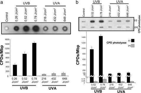
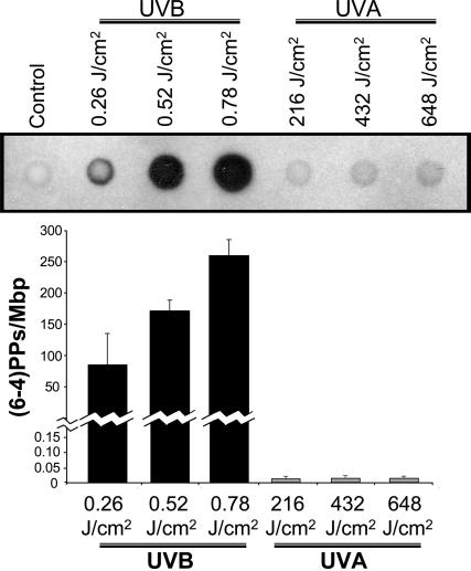
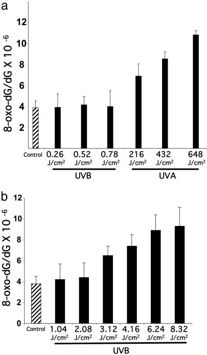
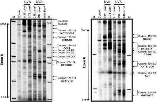
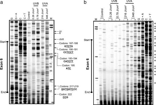
The UV components of sunlight (UVA and UVB) are implicated in the etiology of human skin cancer. The underlying mechanism of action for UVB carcinogenicity is well defined; however, the mechanistic involvement of UVA in carcinogenesis is not fully delineated. We investigated the genotoxicity of UVA1 versus UVB in the overall genome and in the p53 tumor suppressor gene in normal human skin fibroblasts. Immuno-dot blot analysis identified the cis-syn cyclobutane pyrimidine-dimer (CPD) as a distinctive UVB-induced lesion and confirmed its formation in the genomic DNA of UVA1-irradiated cells dependent on radiation dose. HPLC/tandem MS analysis showed an induction of 8-oxo-7,8-dihydro-2'-deoxyguanosine in the genomic DNA of UVA1-irradiated cells only. Mapping of DNA damages by terminal transferase-dependent PCR revealed preferential, but not identical, formation of polymerase-blocking lesions and/or strand breaks along exons 5-8 of the p53 gene in UVB- and UVA1-irradiated cells. The UVB-induced lesions detected by terminal transferase-PCR were almost exclusively mapped to pyrimidine-rich sequences; however, the UVA1-induced lesions were mapped to purine- and pyrimidine-containing sequences along the p53 gene. Cleavage assays with lesion-specific DNA repair enzymes coupled to ligation-mediated PCR showed preferential, but not identical, formation of CPDs along the p53 gene in UVB- and UVA1-irradiated cells. Additionally, dose-dependent formation of oxidized and ring-opened purines and abasic sites was established in the p53 gene in only UVA1-irradiated cells. We conclude that UVA1 induces promutagenic CPDs and oxidative DNA damage at both the genomic and nucleotide resolution level in normal human skin fibroblasts.


Figures

Fig. 1.
Fig. 1.
Quantifying CPDs in the genome by immuno-dot blot assay. (a) Genomic DNAs of UVB-irradiated (0.26, 0.52, and 0.78 J/cm2) or UVA1-irradiated (216, 432, and 648 J/cm2) human fibroblasts were analyzed with immuno-dot blot assay by using TD-M-2 mAb specific for CPDs (19, 20), as described in Materials and Methods. (b) To verify the specificity of CPD detection, the DNAs of UVB- and UVA1-irradiated cells were pretreated with CPD-photolyase, and subsequently subjected to immuno-dot blot analysis. To quantitate the results, we prepared standard controls with known quantities of CPDs, and subsequently established a calibration curve for luminescence signals specific for each assay run (see Materials and Methods). Results are expressed as means of three independent experiments, with each experiment run in triplicate (nine measurements). Error bar, SD. Mbp, megabase pair DNA.
Fig. 2.
Fig. 2.
Quantifying (6-4)PPs in the genome by immuno-dot blot assay. Genomic DNAs of UVB-irradiated (0.26, 0.52, and 0.78 J/cm2) or UVA1-irradiated (216, 432, and 648 J/cm2) human fibroblasts were analyzed with immuno-dot blot assay by using 64-M-2 mAb specific for (6-4)PPs (19, 20), as described in Materials and Methods. To quantitate the results, we prepared standard controls with known quantities of (6-4)PPs, and subsequently established a calibration curve for luminescence signals specific for each assay run (see Materials and Methods). Results are expressed as means of three independent experiments, with each experiment run in triplicate (nine measurements). Error bar, SD. Mbp, megabase pair DNA.
Fig. 3.
Fig. 3.
Quantifying 8-oxo-dG in the genome by HPLC-MS/MS. (a) Genomic DNAs of UVB-irradiated (0.26, 0.52, and 0.78 J/cm2) or UVA1-irradiated (216, 432, and 648 J/cm2) human fibroblasts were assayed with HPLC-MS/MS, as described in Materials and Methods.(b) Effects of high-dose UVB irradiation on the formation of 8-oxo-dG in human fibroblasts. Results are expressed as means of three independent experiments, with each experiment run in triplicate (nine measurements). Error bar, SD.
Fig. 4.
Fig. 4.
Mapping of polymerase-blocking DNA lesions in the p53 gene (nontranscribed strand) by TD-PCR. Genomic DNAs of UVB-irradiated (0.26 and 0.78 J/cm2) or UVA1-irradiated (216, 432, and 648 J/cm2) human fibroblasts were subjected to TD-PCR, as described in Materials and Methods. Representative lesion-mapping data for exons 5 (Left) and 6 (Right) of the p53 gene are shown. Hotspots of lesion formation are indicated by arrows, and the corresponding nucleotide positions, e.g., respective codons, are specified. Sequence contexts of the lesions in introns and exons are written in lowercase and uppercase fonts, respectively. M, sizing standard.
Fig. 5.
Fig. 5.
Mapping of CPDs and Fpg-sensitive sites in exon 6 of the p53 gene (nontranscribed strand) by LM-PCR. Genomic DNAs of UVB-irradiated (0.26 and 0.78 J/cm2) or UVA1-irradiated (216 and 648 J/cm2) human fibroblasts were subjected to T4 endonuclease V cleavage and CPD-photolyase reactivation (to create ligatable ends) (a), or treatment with Fpg (+) or digestion buffer only (-), and subsequently assayed by LM-PCR (b), as described in Materials and Methods. Hotspots of lesion formation are indicated by arrows, and the corresponding nucleotide positions, e.g., respective codons, are specified. Sequence contexts of the lesions in introns and exons are written in lowercase and uppercase, respectively. Underlined bases are the exact positions where the lesions are formed. Major Fpg-sensitive sites are identified by *. To specifically identify G, G + A, C, and C + T, a Maxam/Gilbert sequencing ladder (61) was included in all runs. All samples were processed in parallel and run on the same gel but were separated for better illustration. M, sizing standard.

References

    1. Woodhead, A. D., Setlow, R. B. & Tanaka, M. (1999) J. Epidemiol. 9, S102-S114. - PubMed
    1. de Gruijl, F. R. (1999) Eur. J. Cancer 35, 2003-2009. - PubMed
    1. Jhappan, C., Noonan, F. P. & Merlino, G. (2003) Oncogene 22, 3099-3112. - PubMed
    1. Pfeifer, G. P., You, Y. H. & Besaratinia, A. (2005) Mutat. Res. 571, 19-31. - PubMed
    1. Cadet, J., Sage, E. & Douki, T. (2005) Mutat. Res. 571, 3-17. - PubMed

Publication types