Review
. 2005 May;3(2):102-8.
doi: 10.3121/cmr.3.2.102.

Animal models for acquired bone marrow failure syndromes

Affiliations
Review

Animal models for acquired bone marrow failure syndromes

Jichun Chen. Clin Med Res. 2005 May.

Abstract

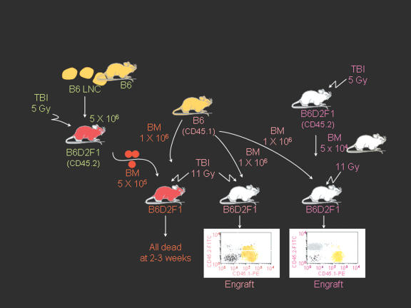
Bone marrow failure is a disease characterized by a drastic decline in the marrow's functional ability to produce mature blood cells. Aplastic anemia, a disease in which patients have essentially empty bone marrow accompanied by severe anemia, neutropenia, and thrombocytopenia, presents a paradigm for bone marrow failure. Damage to the marrow may first result from exposure to toxic chemicals, drug overdose, radiation, and viral infection; however, it is the extended immune-mediated reaction that causes massive destruction of hematopoietic cells and leads to marrow hypoplasia and peripheral pancytopenia. In recent years, animal models of acquired bone marrow failure syndromes have helped to strengthen our understanding of the mechanisms causing bone marrow failure. In this review, animal models for bone marrow failure are summarized by two groups: 1) bone marrow failure induced by toxic chemicals and drugs such as benzene, busulfan, and chloramphenicol, and radiation, and 2) models developed by an immune-related mechanism such as viral infection or foreign lymphocyte infusion.


Figures

Figure 1.
Figure 1.
Marrow lymphocyte infiltration/expansion and marrow hypoplasia in a mouse model of infusion induced BM failure. CByB6F1 mice treated with 5 Gy total body irradiation and 5 x 10⁶ BM cell infusion were euthanized at 14 days after cell infusion along with untreated control mice. BM cells from BM failure mice (A) and control mice (B) were analyzed by flow cytometry. Stern bones from BM failure (C) and control (D) mice were sectioned, HE stained and observed under a light microscope (100x). Marrow cavity was almost empty in BM failure mice (C) while residual BM cells were essentially infiltrated donor lymphocytes (A).
Figure 1.
Figure 1.
Marrow lymphocyte infiltration/expansion and marrow hypoplasia in a mouse model of infusion induced BM failure. CByB6F1 mice treated with 5 Gy total body irradiation and 5 x 10⁶ BM cell infusion were euthanized at 14 days after cell infusion along with untreated control mice. BM cells from BM failure mice (A) and control mice (B) were analyzed by flow cytometry. Stern bones from BM failure (C) and control (D) mice were sectioned, HE stained and observed under a light microscope (100x). Marrow cavity was almost empty in BM failure mice (C) while residual BM cells were essentially infiltrated donor lymphocytes (A).
Figure 2.
Figure 2.
Bystander destruction of marrow hematopoietic cells. BM cells from BM failure mice mixed with BM cells from normal B6-CD45.1 mice could not rescue lethally-irradiated B6D2F1 recipients (left) while B6-CD45.1 marrow cells alone (middle) or a mixture of normal B6D2F1 and B6-CD45.1 marrow cells (right) established effective engraftment and rescued recipients. This experiment shows that residual cells in the marrow of BM failure mice were able to destroy normal hematopoietic cells from B6-CD45.1 donors as bystanders.

Comment in

Similar articles

Cited by

References

    1. Young NS. Acquired aplastic anemia. Ann Intern Med 2002;136:534–546. - PubMed
    1. Young NS. Aplastic anemia, myelodysplasia, and related bone marrow failure syndromes. In: Kasper DL, Braunwald E, Fauci AS, Hauser SL, Longo DL, Jameson JL, eds. Harrison’s Principles of Internal Medicine. New York, NY: McGraw-Hill; 2005. 617–626.
    1. Ehrich P. Ueber einen Fall von Anämie mit Bemerkungen über regenerative Veränderungen des Knochenmarks. Charite-Ann 1888;13:300–309.
    1. Young NS. Drugs and chemicals. In: Young NS, Alter BP, eds. Aplastic Anemia: Acquired and Inherited. Philadelphia, PA: WB Saunders; 1994. 100–132.
    1. Zeng W, Maciejewski JP, Chen G, Young NS. Limited heterogeneity of T cell receptor BV usage in aplastic anemia. J Clin Invest 2001;108:765–773. - PMC - PubMed