

Expression of D-type cyclins in colon cancer and in cell lines from colon carcinomas
- PMID: 16012517
- PMCID: PMC2361572
- DOI: 10.1038/sj.bjc.6602709
Expression of D-type cyclins in colon cancer and in cell lines from colon carcinomas
Abstract
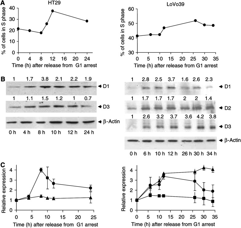
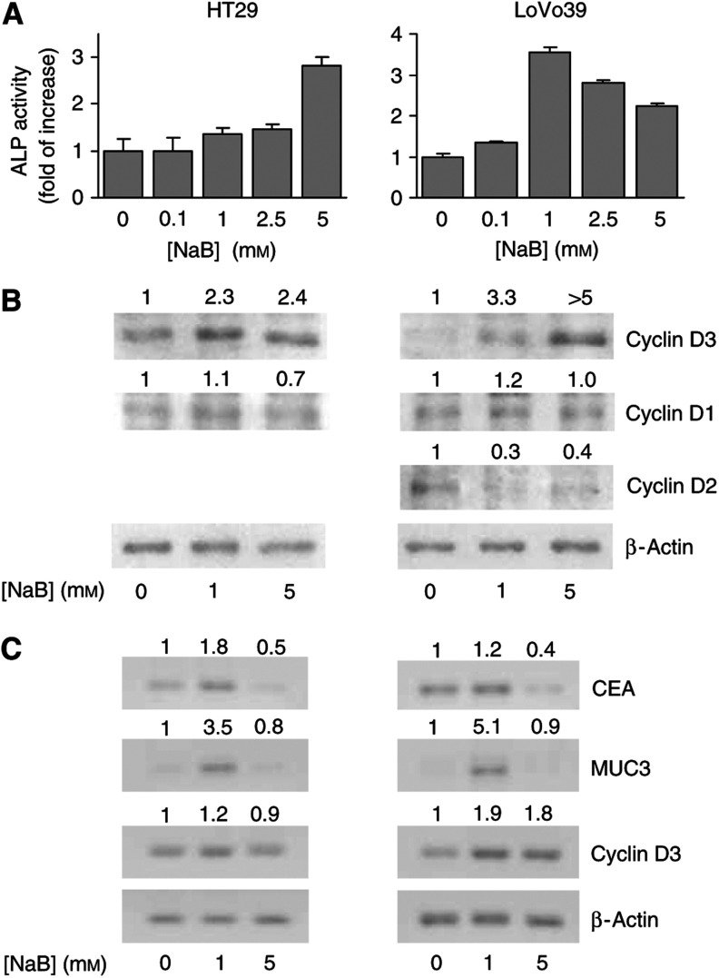
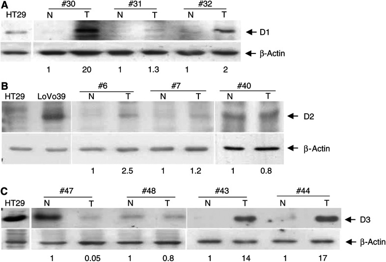
Cyclins D1, D2 and D3 play important roles in cell proliferation and differentiation. Although their abnormal expression has been linked to cancer development and progression in a number of tissues, the expression of cyclin D2 and D3 proteins in colon cancer has not yet been characterised. In this study, we examined cyclin D1, D2 and D3 protein expression by Western blot analysis in tumour and adjacent normal colon tissues of 57 patients. In addition, we examined D-type cyclins protein expression in HT29 and LoVo39 cell lines from colon carcinomas, as a function of induced proliferation and differentiation. In both cell lines, the expression of the three D-type cyclins increased as a result of induced proliferation, whereas the expression of cyclin D3 increased as a result of induced differentiation. In colon tumours, cyclin D1 was overexpressed in 44%, cyclin D2 was overexpressed in 53% and cyclin D3 was overexpressed in 35% of the cases. We also found that in 16% of the cases, cyclin D3 protein expression was reduced in the tumour, as compared to the adjacent normal tissue. Examination of D-type cyclin protein overexpression in relation to the TNM stage of the tumours revealed that overexpression of cyclins D1 and/or D2, but not cyclin D3, is linked to colon carcinogenesis and that overexpression of cyclin D2 may be related to a higher TNM stage of the tumour.
Figures
References
-
- Arber N, Doki Y, Han EK, Sgambato A, Zhou P, Kim NH, Delohery T, Klein MG, Holt PR, Weinstein IB (1997) Antisense to cyclin D1 inhibits the growth and tumorigenicity of human colon cancer cells. Cancer Res 57: 1569–1574 - PubMed
-
- Arber N, Hibshoosh H, Moss SF, Sutter T, Zhang Y, Begg M, Wang S, Weinstein IB, Holt PR (1996) Increased expression of cyclin D1 is an early event in multistage colorectal carcinogenesis. Gastroenterology 110: 669–674 - PubMed
-
- Arber N, Hibshoosh H, Yasui W, Neugut AI, Hibshoosh A, Yao Y, Sgambato A, Yamamoto H, Shapira I, Rosenman D, Fabian I, Weinstein IB, Tahara E, Holt PR (1999) Abnormalities in the expression of cell cycle-related proteins in tumors of the small bowel. Cancer Epidemiol Biomarkers Prev 8: 1101–1105 - PubMed
-
- Baldassarre G, Barone MV, Belletti B, Sandomenico C, Bruni P, Spiezia S, Boccia A, Vento MT, Romano A, Pepe S, Fusco A, Viglietto G (1999) Key role of the cyclin-dependent kinase inhibitor p27kip1 for embryonal carcinoma cell survival and differentiation. Oncogene 18: 6241–6251 - PubMed
-
- Bartkova J, Lukas J, Bartek J (1997) Aberrations of the G1- and G1/S-regulating genes in human cancer. Prog Cell Cycle Res 3: 211–220 - PubMed
Publication types
MeSH terms
Substances
LinkOut - more resources
Full Text Sources
Other Literature Sources
Research Materials
