

Localization of interictal spikes using SAM(g2) and dipole fit
- PMID: 16012648
- PMCID: PMC4041981
Localization of interictal spikes using SAM(g2) and dipole fit
Abstract
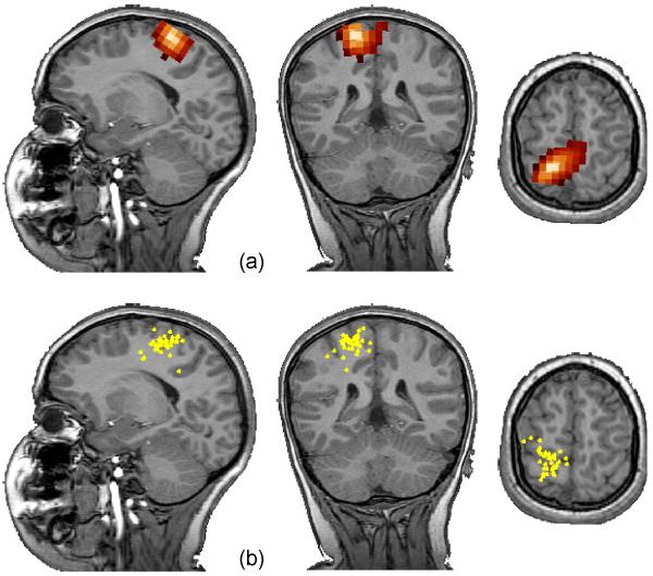
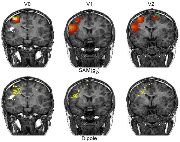
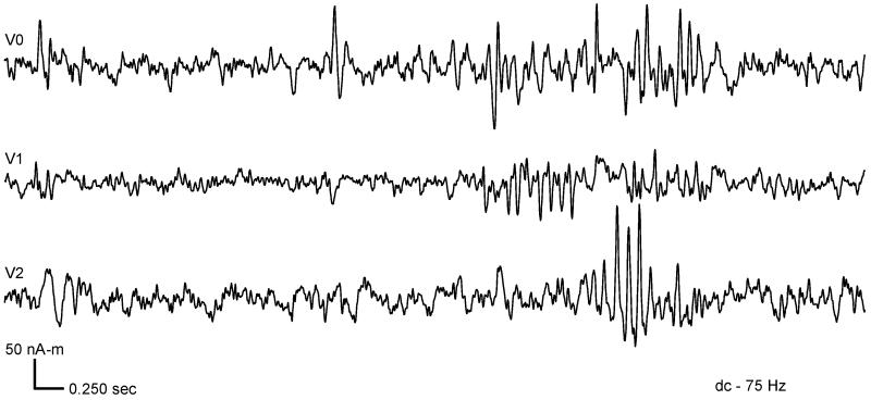
SAM(g2) is an automated analysis that transforms the MEG data into a functional image of spike-like activity, giving the source waveforms for those locations. Since the source waveforms estimated by SAM have higher signal-to-noise ratio (SNR) than does the raw MEG data, it is possible to automatically mark the location and timing of each spike for comparisons with dipole fit procedures. Both SAM(g2) and equivalent current dipole (ECD) fits were used to analyze MEG interictal spike recordings in 10 patients with cortical dysplasias and medial temporal lobe epilepsy. The ECD fit locations obtained by manual spike classification and latency marking were compared with those found by automated SAM(g2) procedures. When the SNR of interictal activity was high (compared to the background) with a clear single focus, there was excellent agreement between the ECD cluster location and the SAM(g2) maximum. However, when the SNR of spikes was low, manual single ECD location scatter was larger than SAM(g2) reconstructions. When multiple independent interictal spike loci were present, there was some disagreement between SAM(g2) and ECD scatter in the cases of low SNR spikes. When SAM(g2) indicated multiple coupled spike loci, the residual variance for the dipole fit was high and its scatter unacceptably large--even for multiple dipole models. This study demonstrates that SAM(g2) is equivalent to ECD fit for localizing interictal spikes when there is a single locus and good SNR. Further studies are required to validate cases in which there are multiple spike loci or poor SNR.
Figures
References
-
- Robinson SE, Vrba J, Otsubo H, Ishii R. In: Nowak H, Haueisein J, Giessler F, Huonker R, editors. Finding epileptic loci by nonlinear parameterization of source waveforms; Proceedings of the 13th International Conference on Biomagnetism; Jena. 2002.Aug, pp. 220–222.
Publication types
MeSH terms
Grants and funding
LinkOut - more resources
Full Text Sources