. 2005 Nov 1;391(Pt 3):481-90.
doi: 10.1042/BJ20050897.

Attempts to characterize the NBD heterodimer of MRP1: transient complex formation involves Gly771 of the ABC signature sequence but does not enhance the intrinsic ATPase activity

Affiliations

Attempts to characterize the NBD heterodimer of MRP1: transient complex formation involves Gly771 of the ABC signature sequence but does not enhance the intrinsic ATPase activity

Odile Ramaen et al. Biochem J. .

Abstract

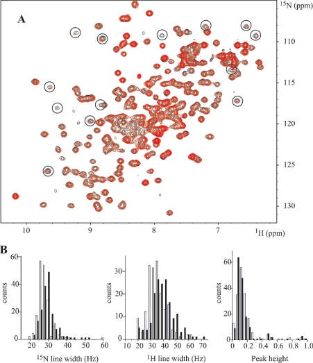
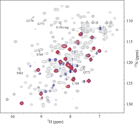
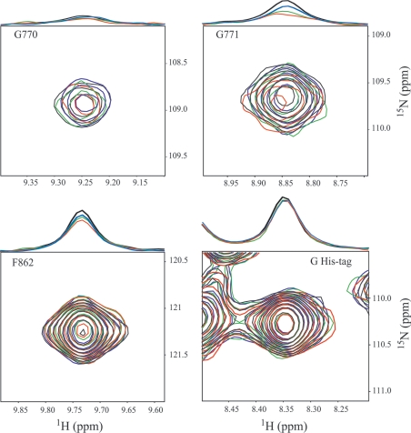
MRP1 (multidrug-resistance-associated protein 1; also known as ABCC1) is a member of the human ABC (ATP-binding cassette) transporter superfamily that confers cell resistance to chemotherapeutic agents. Considering the structural and functional similarities to the other ABC proteins, the interaction between its two NBDs (nucleotide-binding domains), NBD1 (N-terminal NBD) and NBD2 (C-terminal NBD), is proposed to be essential for the regulation of the ATP-binding/ATP-hydrolysis cycle of MRP1. We were interested in the ability of recombinant NBD1 and NBD2 to interact with each other and to influence ATPase activity. We purified NBD1 (Asn642-Ser871) and NBD2 (Ser1286-Val1531) as soluble monomers under native conditions. We measured extremely low intrinsic ATPase activity of NBD1 (10(-5) s(-1)) and NBD2 (6x10(-6) s(-1)) and no increase in the ATP-hydrolysis rate could be detected in an NBD1+NBD2 mixture, with concentrations up to 200 microM. Despite the fact that both monomers bind ATP, no stable NBD1.NBD2 heterodimer could be isolated by gel-filtration chromatography or native-PAGE, but we observed some significant modifications of the heteronuclear single-quantum correlation NMR spectrum of 15N-NBD1 in the presence of NBD2. This apparent NBD1.NBD2 interaction only occurred in the presence of Mg2+ and ATP. Partial sequential assignment of the NBD1 backbone resonances shows that residue Gly771 of the LSGGQ sequence is involved in NBD1.NBD2 complex formation. This is the first NMR observation of a direct interaction between the ABC signature and the opposite NBD. Our study also reveals that the NBD1.NBD2 heterodimer of MRP1 is a transient complex. This labile interaction is not sufficient to induce an ATPase co-operativity of the NBDs and suggests that other structures are required for the ATPase activation mechanism.


Figures

Figure 1
Figure 1. Purification of MRP1-NBD2
(A) Gel-filtration chromatographic profiles of NBD2 preparations. NBD2 samples of three different preparations were applied to a gel-filtration Superdex 75 HR 10/30 column. Protein elution was followed by absorption at A280. Monomeric NBD2 was eluted in the fractions corresponding to peak M, whereas oligomers of NBD2 were recovered in the fractions corresponding to peak P. The Superdex 75 column was calibrated using the low molecular mass gel-filtration kit (Amersham Biosciences, 13.7, 25, 43, 67 and 2000 kDa) and alcohol dehydrogenase (Sigma–Aldrich, Poole, Dorset, U.K., 150 kDa). (B) ATPase activity in Superdex 200 elution fractions. An NBD2 sample purified on cobalt-affinity resin was applied to a gel-filtration Superdex 200HR 10/30 column. Protein elution was followed by A280 (line curve). ATPase activity was followed in 25 μl of aliquots of each fraction by adding 5 μl of [γ-33P]ATP (final concentration 0.3 mM, 22 Bq/pmol) to start the reaction. ATPase activity (shaded bars) was monitored by measuring the amount of [33P]Pi produced during 4 h at 30 °C. (C) SDS/PAGE of NBD1 and NBD2. Purified NBDs were analysed by SDS/PAGE (15% polyacrylamide) and revealed by Coomassie Blue staining. Lane 1, molecular mass standards; lane 2, 5 μg of NBD1; lane 3, 5 μg of NBD2.
Figure 2
Figure 2. NBD2 and NBD1+NBD2 ATPase activity
(A) NBD2 ATPase activity was determined using [γ-33P]ATP as substrate. The reaction mixtures contained NBD2 (66 μM) and ATP (1 mM) in 70 μl of buffer (50 mM Tris/HCl, pH 8 and 100 mM KCl) in the presence of 3 mM MgCl2 (◆) or 1 mM EDTA (▲). Aliquots (5 μl) were withdrawn at indicated times and [33P]Pi produced was counted. (B) ATPase activity of the NBDs was followed in standard buffer (50 mM Tris/HCl, pH 8, 100 mM KCl and 3 mM MgCl2) at 30 °C. The ATPase activity was measured as the amount of [γ-33P]ATP hydrolysed in aliquots (5 μl) at the indicated times and in the presence of 66 μM NBD2 (■); 66 μM NBD1 (◆); 66 μM NBD1 and 66 μM NBD2 (▲).
Figure 3
Figure 3. Gel-filtration chromatography and native PAGE of NBD1+NBD2 mixture
(A) Chromatographic profiles of Superdex 200 column fractions showing the elution of NBD1, 27 kDa; NBD2, 30 kDa; an NBD1+NBD2 mixture; and BSA, 67 kDa. The Superdex 200 column was calibrated with other proteins (indicated by arrows): 1, 43 kDa ovalbumin; 2, 25 kDa chymotrypsinogen A; 3, 13.7 kDa RNase A. Protein elution was recorded at A280. (B) Native PAGE was loaded with NBD2 (3 μg, lane 1), a mixture of NBD1 and NBD2 (3 μg for each NBD, lane 2) and NBD1 (3 μg, lane 3).
Figure 4
Figure 4. 1H-15N HSQC spectra of NBD1 with and without NBD2: distribution of line widths and intensities
(A) Overlaid contour plots of the HSQC spectra of NBD1 (220 μM) at 298 K in the presence of 0:1 (black) and 0.75:1 (red) molar equivalents of NBD2. The solutions contained 4 mM ATP and 8 mM MgCl2. (B) Distribution of 15N and 1H line widths and normalized peak heights in the 15N HSQC spectra without (white bars) and with (black bars) NBD2.
Figure 5
Figure 5. 1H-15N HSQC spectra of NBD1 with different 15N-labelling methods
Overlaid contour plots of the HSQC spectra at 298 K of NBD1 obtained with uniform labelling in E. coli (black, 220 μM), in vivo selective labelling with 15N-Leu in E. coli (blue, 220 μM) and in vitro selective labelling with 15N-Leu by cell-free expression (red, 80 μM). The last HSQC spectrum shows the 23 Leu signals from NBD1 and that of the His tag.
Figure 6
Figure 6. NMR titration experiments of NBD1 by NBD2
The HSQC spectra of NBD1 (220 μM) were titrated in the presence of 1 mM Mg-ATP by addition of 0:1 (black), 0.35:1 (blue), 1:1 (green) and 1.6:1 (red) molar equivalents of NBD2. The selected regions are centred on the backbone correlations of Gly770, Gly771, Phe862 and one Gly of His tag. On the top of each two-dimensional spectrum, the extracted 1H spectrum at the 15N chemical shift of these residues is drawn, which has been normalized relative to the Gly His-tag signal.

Similar articles

Cited by

References

    1. Holland B., Cole S. P., Kuchler K., Higgins C. F., editors. London: Academic Press; 2003. ABC Proteins from Bacteria to M.
    1. Hipfner D. R., Deeley R. G., Cole S. P. Structural, mechanistic and clinical aspects of MRP1. Biochim. Biophys. Acta. 1999;1461:359–376. - PubMed
    1. Cole S. P., Bhardwaj G., Gerlach J. H., Mackie J. E., Grant C. E., Almquist K. C., Stewart A. J., Kurz E. U., Duncan A. M., Deeley R. G. Overexpression of a transporter gene in a multidrug-resistant human lung cancer cell line. Science. 1992;258:1650–1654. - PubMed
    1. Cole S. P., Deeley R. G. Multidrug resistance mediated by the ATP-binding cassette transporter protein MRP. BioEssays. 1998;20:931–940. - PubMed
    1. Leslie E. M., Deeley R. G., Cole S. P. Toxicological relevance of the multidrug resistance protein 1, MRP1 (ABCC1) and related transporters. Toxicology. 2001;167:3–23. - PubMed

MeSH terms

Substances