. 2005 Jul 13:2:54.
doi: 10.1186/1743-422X-2-54.

Use of a novel cell-based fusion reporter assay to explore the host range of human respiratory syncytial virus F protein

Affiliations

Use of a novel cell-based fusion reporter assay to explore the host range of human respiratory syncytial virus F protein

Patrick J Branigan et al. Virol J. .

Abstract

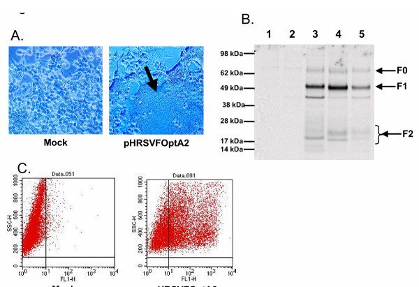
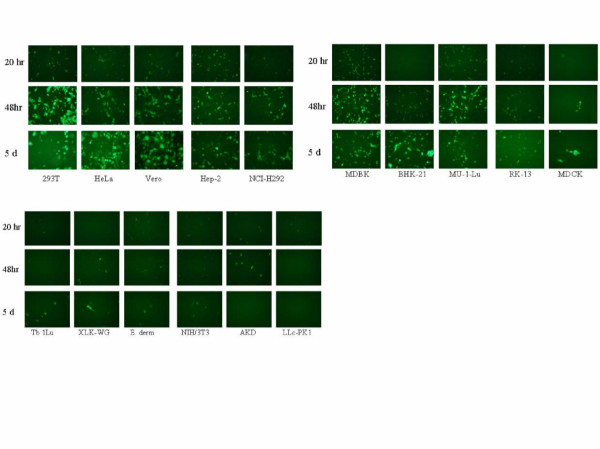
Human respiratory syncytial virus (HRSV) is an important respiratory pathogen primarily affecting infants, young children, transplant recipients and the elderly. The F protein is the only virion envelope protein necessary and sufficient for virus replication and fusion of the viral envelope membrane with the target host cell. During natural infection, HRSV replication is limited to respiratory epithelial cells with disseminated infection rarely, if ever, occurring even in immunocompromised patients. However, in vitro infection of multiple human and non-human cell types other than those of pulmonary tract origin has been reported. To better define host cell surface molecules that mediate viral entry and dissect the factors controlling permissivity for HRSV, we explored the host range of HRSV F protein mediated fusion. Using a novel recombinant reporter gene based fusion assay, HRSV F protein was shown to mediate fusion with cells derived from a wide range of vertebrate species including human, feline, equine, canine, bat, rodent, avian, porcine and even amphibian (Xenopus). That finding was extended using a recombinant HRSV engineered to express green fluorescent protein (GFP), to confirm that viral mRNA expression is limited in several cell types. These findings suggest that HRSV F protein interacts with either highly conserved host cell surface molecules or can use multiple mechanisms to enter cells, and that the primary determinants of HRSV host range are at steps post-entry.


Figures

Figure 1
Figure 1
A) Syncytia formation by RSV-F DNA in transfected cells. 293T cells were mock transfected or transfected with pHRSVFOptA2 and visualized by light microscopy 48 hours post transfection. The arrow indicates giant multinucleated cell formation. B) Processing of RSV F in transfected cells. 293T cells were either mock transfected (lane 1), transfected with pCMV-β-gal (lane 2), transfected with pHRSVFOptA2 (lane 3), pHRSVFOptB18537 (lane 4), or infected with RSV (Long strain, MOI = 1) for 24 hours followed by metabolic labeling for 6 hours with [35S]-cysteine/methionine. Labeled cell lysates were immunoprecipitated with HRSV F specific mAbs, and immunoprecipitates were resolved by SDS-PAGE as described in methods. C) Cell surface expression of RSV F in transfected cells. 293T cells were either mock transfected or transfected with pHRSVFOptA2 for 24 hours followed by flow cytometry using HRSV F specific monoclonal antibodies as described in methods.
Figure 2
Figure 2
A) Dose dependent fusion activity of HRSV F derived. 293T cells were transfected with either pFR-Luc alone (■), pBD-NFκB alone (▲), co-transfected with pFR-Luc and pBD-NFκB (▼), or co-transfected with pHRSVFOptA2 and pBD-NFκB and mixed 24 hours after transfection in various amounts with cells that had been transfected with pFR-Luc alone (◆). Luciferase activity was measured 24 hours post mixing as described in methods and is reported as relative light units. B) Fusion activity of HRSV F derived from subgroups A and B. 293T cells co-transfected with pBD-NFκB and either pHRSVFOptA2, pHRSVFOptB18537, or vector only (NFκB only). Cells were mixed 24 hours later with a separate population of 293T cells transfected with pFR-Luc, and luciferase activity was measured 24 hours post mixing as described in methods. Luciferase activity is reported as relative light units.
Figure 3
Figure 3
A) Comparison of fusion activity of wild-type and a fusion peptide mutant of HRSV F. 293T cells co-transfected with pBD-NFκB and either pHRSVFOptA2 or pL138R were mixed 24 hours later with a separate population of 293T cells that had been transfected with pFR-Luc, and luciferase activity was measured 24 hours post mixing as described in methods. Luciferase activity is reported as relative light units. B) Processing of the wild-type and L138R mutant of HRSV F was determined by metabolic labeling 293T cells transfected with either pHRSVFOptA2 (lane 1), pL138R (lane 2), pCMV-β-gal (lane 3), or mock transfected (lane 4) for 6 hours with [35S]-cysteine/methionine followed by immunoprecipitation of lysates with HRSV F specific mAbs, and analysis of immunoprecipitates by SDS-PAGE as described in methods. C) Cell surface expression of the wild-type and L138R mutant F proteins in 293T cells transfected with either pHRSVFOptA2 or pL138R was compared by flow cytometry as described in methods.
Figure 4
Figure 4
Fusion activity of HRSV F with cell lines derived from various species. Cell lines derived from various species (target cells) were either transfected with pFR-Luc and mixed 24 hours later with 293T cells that had been co-transfected for 24 hours with pHRSVFOptA2 or pVPack-VSV-G and pBD-NFκB (Figs. 4A and 4B), or the target cells were transfected with pBD-NFκB and mixed 24 hours later with 293T cells that had been co-transfected for 24 hours with pHRSVFOptA2 or pVPack-VSV-G together with pFR-Luc (Figs. 4C and 4D). Cell lines derived from various species were transfected with either pFR-Luc or pBD-NFκB only as negative controls. Luciferase activity was measured 24 hours post mixing of the cell populations as described in methods and is reported as relative light units.
Figure 5
Figure 5
Infection of various cell lines by rg224(RSV). Cell lines derived from various species were infected with rgRSV(224) at an MOI = 0.1 and GFP-expressing cells were visualized at 20, 48, and 120 hours post infection by fluorescent microscopy by monitoring fluorescence at 488 nm.

Similar articles

Cited by

References

    1. Shay DKHRCNRDLLLSJWLJA. Bronchiolitis-associated hospitalizations among U.S. children, 1980-1996. JAMA. 1999;282:1440–1446. doi: 10.1001/jama.282.15.1440. - DOI - PubMed
    1. Shay DKHRCRGECMJALJ. Bronchiolitis-associated mortality and estimates of respiratory syncytial virus-associated deaths among U.S. children, 1979-1997. J Infect Dis. 2001;183:16–22. doi: 10.1086/317655. - DOI - PubMed
    1. Han LLJPALJA. Respiratory syncytial virus pneumonia among the elderly: an assessment of disease burden. J Infect Dis. 1999;179:25–30. doi: 10.1086/314567. - DOI - PubMed
    1. Ison MGHFG. Viral infections in immunocompromised patients:what's new with respiratory viruses? Curr Opin Infect Dis. 2002;15:355–367. - PubMed
    1. Collins PLCRMMBR. Respiratory syncytial virus. In: D.M. Knipe HPM, editor. Fields Virology. Vol. 1. Philadelphia, Lippincott, Williams, and Wilkins; 2001. pp. 1443–1485.

MeSH terms

LinkOut - more resources