Comparative Study
. 2005 Jul 19;102(29):10285-90.
doi: 10.1073/pnas.0504578102. Epub 2005 Jul 12.

Suppression of myasthenogenic responses of a T cell line by a dual altered peptide ligand by induction of CD4+CD25+ regulatory cells

Affiliations
Comparative Study

Suppression of myasthenogenic responses of a T cell line by a dual altered peptide ligand by induction of CD4+CD25+ regulatory cells

Badiga Venkata Aruna et al. Proc Natl Acad Sci U S A. .

Erratum in

  • Proc Natl Acad Sci U S A. 2005 Aug 23;102(34):12288

Abstract

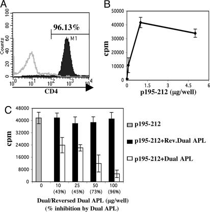
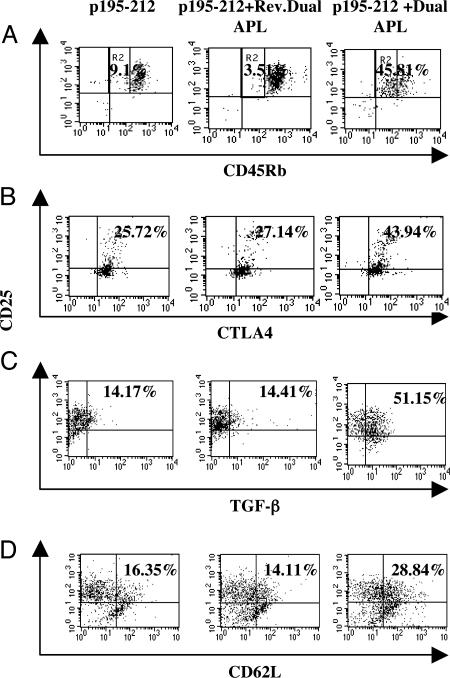
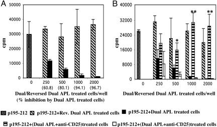
Myasthenia gravis is a T cell-dependent, antibody-mediated autoimmune disease. A dual altered peptide ligand (APL) that is composed of the tandemly arranged two single amino acid analogs of two myasthenogenic peptides, p195-212 and p259-271, was demonstrated to down-regulate in vitro and in vivo myasthenia gravis-associated autoreactive responses. The aims of this study were to demonstrate the suppressive properties and to elucidate the mechanism of action of the dual APL on a T cell line specific to the myasthenogenic peptide p195-212. We demonstrate here that incubation of cells of the line with the dual APL resulted in the inhibition of proliferation and secretion of IL-2 and IFN-gamma triggered by p195-212. In contrast, secretion of TGF-beta and IL-10 was upregulated. The dual APL induced the generation of CD4+CD25+ cells that were characterized by the expression of CD45Rb(low), cytotoxic T lymphocyte-associated antigen-4, TGF-beta, CD62L, Foxp3, and neuropilin. In addition, the dual APL-treated cells were capable of inhibiting the proliferation response of the line when the two sets of cells were cocultured. The role of CD4+CD25+ cells was further confirmed by demonstrating that the suppression was abrogated by blocking/neutralization of CD25. Thus, the dual APL acts by inducing the formation of CD4+CD25+ regulatory cells. By using a T cell line, we could show that the immunosuppressive CD4+CD25+ cells were indeed induced by the dual APL and are not part of the naturally occurring regulatory cells.


Figures

Fig. 1.
Fig. 1.
Characterization of the p195–212-specific T cell line. (A) SJL mice were immunized with p195–212 (10 μg per mouse in complete Freund's adjuvant), and LN cells were harvested 10 days later. p195–212-specific T cell line was established and maintained as described in Materials and Methods. Cells were stained with PE-coupled anti-CD4 specific antibody 7 days after antigenic stimulation and analyzed by FACS. (B) Cells of the line (n = 104 per well) were stimulated with various concentrations of p195–212 in the presence of irradiated syngeneic splenocytes (n = 0.5 × 10⁶ per well). (C) Cells were incubated with p195–212 (1 μg per well) in the presence or absence of various doses of the dual APL or the reversed dual APL. (B and C) The proliferation assay was performed as described in Materials and Methods. Results are expressed as mean cpm of triplicates ± SD values, and they represent one experiment of four performed.
Fig. 2.
Fig. 2.
The effect of the dual APL on cytokine secretion by the p195–212-specific T cells. Cells of the line (n = 106 per ml) were stimulated with p195–212 (5 μg per ml) with or without the dual APL or the reversed dual APL (500 μg per ml) for 24–48 h. Supernatants were collected, and concentrations of IFN-γ (A), IL-2 (B), TGF-β (C), and IL-10 (D) were determined by ELISA. Results are mean concentrations of triplicates ± SD values. *, P < 0.005; **, P < 0.0001; as compared with supernatants of cells stimulated with p195–212 alone.
Fig. 3.
Fig. 3.
The dual APL down-regulates the expression of CD28. Cells (n = 106 per ml) of the line were stimulated with p195–212 (5 μg per ml) with or without the dual or the reversed dual APL (500 μg per ml). After 24 h, cells were stained with FITC-coupled anti-CD28 antibody and analyzed by FACS. Results are presented as the percentage of CD28 cells, and they represent one of two experiments performed.
Fig. 4.
Fig. 4.
The dual APL up-regulates regulatory markers on p195–212-specific T cells. Cells (n = 106 per ml) of the line were stimulated with p195–212 (5 μg per ml) with or without the dual or reversed dual APL (500 μg per ml) for 24 h. Cells were double-stained for CD25 and CD45Rb (A), intracellular CTLA-4 (B), intracellular TGF-β (C), or CD62L (D). The results in A are presented as the percentage of CD45Rblow gated as R2. Results represent one of two experiments performed.
Fig. 5.
Fig. 5.
Foxp3 and neuropilin are up-regulated in p195–212-specific T cells by the dual APL. T cells (n = 106 per ml) of the line were stimulated with p195–212 (5 μg per ml) with or without the dual or the reversed dual APL (500 μg per ml) for 24 h. (A) Total RNA was extracted, and real-time PCR was performed for Foxp3 expression as described in Materials and Methods. Results are expressed as the mean percentage of the gene expression of triplicates ± SD values. *, P < 0.0005 when gene expression in the presence of the dual APL was compared with that in the absence of the dual APL. (B) Whole-cell lysates were prepared, separated on SDS/PAGE, and transferred to a nitrocellulose membrane that was later blotted with anti-neuropilin or anti-β-actin. Results are expressed as densitometry units (measured with National Institutes of Health image software), and they represent one of two experiments performed.
Fig. 6.
Fig. 6.
Dual APL-treated cells inhibit the proliferation of p195–212-specific T cell line. (A) Cells (n = 104 per well) of the line were cocultured with various numbers of reversed dual APL or dual APL-treated cells (described in Materials and Methods) in the presence of p195–212 (1 μg per well) and antigen-presenting cells. (B) Cells of the line were cocultured with reversed dual APL-treated cells or dual APL (in the absence or presence of anti-CD25 or its matched isotype control) treated cells with p195–212 and antigen-presenting cells. A proliferation assay was performed as described in Materials and Methods. Results are expressed as mean cpm of triplicates ± SD values, and they represent one of four experiments performed. *, P < 0.05; **, P < 0.0001; proliferation with dual APL plus anti-CD25 treated cells was compared with that of dual APL treated cells.

Similar articles

Cited by

References

    1. Lindstrom, J. (1985) Annu. Rev. Immunol. 3, 109-131. - PubMed
    1. Drachman, D. B. (1994) N. Engl. J. Med. 25, 1797-1810. - PubMed
    1. Lindstrom, J. D., Shelton, D. & Fuji, Y. (1988) Adv. Immunol. 42, 233-284. - PubMed
    1. Brocke, S., Brautbar, C., Steinman, L., Abramsky, O., Rothbard, J., Neumann, D., Fuchs, S. & Mozes, E. (1988) J. Clin. Invest. 82, 1894-1900. - PMC - PubMed
    1. Brocke, S., Dayan, M., Rothbard, J., Fuchs, S. & Mozes, E. (1990) Immunology 69, 495-500. - PMC - PubMed

Publication types