Comparative Study
. 2005 Jul 19;102(29):10339-44.
doi: 10.1073/pnas.0501866102. Epub 2005 Jul 12.

Intrinsically photosensitive retinal ganglion cells detect light with a vitamin A-based photopigment, melanopsin

Affiliations
Comparative Study

Intrinsically photosensitive retinal ganglion cells detect light with a vitamin A-based photopigment, melanopsin

Yingbin Fu et al. Proc Natl Acad Sci U S A. .

Abstract

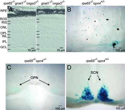
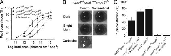
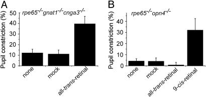
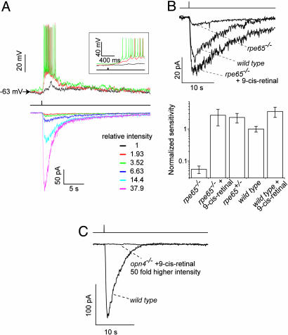
In mammals, intrinsically photosensitive retinal ganglion cells (ipRGCs) mediate non-image-forming visual functions such as pupillary light reflex (PLR) and circadian photoentrainment. This photosensitivity requires melanopsin, an invertebrate opsin-like protein expressed by the ipRGCs. The precise role of melanopsin remains uncertain. One suggestion has been that melanopsin may be a photoisomerase, serving to regenerate an unidentified pigment in ipRGCs. This possibility was echoed by a recent report that melanopsin is expressed also in the mouse retinal pigment epithelium (RPE), a key center for regeneration of rod and cone pigments. To address this question, we studied mice lacking RPE65, a protein essential for the regeneration of rod and cone pigments. Rpe65-/- ipRGCs were approximately 20- to 40-fold less photosensitive than normal at both single-cell and behavioral (PLR) levels but were rescued by exogenous 9-cis-retinal (an 11-cis-retinal analog), indicating the requirement of a vitamin A-based chromophore for ipRGC photosensitivity. In contrast, 9-cis-retinal was unable to restore intrinsic photosensitivity to melanopsin-ablated ipRGCs, arguing against melanopsin functioning merely in photopigment regeneration. Interestingly, exogenous all-trans-retinal was also able to rescue the low sensitivity of rpe65-/- ipRGCs, suggesting that melanopsin could be a bistable pigment. Finally, we detected no melanopsin in the RPE and no changes in rod and cone sensitivities due to melanopsin ablation. Together, these results strongly suggest that melanopsin is the photopigment in the ipRGCs.


Figures

Fig. 1.
Fig. 1.
Retinal morphology and axonal projections of ipRGCs in the absence of RPE65. (A) Retinal cross sections from gnat1-/-cnga3-/- and rpe65-/- gnat1-/-cnga3-/- mice. ROS, rod outer segment; RIS, rod inner segment; ONL, outer nuclear layer; OPL, outer plexiform layer; INL, inner nuclear layer; IPL, inner plexiform layer; GCL, ganglion cell layer. (B) Flat-mounted retina from an rpe65-/-opn4+/- mouse stained with X-Gal (blue). (C and D) Coronal sections of rpe65-/-opn4-/- (C) and rpe65-/-opn4+/- (D) mouse brains showing the X-Gal-labeled ipRGC axons projecting to the OPN and SCN. Dorsal side is up in each case. The rpe65-/-opn4-/- mouse instead of the rpe65-/-opn4+/- mouse was used in C to give a more intense X-Gal labeling of the OPN.
Fig. 2.
Fig. 2.
PLR of mice with different genotypes. (A) Irradiance–response relations for the PLR of gnat1-/-cnga3-/-, rpe65-/-gnat1-/-cnga3-/-, and 9-cis-retinal-treated rpe65-/-gnat1-/-cnga3-/- mice with steady light (480 nm). Fractional pupil constriction was calculated as 1 - (pupil area in light/dark-adapted pupil area). The best-fit curves fitted to the data were calculated from formula image, where FL is the maximal percentage pupil constriction in bright light (a parameter in the fit), I is irradiance, Io is a constant, and n is the Hill coefficient. Note that FL is generally not 100% because the pupil area does not go down to zero even in very bright light. The parameters for the fits are as follows: FL = 88.0%, Io = 1012.71, n = 0.68 (gnat1-/-cnga3-/-); FL = 79.5%, Io = 1014.0, n = 0.78 (rpe65-/-gnat1-/-cnga3-/-); FL = 79.7%, Io = 1013.3, n = 0.78 (rpe65-/-gnat1-/-cnga3-/- plus 9-cis-Retinal). (B) 9-cis-Retinal had no effect on the PLR of opn4-/-gnat1-/-cnga3-/- mice, whereas topical application of 100 mM carbachol was able to produce a complete PLR (arrowhead indicates pupil). (C) Collected results on PLR for different genetic lines in bright light. Note that 9-cis-retinal was unable to rescue the PLR of opn4-/-gnat1-/-cnga3-/- mice (n = 6). Data on rpe65-/-gnat1-/-cnga3-/- and gnat1-/-cnga3-/- mice were from A. Irradiance was 480 nm and 1.6 × 10¹⁴ photons·cm-2·sec-1. For opn4-/-gnat1-/-cnga3-/- mice plus 9-cis-retinal, a light 10-fold brighter still produced no improvement in PLR over untreated opn4-/-gnat1-/-cnga3-/- mice (data not shown). Data are mean ± SEM (n = 4–7).
Fig. 3.
Fig. 3.
Effect of all-trans-retinal on the PLR. i.p. injection of all-trans-retinal rescued the PLR of rpe65-/-gnat1-/-cnga3-/- mice (A) but not of rpe65-/- opn4-/- mice (B). As a positive control, 9-cis-retinal improved the PLR of rpe65-/-opn4-/- mice. Irradiance was 480 nm and 2.4 × 10¹³ and 3.2 × 10¹² photons·cm-2·sec-1 in A and B, respectively. Data are mean ± SEM (n = 12 in A, and n = 4 in B).
Fig. 4.
Fig. 4.
Light responses and sensitivities of single rpe65+/-, rpe65-/-, and opn4-/- ipRGCs. (A) Flash responses recorded from a representative ipRGC in an rpe65+/- retina under current-clamp (Upper) and voltage-clamp (Lower, -70 mV) modes, respectively. Single flash trials are shown. The same stimulus was used in both cases: a 20-msec, 480-nm flash (middle trace) of the indicated relative intensities focused on a 300-μm spot centered at the soma. Not all indicated intensities were used in the current-clamp and voltage-clamp recordings. (Inset) The same current-clamp records on a faster time base. The light spot did not cover all distal dendrites of the cell. (B) Rescue of photosensitivity of rpe65-/- ipRGC by exogenous 9-cis-retinal. (Upper) Representative flash responses of WT, rpe65-/-, and 9-cis-retinal-treated rpe65-/- ipRGCs. The same flash intensity and single trials were used in all three cases. (Lower) Collected results on relative flash sensitivity from various mouse lines and manipulations. Values relative to WT are as follows (mean ± SEM): 0.056 ± 0.015 (rpe65-/-, n = 12); 2.7 ± 1.4 (rep65-/- plus 9-cis-retinal, n = 5); 2.3 ± 0.8 (rpe65+/-, n = 7); 1.0 ± 0.2 (WT, n = 12); 3.5 ± 1.2 (WT plus 9-cis-retinal, n = 6). See Materials and Methods. (C) 9-cis-Retinal was unable to restore the sensitivity of opn4-/- ipRGCs. Representative recordings from opn4-/- and WT ipRGCs are shown. Light responses by opn4-/- ipRGCs (n = 7) were never observed with or without 9-Cis-retinal, even for the strongest light.
Fig. 5.
Fig. 5.
Immunocytochemistry and X-Gal labeling of retinal cross sections from albino WT and albino opn4-/- mice. (A) Double labeling with an antibody against the N terminus of melanopsin (3) (green) and the anti-rhodopsin 1D4 monoclonal antibody (red). The rhodopsin immunofluorescence helped in identifying the adjacent RPE layer by showing the rod outer segments. Upon normal frame exposure, melanopsin-expressing ipRGC is visible in WT inner retina, but no melanopsin signal is detectable in the RPE layer. Upon overexposure, punctate green signal is present in the RPE layer (and elsewhere) of WT and opn4-/- mice, suggesting that the signal is nonspecific. (B) X-Gal labeling of albino opn4-/- retinal cross sections. The blue labeling is absent in the RPE but present in the ipRGC (arrow), and its processes are present in the inner plexiform layer (arrowheads). The abbreviations for retinal layers are as in Fig. 1.
Fig. 6.
Fig. 6.
ERG responses of opn4-/- and WT mice. (A) Light-adapted ERGs measured in a steady background of 2.5 log scotopic troland, sufficient to saturate the rod response. (B) Intensity–response relations for the light-adapted b-wave. The b-wave amplitude was measured at 45 msec after the flash, when the positive-going wave was maximal over most of the stimulus range (see A). The eight WT mice indicated included seven C57BL6 animals, owing to the reduced sample of WT B6/129 mice.

References

    1. Provencio, I., Jiang, G., De Grip, W. J., Hayes, W. P. & Rollag, M. D. (1998) Proc. Natl. Acad. Sci. USA 95, 340-345. - PMC - PubMed
    1. Berson, D. M., Dunn, F. A. & Takao, M. (2002) Science 295, 1070-1073. - PubMed
    1. Hattar, S., Liao, H. W., Takao, M., Berson, D. M. & Yau, K. W. (2002) Science 295, 1065-1070. - PMC - PubMed
    1. Lucas, R. J., Hattar, S., Takao, M., Berson, D. M., Foster, R. G. & Yau, K. W. (2003) Science 299, 245-247. - PubMed
    1. Ruby, N. F., Brennan, T. J., Xie, X., Cao, V., Franken, P., Heller, H. C. & O'Hara, B. F. (2002) Science 298, 2211-2213. - PubMed

Publication types

LinkOut - more resources