. 2005 Sep;16(9):4410-22.
doi: 10.1091/mbc.e05-01-0081. Epub 2005 Jul 12.

PAK5 kinase is an inhibitor of MARK/Par-1, which leads to stable microtubules and dynamic actin

Affiliations

PAK5 kinase is an inhibitor of MARK/Par-1, which leads to stable microtubules and dynamic actin

Dorthe Matenia et al. Mol Biol Cell. 2005 Sep.

Abstract

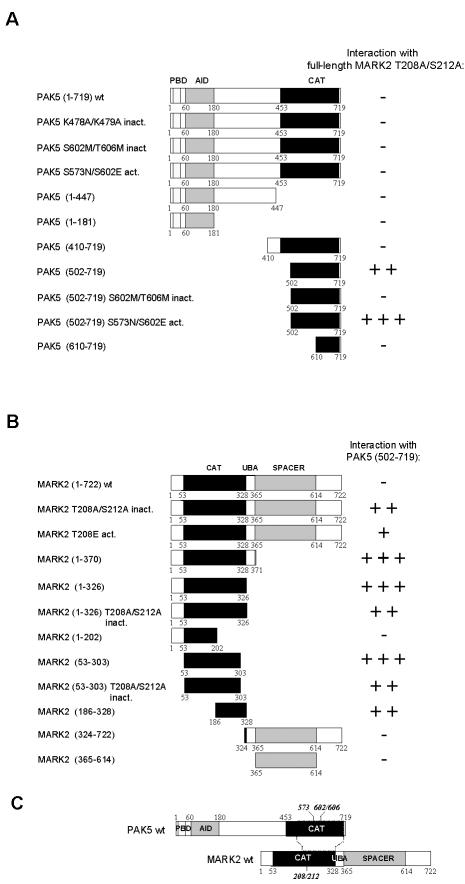
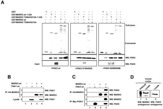
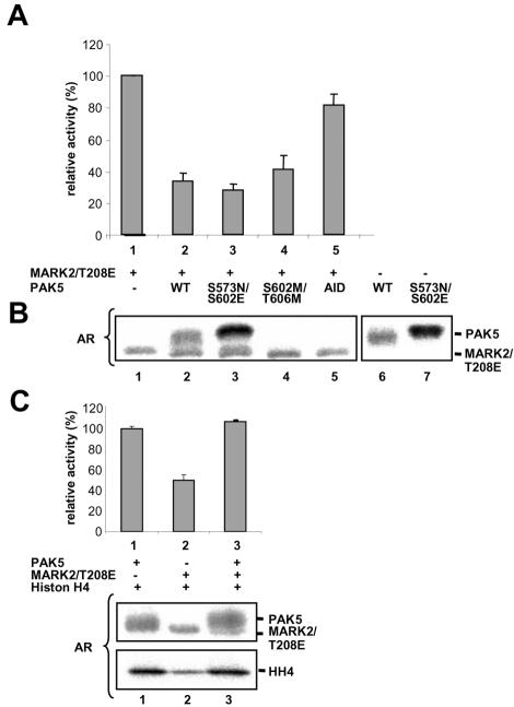
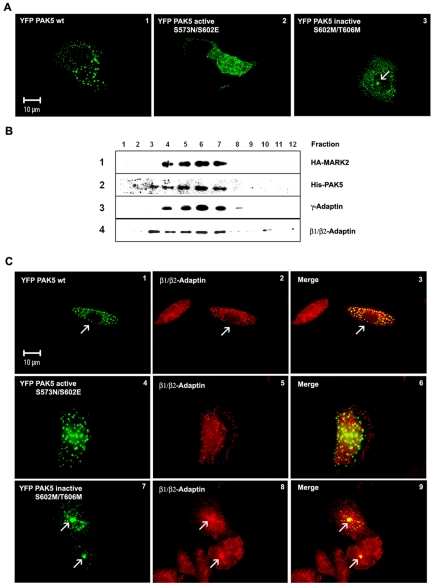
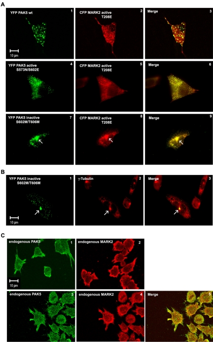
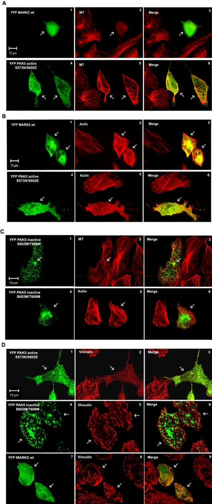
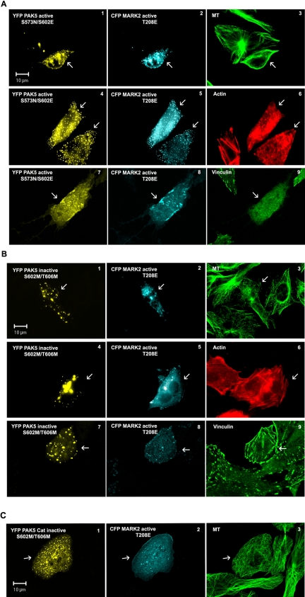
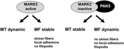
MARK/Par-1 is a kinase involved in development of embryonic polarity. In neurons, MARK phosphorylates tau protein and causes its detachment from microtubules, the tracks of axonal transport. Because the target sites of MARK on tau occur at an early stage of Alzheimer neurodegeneration, we searched for interaction partners of MARK. Here we report that MARK2 is negatively regulated by PAK5, a neuronal member of the p21-activated kinase family. PAK5 suppresses the activity of MARK2 toward its target, tau protein. The inhibition requires the binding between the PAK5 and MARK2 catalytic domains, but does not require phosphorylation. In transfected Chinese hamster ovary (CHO) cells both kinases show a vesicular distribution with partial colocalization on endosomes containing AP-1/2. Although MARK2 transfected alone destabilizes microtubules and stabilizes actin stress fibers, PAK5 keeps microtubules stable through the down-regulation of MARK2 but destabilizes the F-actin network so that stress fibers and focal adhesions disappear and cells develop filopodia. The results point to an inverse relationship between actin- and microtubule-related signaling by the PAK5 and MARK2 pathways that affect both cytoskeletal networks.


Figures

Figure 1.
Figure 1.
Interactions of MARK2 and PAK5 mapped by yeast two-hybrid screening. (A) Schematic diagrams of PAK5-constructs used to map the MARK2-binding site. Amino acid residue numbers at boundaries of deletion constructs are indicated below the schematics. Yellow, blue, and red boxes indicate the p21-binding domain (PBD), the autoinhibitory domain (AID), and the kinase domain (CAT). Results of two-hybrid analysis are shown on the right (-, no interaction; +, weak; ++, strong; +++, very strong interaction). (B) Schematic diagrams of MARK2-constructs used to map the PAK5 binding site. Red and blue boxes indicate the kinase domain (CAT) and the SPACER domain, respectively. (C) Model of interaction of MARK2 and PAK5. The catalytic domain of PAK5 binds to the kinase domain (activation loop) of MARK2.
Figure 2.
Figure 2.
Interaction of PAK5 with MARK2 determined by GST pulldown assay and coprecipitation. (A) His-tagged PAK5 expressed in Sf9 cells (wt 75 nM, active 30 nM, or inactive mutant 10 nM) was pulled down with equal amounts (1.5 μM) of different bacterially purified GST-MARK2 constructs. The pulldown fractions were analyzed by SDS-PAGE followed by Coomassie staining (GST-constructs) or immunoblotting with anti-PAK5 antibody. Quantification shows that typically 2-3% of PAK5 was pulled down. (B and C) HEK293 cells were transfected with plasmids encoding CFP-PAK1 (B), myc-tagged PAK5 (C) and HA-tagged MARK2 either singly (lanes 2 and 3) or in combination (lanes 4). Empty pEU vector (lane 1) was used to make the total amount of transfected DNA equivalent. Cell lysates were immunoprecipitated with anti-HA antibody (B, row 2, and C, row 2 and 3) and immunoblotted either with anti-PAK1 antibody (B, row 1) or anti-myc antibody (C, row 1). Expression of PAK1 and myc-tagged PAK5 was analyzed by immunoblotting with anti-PAK1 antibody (B, row 3) or anti-myc-antibody (C, row 3). (D) The cortex of an adult mouse was lysed and endogenous PAK5 was immunoprecipitated with anti-PAK5 antibody. The same probe was immunoblotted with anti-MARK2 antibody (lane 1). Endogenous expression of both proteins in the lysate was analyzed with the corresponding antibodies (lanes 2 and 3). IP, immunoprecipitation; WB, Western blotting.
Figure 3.
Figure 3.
PAK5 inhibits the kinase activity of MARK2 but not vice versa. (A) The inhibition of constitutively active MARK2T208E by recombinant PAK5 was measured via the phosphorylation of the tau peptide TR1 by MARK2. The kinase activity of MARK2T208E alone was normalized to 100% (lane 1). PAK5 wild-type and different mutants reduce the kinase activity of MARK2 about threefold (lanes 2-4). The N-terminal domain of PAK5 (1-181) is only marginally inhibitory, consistent with the lack of interaction with MARK2 (lane 5). Triplicate experiments showing mean ± SE. (B) Autoradiograms of combinations of PAK5 and MARK2 in the presence of TR-1 peptide. Lane 1, MARK2 alone; lanes 2-5, combinations of PAK5 and MARK2 mutants; and lanes 6-7, PAK5wt or PAK5NE alone. Each kinase shows some autophosphorylation, but the phosphorylation of MARK2 does not depend on the activity of PAK5 (see lanes 3 and 4). (C) Autoradiograms of PAK5wt and active MARK2T208E in the presence of histone H4. Lane 1, PAK5 shows some autophosphorylation (top) and strongly phosphorylates histone H4 (bottom). Lane 2, MARK2T208E only weakly phosphorylates histone; lane 3, the presence of MARK2T208E does not alter the phosphorylation of histone by PAK5 and shows that PAK5 is not inhibited. AR, autoradiogram.
Figure 4.
Figure 4.
Subcellular distribution of PAK5. (A) Cytoplasmic distribution of different PAK5 constructs in CHO cells. The cells transfected with different YFP-PAK5 plasmids (wild-type, active PAK5NE, and inactive PAK5MM mutants) were cultured for 16 h and fixed, and the intracellular distribution of the proteins was visualized by fluorescence microscopy. PAK5 wild-type and kinase dead mutant are distributed in vesiclelike dots (A1 and A3), whereas the constitutively active mutant shows vesicular and cytosolic distribution throughout the cell (A2; PAK5 shown in green). (B) Total vesicle proteins from Sf9 cells expressing MARK2 wt and PAK5 wt were fractionated on 2.5-30% discontinuous iodixanol gradients. Twenty microliters of each fraction was separated by SDS-PAGE and immunoblotted with anti-HA (row 1), anti-PAK5 (row 2), anti-γ-adaptin (Golgi/TGN marker) (row 3), and anti-β1/β2-adaptin antibodies (row 4). Fractions are numbered from top of gradient (left) to bottom (right). (C) (1-3) Colocalization of transfected YFP-PAK5 wt (green, 1) with endogenous β-adaptin visualized by fixing and staining with an antibody against β1/β2-adaptin (red, 2) which shows a vesicular distribution with a cytosolic background. PAK5 and β1/β2-adaptin colocalize on the vesicles (3, yellow merge). (4-6) Similar experiment with constitutively active YFP-PAK5NE showing a vesicular and cytosolic distribution throughout the cell and is no longer colocalized with β1/β2-adaptin. (7-9) Similar experiment with inactive YFP-PAK5MM. The colocalization is concentrated on the pericentriolar region (merge, 9, arrows), reminiscent of MARK4 (Trinczek et al., 2004). The highest coincidence is observed with wild-type PAK5.
Figure 5.
Figure 5.
Subcellular localization of MARK2 and PAK5. (A) Colocalization of different YFP-PAK5 constructs with transfected CFP-MARK2 (red). (A1-3) Cotransfection of PAK5wt and active MARK2E shows colocalization of both kinases on vesicles and a diffuse background of MARK2E. (A4-6) Cotransfection of active PAK5NE and active MARK2E shows colocalization on vesicles and a diffuse background of both kinases. (A7-9) Cotransfection of inactive PAK5MM and active MARK2E shows colocalization on vesicles and accumulation of both kinases around the centrosome (arrow). (B) Colocalization of transfected inactive YFP-PAK5MM (green) with centrosomes, visualized by fixation and labeling with an antibody against γ-tubulin (red). The merge (yellow, B3) confirms that inactive PAK5 preferentially localizes on the centrosome (arrow). (C) Localization of endogenous PAK5 (C1) or endogenous MARK2 (C2) stained with specific PAK5 or MARK2 antibodies, and TRITC secondary antibody in differentiated LAN5 cells. Colocalization of endogenous PAK5 (C3, green) visualized by first staining with a PAK5-specific antibody plus Cy5 secondary antibody and then staining endogenous MARK2 in differentiated LAN5 cells (C4, red) with a MARK2 antibody (SA 2117) directly coupled to Alexa 488.
Figure 6.
Figure 6.
Effects of PAK5 and MARK2 on the stability of microtubules and actin filament networks. CHO cells transfected with different YFP-PAK5 and YFP-MARK2 (green) constructs were cultured for 16 h, fixed, and costained with YL1/2- and TRITC-secondary antibody (MT-staining, red) or with anti-vinculin antibody and Cy5-secondary antibody, respectively (red). Actin was stained using rhodamine-conjugated phalloidin (red). Transfected cells are indicated by arrows. (A and B) In cells expressing wild-type YFP-MARK2 (A1 and B1), microtubules disappear (A2, arrow) and actin stress fibers are stabilized (B2). In contrast, constitutively active PAK5 (A4 and B4) stabilizes MT (A5), but the actin stress fibers are dissolved (B5). (C) Inactive PAK5 has no effect on stability of microtubules and actin networks (C1-6). (D) Active PAK5 causes a dissolution of focal adhesions (D1 and D2), whereas cells expressing inactive PAK5 or MARK2 show normal vinculin staining (D4, D5, D7, and D8).
Figure 7.
Figure 7.
PAK5 inhibits the MARK2 effect on the microtubule and actin networks. (A) CHO cells coexpressing the constitutively active form of PAK5 (A1, A4, and A7, yellow) and MARK2 (A2, A5, and A8, cyan) show a stabilized microtubule network (A3, green) and a dynamic actin cytoskeleton discernible by loss of actin stress fibers (A6, red) and focal adhesions (A9, green). (B) Coexpression of inactive PAK5 (B1, B4, and B7, yellow) and active MARK2 (B2, B5, and B8, cyan) results in an inhibition of MARK2 and stabilization of microtubules (B3). Actin stress fibers (B6, red) and focal adhesions (vinculin, B9, green) also remain stable because only active PAK5 makes the actin organization dynamic. Transfected cells labeled by arrows. (C) Coexpression of inactive catalytic domain of PAK5MM (residues 502-719) and active MARK2E shows partial colocalization (C1 and C2) and inhibition of MARK2 activity, as seen by the intact microtubule network (C3).
Figure 8.
Figure 8.
Summary of effects of PAK5 and MARK2 on cytoskeleton. For details see text.

Similar articles

Cited by

References

    1. Augustinack, J. C., Schneider, A., Mandelkow, E.-M., and Hyman, B. (2002). Specific tau phosphorylation sites correlate with severity of neuronal cytopathology in Alzheimer's disease. Acta Neuropathol. 103, 26-35. - PubMed
    1. Benton, R., Palacios, I. M., St. Johnston, D. (2002). Drosophila 14-3-3/PAR-5 is an essential mediator of PAR-1 function in axis formation. Dev. Cell 3, 659-671. - PubMed
    1. Biernat, J., and Mandelkow, E.-M. (1999). The development of cell processes induced by tau protein requires phosphorylation of serine 262 and 356 in the repeat domain and is inhibited by phosphorylation in the proline-rich domains. Mol. Biol. Cell 10, 727-740. - PMC - PubMed
    1. Biernat, J., Wu, Y. Z., Timm, T., Zheng-Fischhofer, Q., Mandelkow, E., Meijer, L., and Mandelkow, E.-M. (2002). Protein kinase MARK/par-1 is required for neurite outgrowth and establishment of neuronal polarity. Mol. Biol. Cell 13, 4013-4028. - PMC - PubMed
    1. Bokoch, G. M. (2003). Biology of the p21-activated kinases. Annu. Rev. Biochem. 72, 743-781. - PubMed

Publication types

MeSH terms