. 2005 Oct;16(10):4473-84.
doi: 10.1091/mbc.e05-01-0061. Epub 2005 Jul 12.

Cdc42 is not essential for filopodium formation, directed migration, cell polarization, and mitosis in fibroblastoid cells

Affiliations

Cdc42 is not essential for filopodium formation, directed migration, cell polarization, and mitosis in fibroblastoid cells

Aleksandra Czuchra et al. Mol Biol Cell. 2005 Oct.

Erratum in

  • Mol Biol Cell. 2005 Dec;16(12):5901

Abstract

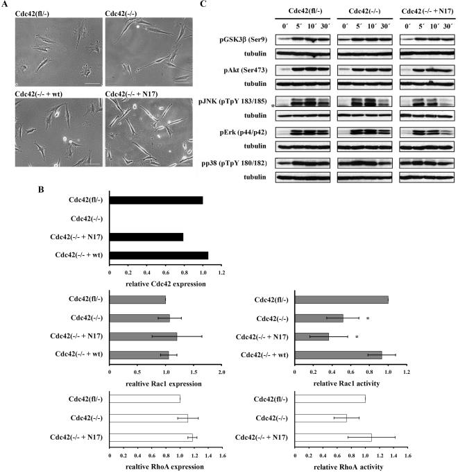
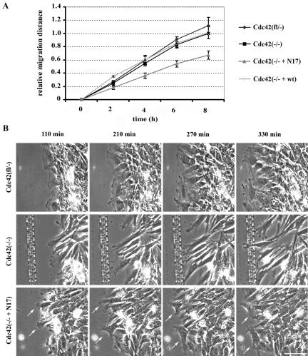
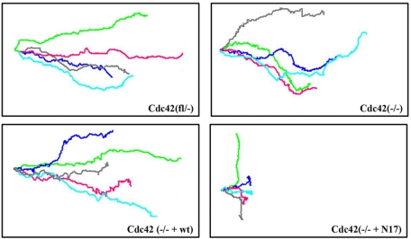
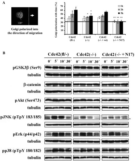
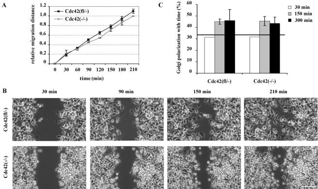
Cdc42 is a small GTPase involved in the regulation of the cytoskeleton and cell polarity. To test whether Cdc42 has an essential role in the formation of filopodia or directed cell migration, we generated Cdc42-deficient fibroblastoid cells by conditional gene inactivation. We report here that loss of Cdc42 did not affect filopodium or lamellipodium formation and had no significant influence on the speed of directed migration nor on mitosis. Cdc42-deficient cells displayed a more elongated cell shape and had a reduced area. Furthermore, directionality during migration and reorientation of the Golgi apparatus into the direction of migration was decreased. However, expression of dominant negative Cdc42 in Cdc42-null cells resulted in strongly reduced directed migration, severely reduced single cell directionality, and complete loss of Golgi polarization and of directionality of protrusion formation toward the wound, as well as membrane blebbing. Thus, our data show that besides Cdc42 additional GTPases of the Rho-family, which share GEFs with Cdc42, are involved in the establishment and maintenance of cell polarity during directed migration.


Figures

Figure 1.
Figure 1.
Cdc42 mutant cell lines display morphological alterations and reduced Rac1 activity. (A) Phase-contrast images of fibroblastoid cell lines studied. Note the spindle-shape morphology of Cdc42(–/–) and Cdc42(–/– + N17) mutants and rescue of the mutant morphology in Cdc42(–/– + wt) cells (scale bar, 100 μm). (B) Top: quantification of Western blot analyses confirming loss of Cdc42 expression in Cdc42(–/–) cells and reexpression of Cdc42 in Cdc42(–/– + N17) and Cdc42(–/– + wt) cell lines. Middle: Rac1 activity relative to total Rac1 as determined by pulldown experiments demonstrating decreased Rac1 activity in Cdc42(–/–) cells (n = 5), a further reduction in Cdc42(–/– + N17) cells (n = 3), and normal levels in Cdc42(–/– + wt) cells (n = 2), relative to Cdc42(fl/–) cells (n = 5). Expression levels of Rac1 were similar in all these cell lines (n = 3). *p < 0.05, compared with control cells. Bottom: RhoA activity as assessed by pulldown assay is similar in control and mutant cell lines and thus does not correlate with observed morphological alterations (n = 3). Total RhoA levels are not significantly different (n = 3). (C) Western blot analysis of starved confluent cells, stimulated with serum as indicated. Representative results of three independent experiments are shown. Asterisk denotes an unspecific band.
Figure 2.
Figure 2.
Comparable adhesion and spreading kinetics and normal integrin expression in the absence of Cdc42. (A) Cell adhesion to different ECM components normalized to the adhesion of the Cdc42(fl/–) cells (n = 2). (B) Integrin expression is not changed in the absence of Cdc42; the background staining is identical for both cell lines.
Figure 3.
Figure 3.
Differentially organized cytoskeleton in Cdc42 mutant cells. Cdc42(–/–) and Cdc42(–/– + N17) mutants form focal contacts but display actin organization rather different from the control (fl/–) and reconstituted (–/– + wt) cell lines as determined by paxillin (red) and phalloidin (green) staining (scale bar, 10 μm). Insets show enlargements of the focal contacts at the tips of actin filaments (scale bar, 5 μm).
Figure 4.
Figure 4.
Loss of Cdc42 does not abrogate lamellipodia and filopodia protrusions. (A) Fluorescence and phase-contrast videomicroscopy (min:sec) taken from a EGFP-VASP-expressing Cdc42-null cell revealing normal recruitment of this protein to the tips of lamellipodia (arrowheads) and filopodia (arrows; scale bar, 5 μm). (B) Phase-contrast videomicroscopy (min:sec) demonstrating formation of a filopodium (arrows) in Cdc42 null ES cells (scale bar, 10 μm). (C) Phase-contrast videomicroscopy of cells expressing dnCdc42 displaying prominent membrane blebbing (small arrows). Note also the lack of proper retraction of the trailing edge, which is commonly left behind (arrows), causing the frequent formation of a-nucleated cytoplasts (arrowheads; scale bar, 10 μm).
Figure 5.
Figure 5.
Cdc42 is not essential for directed cell migration. (A) Relative migration of Cdc42(fl/–), Cdc42(–/–), Cdc42(–/– + wt), and Cdc42(–/– + N17) cells in a wounding assay (n = 5). Only Cdc42(–/– + N17) cells show a significantly reduced directed migration. (B) Phase-contrast time-lapse microscopy of Cdc42(fl/–), Cdc42(–/–), and Cdc42(–/– + N17) cells during a scratch assay, demonstrating stable polarization of lamellipodial activity oriented toward the scratch in Cdc42(fl/–) and Cdc42(–/–) cells and instable polarization in the Cdc42(–/–+ N17) cells with lamellipodial protrusions also pointing into other directions than the scratch (arrows; scale bar, 100 μm).
Figure 6.
Figure 6.
Cdc42 influences single-cell directionality. Single-cell tracking of migrating cells revealed stable directionality of Cdc42(fl/–) and Cdc42(–/– + wt) rescued cells, reduced directionality of Cdc42(–/–) cells and further reduction in Cdc42(–/– + N17) cells. Lines represent individual cell tracks; five representative tracks per cell line are shown (n = 60).
Figure 7.
Figure 7.
Golgi reorientation is partially dependent on Cdc42, but independent of Gsk3β phosphorylation. (A) Percentage of cells with their Golgi apparatus oriented to the direction of migration (random polarization of 33.3% is indicated by solid line). Cdc42(–/–) cells showed a 50% reduction of nonrandomly polarized cells at 5 h of migration, whereas Cdc42(–/–+ N17) cells showed no nonrandom polarization at all (n = 5). Results significantly different from control cells; *p < 0.05); results significantly different from control and null cells; **p < 0.05). (B) Western blot analysis of signal transduction in migrating Cdc42(fl/–), Cdc42(–/–), and Cdc42(–/– + N17) cells at different time points after wounding. Representative results of three independent experiments are shown. Asterisk denotes an unspecific band.
Figure 8.
Figure 8.
Cdc42 endodermal cells display migration and polarization capacity similar to control cells. (A) Similar relative migration of Cdc42(fl/–) and Cdc42(–/–) endodermal cells in a wounding assay (n = 3). (B) Phase-contrast pictures of migrating endodermal cells. Note reduced cell-cell contacts in the migrating front of both cell lines (scale bar, 100 μm). (C) Percentage of cells with their Golgi apparatus oriented in the direction of migration. Cdc42 null cells show polarization ability not significantly different from control cells at 150 and 300 min of migration. Initial polarization was assessed 30 min after wounding (n = 1) and was close to random polarization of 33.3%, indicated by solid line.

References

    1. Abe, T., Kato, M., Miki, H., Takenawa, T., and Endo, T. (2003). Small GTPase Tc10 and its homologue RhoT induce N-WASP-mediated long process formation and neurite outgrowth. J. Cell Sci. 116, 155–168. - PubMed
    1. Aspenström, P., Fransson, A., and Saras, J. (2004). Rho GTPases have diverse effects on the organization of the actin filament system. Biochem. J. 377, 327–337. - PMC - PubMed
    1. Bishop, A. L., and Hall, A. (2000). Rho GTPases and their effector proteins. Biochem. J. 348, 241–255. - PMC - PubMed
    1. Bokoch, G. M. (2003). Biology of the p21-activated kinases. Annu. Rev. Biochem. 72, 743–781. - PubMed
    1. Braga, V. M., Betson, M., Li, X., and Lamarche-Vane, N. (2000). Activation of the small GTPase Rac is sufficient to disrupt cadherin-dependent cell-cell adhesion in normal human keratinocytes. Mol. Biol. Cell. 11, 3703–3721. - PMC - PubMed

Publication types

Substances