. 2005 Jul 13;25(28):6594-600.
doi: 10.1523/JNEUROSCI.0970-05.2005.

Ablation of the inflammatory enzyme myeloperoxidase mitigates features of Parkinson's disease in mice

Affiliations

Ablation of the inflammatory enzyme myeloperoxidase mitigates features of Parkinson's disease in mice

Dong-Kug Choi et al. J Neurosci. .

Abstract

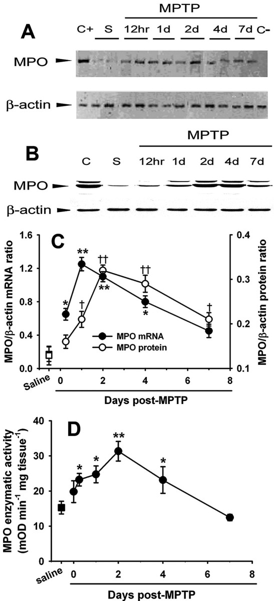
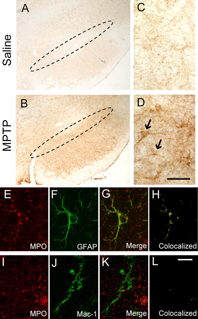
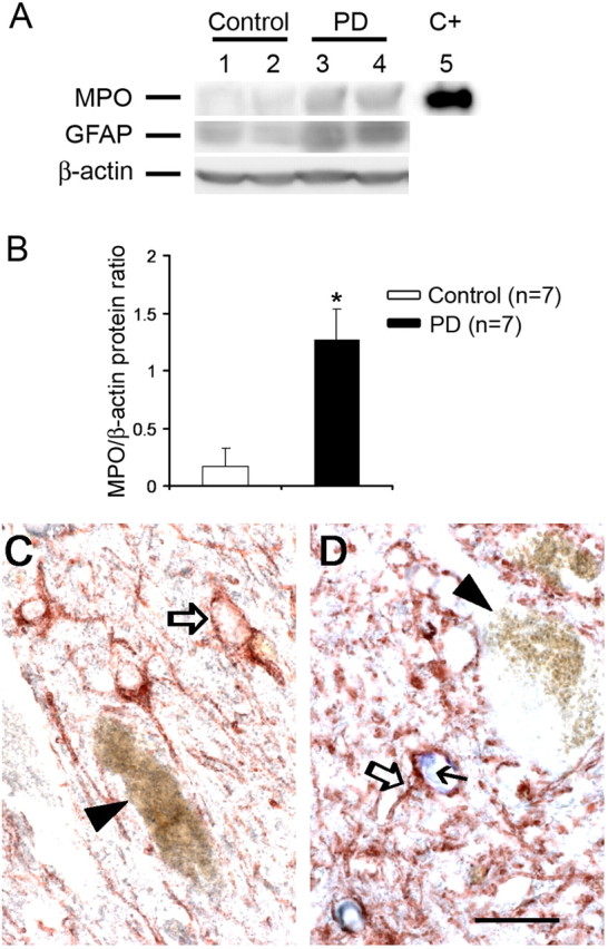
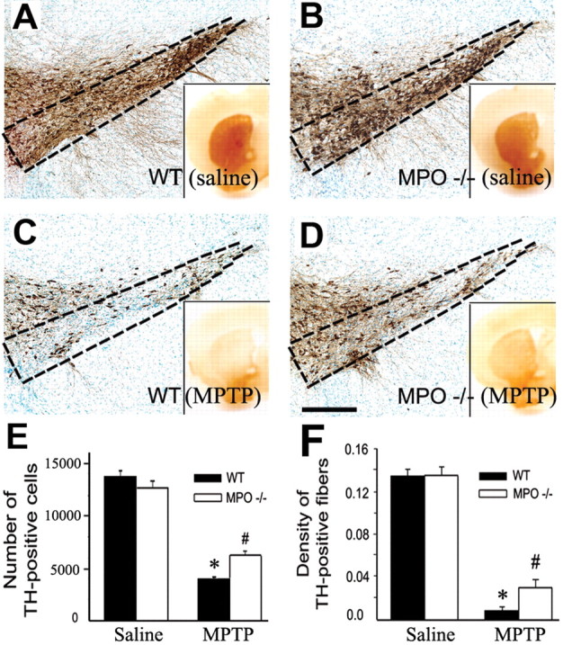
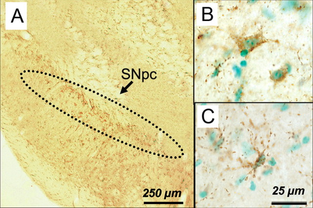
Parkinson's disease (PD) is characterized by a loss of ventral midbrain dopaminergic neurons, which can be modeled by the neurotoxin 1-methyl-4-phenyl-1,2,3,6-tetrahydropyridine (MPTP). Inflammatory oxidants have emerged as key contributors to PD- and MPTP-related neurodegeneration. Here, we show that myeloperoxidase (MPO), a key oxidant-producing enzyme during inflammation, is upregulated in the ventral midbrain of human PD and MPTP mice. We also show that ventral midbrain dopaminergic neurons of mutant mice deficient in MPO are more resistant to MPTP-induced cytotoxicity than their wild-type littermates. Supporting the oxidative damaging role of MPO in this PD model are the demonstrations that MPO-specific biomarkers 3-chlorotyrosine and hypochlorous acid-modified proteins increase in the brains of MPTP-injected mice. This study demonstrates that MPO participates in the MPTP neurotoxic process and suggests that inhibitors of MPO may provide a protective benefit in PD.


Figures

Figure 1.
Figure 1.
MPTP injections are associated with a time-dependent increase in ventral midbrain MPO mRNA (A, C), protein expression (B, C), and enzymatic activity (D) relative to saline injections. Data are means ± SEM for 3-11 mice per group. *,p < 0.05, **,††p < 0.01 compared (Newman-Keuls post hoc test) with saline-injected control animals. S, Saline; C+, positive control (bone marrow); C-, negative control (absence of reverse transcriptase); mOD, millioptical density.
Figure 2.
Figure 2.
A, C, Immunochemical studies revealed no specific MPO immunoreactivity in the ventral midbrain of saline-injected control mice. The dashed oval delineates the SNpc. B, D, However, a dense network of fibers and scattered cell bodies positive for MPO are seen at the level of the SNpc after MPTP injections. Black arrows in D show the MPO-positive cellular elements. E-H, Confocal microscopy demonstrates that ventral midbrain MPO-positive structures (E, red) are also GFAP positive (F, green), as evidenced by the overlay of the two fluorochromes (G) and by the computed mask of the colocalized pixels (H). J-L, In contrast, ventral midbrain MPO-positive structures (I, red) are not MAC-1 positive (J, green), as evidenced by the overlay (K) and the mask of colocalized pixels (L). Tissue sections are from mice at 24 and 48 h after saline or MPTP injections. Scale bars: (in D) A, B, 250 μm; C, D, 25 μm; (in L) E-L,10 μm.
Figure 3.
Figure 3.
A, B, Ventral midbrain MPO tissue content is increased in postmortem tissue from PD patients compared with controls, as well as GFAP tissue content. C+, Positive control (purified MPO). C, Inventral midbrain sections, MPO (blue) is not detected in control tissues, neither in GFAP-positive cells (open arrow) nor in or around neuromelanized dopaminergic neurons (arrowhead). D, Conversely, MPO immunoreactivity (blue, small black arrow) is found in GFAP-positive cells (open arrow) in PD tissue but not in the rare remaining neuromelanized dopaminergic neurons (arrowhead). Scale bar, 20 μm. Data are means ± SEM for seven samples per group. *p < 0.05 compared with normal controls (Newman-Keuls post hoc test).
Figure 4.
Figure 4.
A-D, Ablation of MPO in mutant mice attenuates MPTP-induced striatal TH fibers and SNpc TH neuronal loss, as assessed 7 d after either saline or MPTP injections. E, F, Quantification of neuronal (E) and fiber (F) loss. Data are means ± SEM for four to six mice per group. *p < 0.05 compared with saline-injected animals; #p < 0.05 compared with saline- and MPTP-injected MPO +/+ mice.
Figure 5.
Figure 5.
Immunohistochemical localization of HOCl-modified proteins with the HOP-1 antibody in ventral midbrain sections. Twenty-four hours after MPTP injections, HOP-1-positive immunoreactive material is seen mainly at the level of the SNpc (A) and within or around cellular elements (B, C). Scale bars: A, 250 μm; B, C, 25 μm.

Similar articles

Cited by

References

    1. Andrews PC, Krinsky NI (1982) Quantitative determination of myeloperoxidase using tetramethylbenzidine as substrate. Anal Biochem 127: 346-350. - PubMed
    1. Ara J, Przedborski S, Naini AB, Jackson-Lewis V, Trifiletti RR, Horwitz J, Ischiropoulos H (1998) Inactivation of tyrosine hydroxylase by nitration following exposure to peroxynitrite and 1-methyl-4-phenyl-1,2,3,6-tetrahydropyridine (MPTP). Proc Natl Acad Sci USA 95: 7659-7663. - PMC - PubMed
    1. Brennan Jr WA, Bird ED, Aprille JR (1985) Regional mitochondrial respiratory activity in Huntington's disease brain. J Neurochem 44: 1948-1950. - PubMed
    1. Chen H, Zhang SM, Hernan MA, Schwarzschild MA, Willett WC, Colditz GA, Speizer FE, Ascherio A (2003) Nonsteroidal anti-inflammatory drugs and the risk of Parkinson disease. Arch Neurol 60: 1059-1064. - PubMed
    1. Dauer W, Przedborski S (2003) Parkinson's disease: mechanisms and models. Neuron 39: 889-909. - PubMed

Publication types

MeSH terms