Comparative Study
. 2005 Jul 13;25(28):6610-20.
doi: 10.1523/JNEUROSCI.5009-04.2005.

The zebrafish shocked gene encodes a glycine transporter and is essential for the function of early neural circuits in the CNS

Affiliations
Comparative Study

The zebrafish shocked gene encodes a glycine transporter and is essential for the function of early neural circuits in the CNS

Wilson W Cui et al. J Neurosci. .

Abstract

shocked (sho) is a zebrafish mutation that causes motor deficits attributable to CNS defects during the first2dof development. Mutant embryos display reduced spontaneous coiling of the trunk, diminished escape responses when touched, and an absence of swimming. A missense mutation in the slc6a9 gene that encodes a glycine transporter (GlyT1) was identified as the cause of the sho phenotype. Antisense knock-down of GlyT1 in wild-type embryos phenocopies sho, and injection of wild-type GlyT1 mRNA into mutants rescues them. A comparison of glycine-evoked inward currents in Xenopus oocytes expressing either the wild-type or mutant protein found that the missense mutation results in a nonfunctional transporter. glyt1 and the related glyt2 mRNAs are expressed in the hindbrain and spinal cord in nonoverlapping patterns. The fact that these regions are known to be required for generation of early locomotory behaviors suggests that the regulation of extracellular glycine levels in the CNS is important for proper function of neural networks. Furthermore, physiological analysis after manipulation of glycinergic activity in wild-type and sho embryos suggests that the mutant phenotype is attributable to elevated extracellular glycine within the CNS.


Figures

Figure 1.
Figure 1.
Spontaneous coiling behavior is defective in sho embryos. A, Individual frames from a time-lapse video showing two consecutive coils by a wild-type sibling embryo (22 hpf). Each coil lasted ∼500 ms and is denoted by an arrow. Time of each frame is shown at the top right corner. Scale bar, 0.5 mm. B, The frequency of coiling is reduced at all stages in sho embryos (circles) compared with wild-type siblings (wtsib; squares). *p < 0.001 by Student's t test. Error bars are SDs.
Figure 2.
Figure 2.
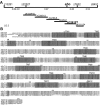
Molecular identification of the sho gene. A, The sho locus meiotically mapped to a region on LG 2 flanked by several zebrafish microsatellite markers. The relative meiotic distance between these markers and the sho locus are shown. Six contiguous, ordered BACs (shown below the meiotic map) spanned the entire region containing the two closest markers, z22527 and z7632. BAC zC261P7 contained an ORF homologous to the mammalian gene slc6a9 that encodes GlyT1A. B, The amino acid sequence alignment of three isoforms of zebrafish GlyT1 (zGlyT1A, zGlyT1B, and zGlyT1Z) with human GlyT1A. Identical sequences are lightly shaded, and the highly conserved transmembrane domains (TM1-TM12) are darkly shaded. The position of point mutation G81D in the sho allele is denoted by an asterisk.
Figure 3.
Figure 3.
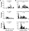
GlyT1 function is required for normal spontaneous coiling behavior. A, GlyT1 antisense MO injections phenocopy sho. Histogram showing the percentage (y-axis) of uninjected wild-type (wt; top), control (ctrl) MO-injected wild-type (middle), and GlyT1 E2I2 MO-injected wild-type (bottom) embryos exhibiting spontaneous coiling at various frequencies (x-axis). Knock-down of GlyT1 function severely reduced the frequency of spontaneous coiling by wild-type embryos. B, Injection of wild-type GlyT1A mRNA but not G81D mutant mRNA rescues sho embryos. Approximately one-quarter of uninjected embryos (top) and embryos injected with G81D mRNA (middle) from a cross of sho carriers coiled with reduced frequencies, but nearly all embryos from a cross of carriers injected with wild-type mRNA coiled at frequencies within the normal range (bottom).
Figure 4.
Figure 4.
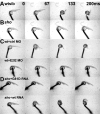
GlyT1 function is required for normal escape responses at 26 hpf. A, A wild-type sibling (wtsib) embryo responded to tactile stimulation with fast, alternating trunk contractions. Shown is the first of two contractions. Time of each frame is noted in the top right corner. B, A sho embryo failed to respond to tactile stimulation. C, Apparent knock-down of GlyT1 phenocopied sho. A control (ctrl) MO-injected wild-type embryo responds to tactile stimulation with fast, alternating trunk contractions (top), whereas an antisense E2I2 MO-injected wild-type embryo failed to respond (bottom). D, Apparent restoration of GlyT1 activity in sho embryos rescued the mutant phenotype. A sho embryo injected with G81D mutant mRNA failed to respond to tactile stimulation (top), whereas a sho embryo injected with wild-type GlyT1 mRNA responded normally (bottom). The wild-type mRNA-injected embryo was genotyped based on its mutant response to tactile stimulation at 48 hpf (for details, see Results).
Figure 5.
Figure 5.
The G81D mutation in GlyT1 results in a nonfunctional glycine transporter. A, Current responses in Xenopus oocytes injected with wild-type zebrafish GlyT1A mRNA, G81D mutant, and control oocytes, to bath application of glycine. The shaded bars at the top represent different concentrations of glycine in the bath (in order: 2, 5, 10, 50, 100, 300, and 600 μm). B, A plot of the average relative current responses of oocytes expressing wild-type GlyT1A to different concentrations of glycine (n = 10). The current measurements were normalized to the maximum response of each oocyte and expressed as percentage of the maximum current. C, The G81D mutant GlyT1 does not generate a glycine-dependent inward current. Shown are the average maximum inward currents induced by 600 μm glycine in wild-type GlyT1 mRNA-injected, G81D mRNA-injected, and uninjected control oocytes. Error bars represent SEs.
Figure 6.
Figure 6.
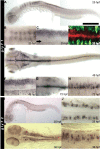
glyT1 and glyT2 are predominantly expressed by the hindbrain and spinal cord. In all panels, anterior is to the left, and, in the lateral views, dorsal is up. A, Lateral view of a whole-mounted embryo showing that glyT1 is expressed in hindbrain stripes, dorsal spinal cord, and the tectum at 25 hpf. B, Dorsal view showing the stripes of glyT1-positive cells in the dorsal hindbrain at 25 hpf. C, Lateral view focused at the midline focal plane showing that glyT1 is expressed by cells in the medial dorsal spinal cord. The row of midline floor-plate cells can be seen at the ventral midline (arrow). D, A confocal image of the 25 hpf spinal cord seen in a dorsal view demonstrating that glyT1-positive dorsal cells (red) at the midline are not postmitotic neurons. Neurons are labeled with anti-Hu (green) and located more laterally. E, Dorsal view of a whole-mounted 48 hpf embryo showing that glyT1 is expressed at the midline of the CNS, stripes in the hindbrain, and scattered cells in the lateral spinal cord. F, Lateral view of the 48 hpf spinal cord showing that glyT1 is expressed in longitudinal dorsal and ventral stripes in the spinal cord but not in between. G, Dorsal view of the 72 hpf hindbrain showing expression of glyT1 by cells at the superficial surface near the ventricle. H, Lateral view of the 72 hpf spinal cord showing that glyT1 is now expressed by a longitudinal stripe of cells that occupies the intermediate region that was glyT1 negative at 48 hpf. glyT1 is no longer expressed in the dorsal and ventral stripes. I, Lateral view of a whole-mounted 25 hpf embryo showing that glyT2 is expressed by apparent neurons in the spinal cord. J, Dorsal view of the 25 hpf spinal cord showing that the glyT2-positive cells are located at the margin of the spinal cord and are likely to be neurons. K, Dorsal view of a 36 hpf embryo showing an increased number of glyT2-positive cells in the spinal cord and many glyT2-positive cells in the hindbrain. L, Lateral view of the spinal cord showing that the glyT2-positive cells are located in the position occupied by commissural neurons at 36 hpf. Scale bar: A, E, K, 300 μm; B, 210 μm; C, F, G, J, L, 60 μm; D, 70 μm; H, 160 μm; I, 450 μm.
Figure 7.
Figure 7.
Partial block of glycinergic inhibitory synaptic transmission can restore swimming in sho embryos, but the response to mechanosensory stimulation is still aberrant. A, A series of muscle voltage recordings from a wild-type embryo in response to mechanosensory stimulation (arrow) after exposure to different concentrations of strychnine. i, Rhythmic depolarizations are elicited in the absence of strychnine. ii, An irregular, large depolarization is elicited in 10 μm strychnine. iii, An irregular, large depolarization is elicited in 50 μm strychnine. B, A series of muscle recordings from a sho embryo in response to mechanosensory stimulation (arrow) after exposure to different concentrations of strychnine. i, A typical sho-like depolarization is elicited. ii, An irregular, large depolarization followed by rhythmic depolarizations (bracket) similar to swimming is elicited in 10 μm strychnine. iii, Only an irregular, large depolarization is elicited in 50 μm strychnine. wtsib, Wild-type sibling.
Figure 8.
Figure 8.
The sho response to mechanosensory stimulation is phenocopied by pharmacologically blocking GlyT1 in wild-type embryos, and putative dilution of high extracellular glycine in sho embryos restores swimming responses. A, Muscle voltage recording from a wild-type (wt) embryo (36hpf) 3 h after a control injection showed a normal fictive swimming in response to mechanosensory stimulation (arrow). B, Muscle recording from a wild-type embryo (36 hpf) 3 h after a NFPS injection showing showed a large, nonrhythmic depolarization resembling the sho in response to touch. C, Muscle recording from a sho embryo (42 hpf) after the hindbrain was exposed and perfused for 30 min with glycine-free solution showed rhythmic depolarizations similar to fictive swimming in response to touch. D, Muscle recording from the same embryo as in C after switching from perfusion with glycine-free solution to perfusion with solution containing 200 μm glycine showed the large, nonrhythmic depolarization characteristic of sho in response to touch.

Similar articles

Cited by

References

    1. Adams RH, Sato K, Shimada S, Tohyama M, Puschel AW, Betz H (1995) Gene structure and glial expression of the glycine transporter GlyT1 in embryonic and adult rodents. J Neurosci 15: 2524-2532. - PMC - PubMed
    1. Applegarth DA, Toone JR (2004) Glycine encephalopathy (nonketotic hyperglycinaemia): review and update. J Inherit Metab Dis 27: 417-422. - PubMed
    1. Aragon C, Lopez-Corcuera B (2003) Structure, function and regulation of glycine neurotransporters. Eur J Pharmacol 479: 249-262. - PubMed
    1. Aubrey KR, Vandenberg RJ (2001) N[3-(4′-fluorophenyl)-3-(4′-phenyl-phenoxy)propyl]sarcosine (NFPS) is a selective persistent inhibitor of glycine transport. Br J Pharmacol 134: 1429-1436. - PMC - PubMed
    1. Ben-Ari Y (2001) Developing networks play a similar melody. Trends Neurosci 24: 353-360. - PubMed

Publication types

MeSH terms

Substances