. 2005 Jul 13;25(28):6631-40.
doi: 10.1523/JNEUROSCI.1139-05.2005.

Rat adrenal chromaffin cells are neonatal CO2 sensors

Affiliations

Rat adrenal chromaffin cells are neonatal CO2 sensors

Ana M Muñoz-Cabello et al. J Neurosci. .

Abstract

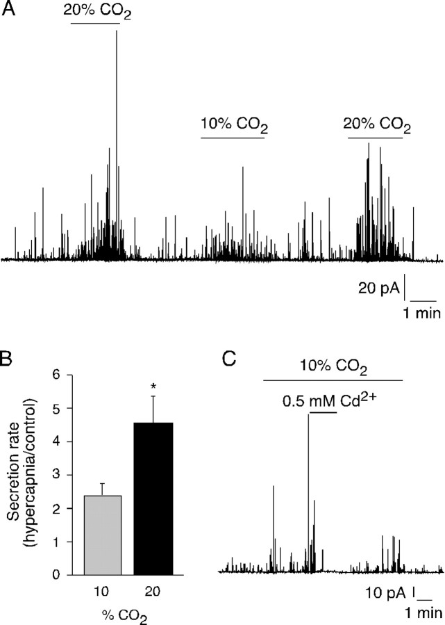
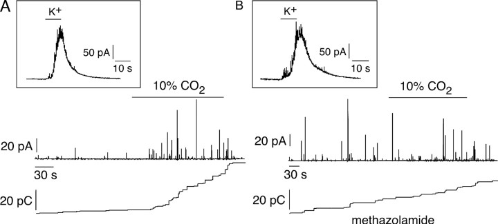
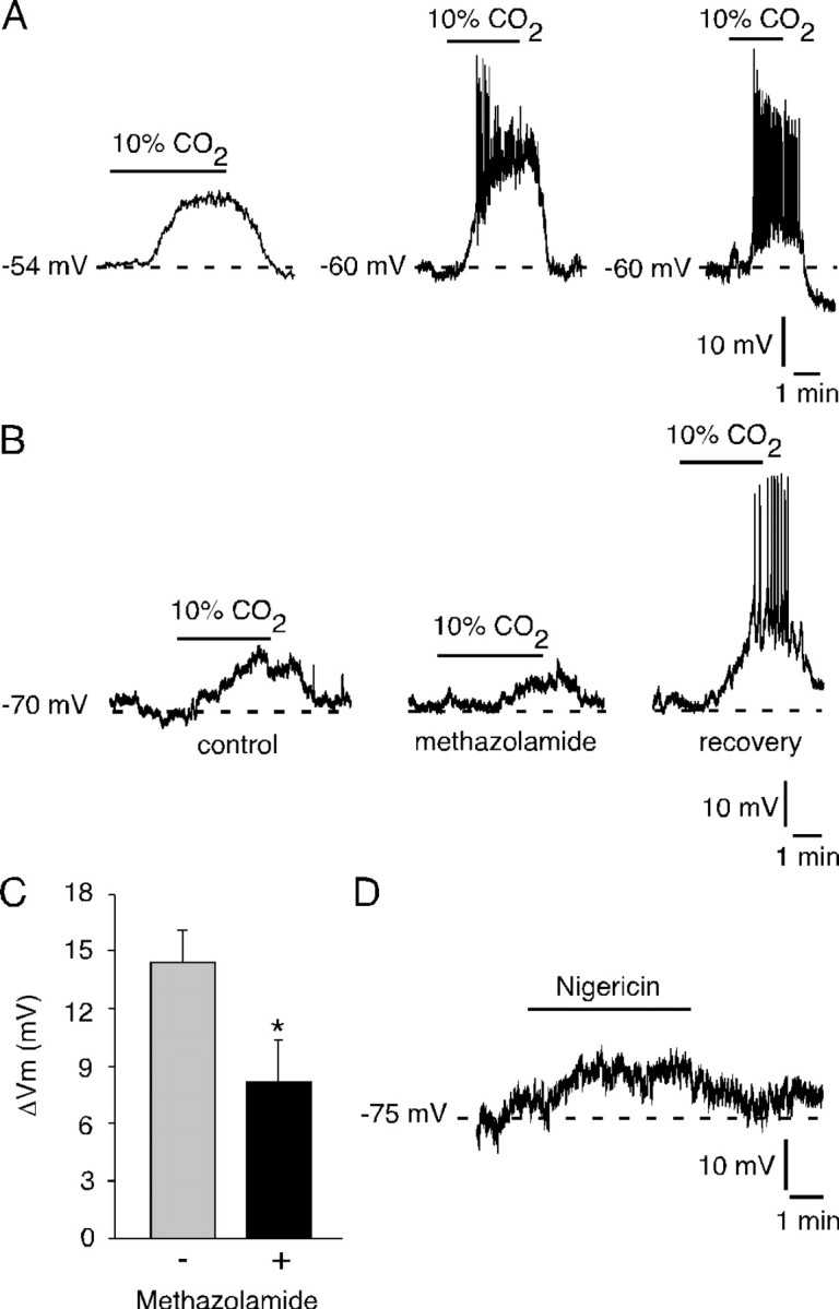
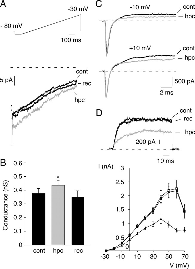
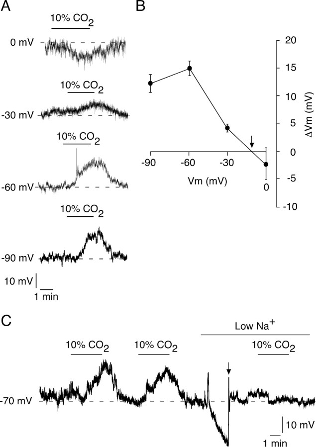
We studied the participation of adrenal medulla (AM) chromaffin cells in hypercapnic chemotransduction. Using amperometric recordings, we measured catecholamine (CAT) secretion from cells in AM slices of neonatal and adult rats perfused with solutions bubbled with different concentrations of CO2. The secretory activity augmented from 1.74 +/- 0.19 pC/min at 5% CO2 to 6.36 +/- 0.77 pC/min at 10% CO2. This response to CO2 was dose dependent and appeared without changes in extracellular pH, although it was paralleled by a drop in intracellular pH. Responsiveness to hypercapnia was higher in neonatal than in adult slices. The secretory response to hypercapnia required extracellular Ca2+ influx. Both the CO2-induced internal pH drop and increase in CAT secretion were markedly diminished by methazolamide (2 microm), a membrane-permeant carbonic anhydrase (CA) inhibitor. We detected the presence of two CA isoforms (CAI and CAII) in neonatal AM slices by in situ hybridization and real-time PCR. The expression of these enzymes decreased in adult AM together with the disappearance of responsiveness to CO2. In patch-clamped chromaffin cells, hypercapnia elicited a depolarizing receptor potential, which led to action potential firing, extracellular Ca2+ influx, and CAT secretion. This receptor potential (inhibited by methazolamide) was primarily attributable to activation of a resting cationic conductance. In addition, voltage-gated K+ current amplitude was also decreased by high CO2. The CO2-sensing properties of chromaffin cells may be of physiologic relevance, particularly for the adaptation of neonates to extrauterine life, before complete maturation of peripheral and central chemoreceptors.


Figures

Figure 1.
Figure 1.
Secretory response to hypercapnia of rat chromaffin cells. A, Top, Amperometric recording showing the secretory response to hypercapnia (10% CO2) of a neonatal chromaffin cell in an adrenal gland slice. Bottom, Cumulative secretion signal. B, Quantification of the secretory response of neonatal chromaffin cells during normocapnia (5% CO2), acidic hypercapnia [10% CO2, extracellular pH (pHe) 7.1], and isohydric hypercapnia (10% CO2, pHe 7.4). Secretion rate was expressed as picocoulombs per minute (mean ± SEM; n = 7 cells), *p < 0.05 (one-way ANOVA). C, Comparison of the secretion rate (picocoulombs per minute) during 5 and 10% CO2 exposure in responsive chromaffin cells from neonatal and adult rats (mean ± SEM; n = 33 for neonatal and n = 5 for adult rats). C, Inset, Secretory response to hypercapnia of an adult chromaffin cell in an adrenal gland slice. **p < 0.005 (Student's t test).
Figure 2.
Figure 2.
Dose and Ca2+ dependence of the secretory response of adrenal chromaffin cells to hypercapnia. A, Amperometric recording of a neonatal chromaffin cell from an adrenal gland slice illustrating the secretory activity evoked by increasing CO2 levels from control (5%) to hypercapnia (10 and 20%). B, Quantification of the secretory dose-response to hypercapnia. Data are expressed as the secretion rate in hypercapnia (PCO2, 10 or rate 20%) divided by the secretion rate in control (PCO2, 5%) (mean ± SEM; n = 9), *p < 0.05 (Student's t test). C, Inhibition of the secretory response to 10% CO2 by extracellular cadmium chloride (0.5 mm).
Figure 3.
Figure 3.
Intracellular acidification induced by hypercapnia and inhibition of the response by methazolamide. A, Microfluorimetric measurements of changes in intracellular pH [arbitrary units (a.u.)] of isolated neonatal chromaffin cells using the acetoxymethyl ester of BCECF at a concentration of 2 μm. Relative intracellular pH expressed as the ratio of fluorescence intensities (R) measured after excitation at two wavelengths (F490 and F450). CO2 in the external solution was changed from 5% (control) to either 10 or 20%. B, Reversible inhibition of hypercapnia-induced intracellular acidification by methazolamide (2 μm). Cells were preincubated for 5 min in methazolamide before application of 10% CO2.
Figure 4.
Figure 4.
Participation of CA in the secretory response to hypercapnia of neonatal chromaffin cells. A, Amperometric recording from a neonatal chromaffin cell during exposure to hypercapnia (10% CO2). A, Inset, Secretory response to high extracellular K+ (40mm)in the same conditions. B,Effect of CAs inhibition by 2 μm methazolamide on the secretory response to hypercapnia of the same cell. B, Inset, Secretory response to high extracellular K+ in the presence of methazolamide.
Figure 5.
Figure 5.
Expression of CA isoforms (CAI and CAII) in the AM. A-B, Comparison by in situ hybridization of CAI, CAII, and TH mRNA levels in slices of neonatal (A) and adult (B) adrenal glands. Note the disappearance of CAI and CAII expression in adult adrenal glands. In both cases, two series of microphotographs with different magnification are shown.
Figure 6.
Figure 6.
Hypercapnia- and acid load-elicited membrane depolarization in isolated neonatal chromaffin cells. A, Examples of membrane potential changes induced by exposure to 10% CO2 in three different isolated current-clamped neonatal chromaffin cells. The recordings were done with the perforated patch configuration of the patch-clamp technique. Broken lines indicate the spontaneous resting potential level. B, C, Inhibition of hypercapnia-induced membrane depolarization by methazolamide (2 μm). Cells were preincubated for 5 min in methazolamide before application of 10% CO2. The external solution contained TTX (1 μm). Resting membrane potential was held at -70 mV with hyperpolarizing current. Data in C represent the peak amplitude of the depolarizing response to hypercapnia (ΔVm) before and during application of methazolamide (n = 5 cells; *p < 0.05; Student's t test). D, Membrane depolarization induced by exposure to nigericin (4 μm). Resting membrane potential was held at -75 mV with hyperpolarizing current. A representative example of three experiments is shown.
Figure 7.
Figure 7.
Changes in membrane ionic conductances induced by hypercapnia. A, Representative example of whole-cell membrane currents recorded during a ramp depolarization from -90 to -30 mV in a voltage-clamped (perforated patch) neonatal chromaffin cell treated with TTX(1 μm) and exposed to hypercapnia (10%CO2). The broken line indicates the 0 current level. B, Reversible increase of slope membrane conductance (measured from -90 to -60 mV) induced by high PCO2 in voltage-clamped chromaffin cells (13 measurements in 6 cells; *p < 0.05; Student's t test). C, Lack of effect of hypercapnia on the voltage-dependent inward currents and inhibition of outward currents in a voltage-clamped neonatal chromaffin cell. Superimposed recordings of the currents elicited by depolarizing pulses from -80 mV to the potential indicated are illustrated. Broken lines indicate the 0 current level. D, Top, Reduction of the outward K+ current by hypercapnia elicited by a depolarizing pulse from -80 to +20 mV in a neonatal voltage-clamped chromaffin cell treated with 1 μm TTX. D, Bottom, I-V relation for the peak outward current. Voltage steps were applied from the holding potential (-80 mV) to different test potentials (from -30 to +70 mV) in neonatal chromaffin cells treated with 1μm TTX. Average of the peak outward current is plotted against the test potential in normocapnia (□), hypercapnia (▴), and after removal of the hypercapnic stimulus (•). Data were collected from n = 16 cells. cont, Control (5% CO2); hpc, hypercapnia (10% CO2); rec, recovery (5% CO2).
Figure 8.
Figure 8.
Cationic conductance induced by high PCO2. A, Hypercapnia-induced receptor potential at various resting membrane voltage levels in cells bathed with TTX (1 μm)-containing solutions to block voltage-gated Na+ channels. The broken lines indicate the potential level before exposure to 10% CO2. B, Amplitude of the hypercapnia-induced receptor potential (ΔVm) and reversal at approximately -10 mV (arrow) (each point represents the average of 3-9 cells tested).C,Abolishment of the hypercapnia-induced depolarizing receptor potential by removal of extracellular Na+. Resting membrane potential (broken line) was held at -70 mV with hyperpolarizing current. Hyperpolarization induced by removal of Na+ was also compensated by application of depolarizing current (arrow). A representative example of three other experiments is shown.

Similar articles

Cited by

References

    1. Andersson DA, Chase HW, Bevan S (2004) TRPM8 activation by menthol, icilin, and cold is differentially modulated by intracellular pH. J Neurosci 24: 5364-5369. - PMC - PubMed
    1. Augustine GJ, Neher E (1992) Calcium requirements for secretion in bovine chromaffin cells. J Physiol (Lond) 450: 247-271. - PMC - PubMed
    1. Buckler KJ, Vaughan-Jones RD (1994) Effects of hypercapnia on membrane potential and intracellular calcium in rat carotid body type I cells. J Physiol (Lond) 478: 157-171. - PMC - PubMed
    1. Buckler KJ, Vaughan-Jones RD, Peers C, Nye PC (1991a) Intracellular pH and its regulation in isolated type I carotid body cells of the neonatal rat. J Physiol (Lond) 436: 107-129. - PMC - PubMed
    1. Buckler KJ, Vaughan-Jones RD, Peers C, Lagadic-Gossmann D, Nye PC (1991b) Effects of extracellular pH, PCO2 and HCO-3 on intracellular pH in isolated type-I cells of the neonatal rat carotid body. J Physiol (Lond) 444: 703-721. - PMC - PubMed

Publication types

MeSH terms

LinkOut - more resources