Comparative Study
. 2005 Jul 13;25(28):6676-86.
doi: 10.1523/JNEUROSCI.1079-05.2005.

Structural determinants of alpha4beta2 nicotinic acetylcholine receptor trafficking

Affiliations
Comparative Study

Structural determinants of alpha4beta2 nicotinic acetylcholine receptor trafficking

Xiao-Qin Ren et al. J Neurosci. .

Abstract

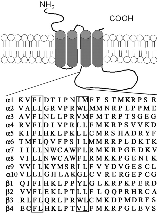
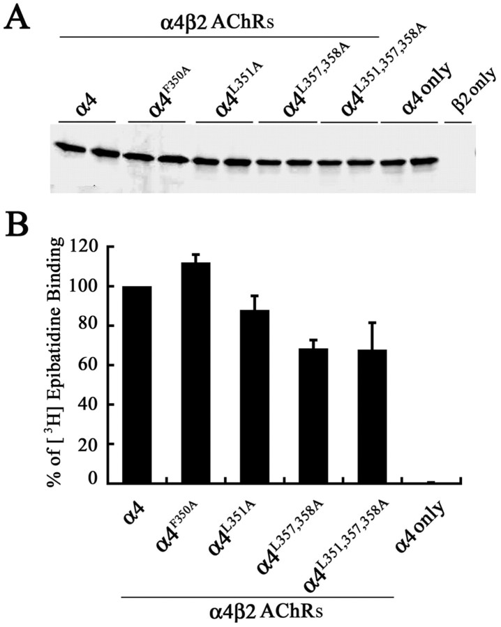
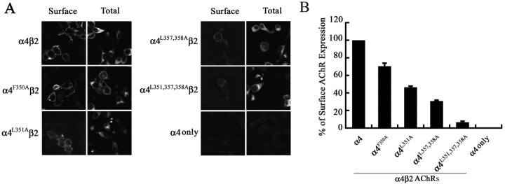
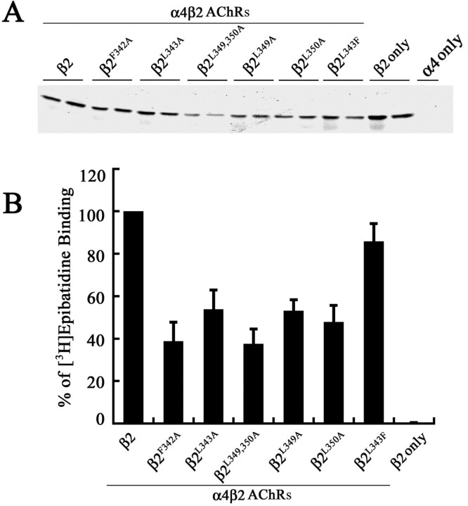
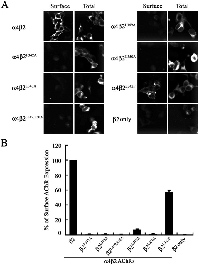
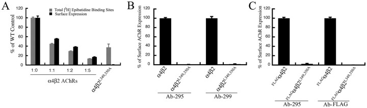
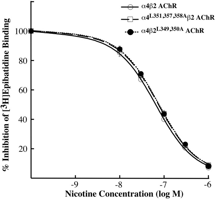
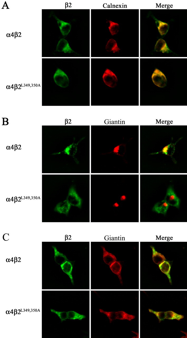
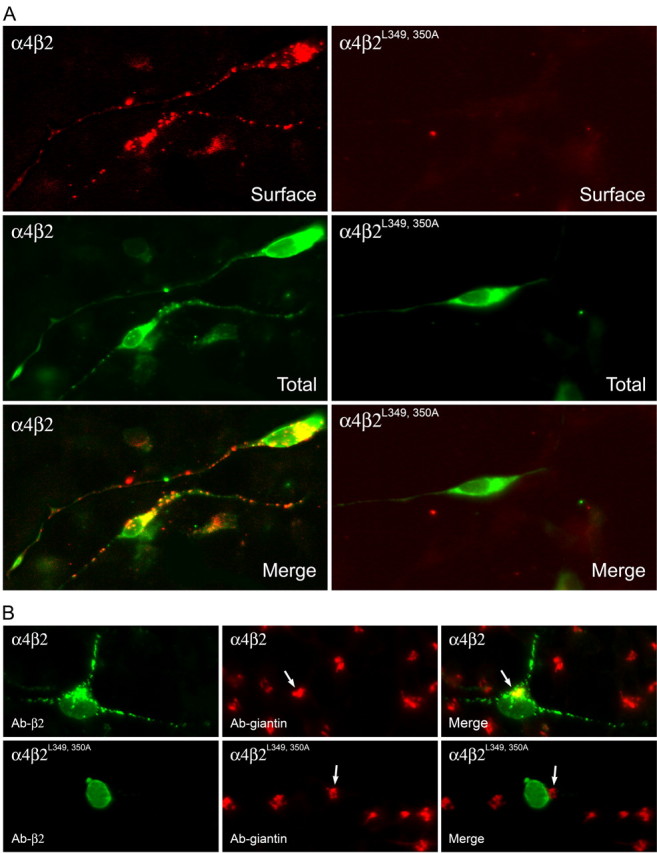
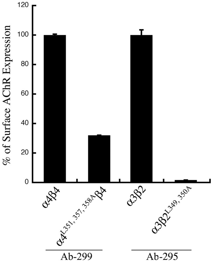
The structural determinants of nicotinic acetylcholine receptor (AChR) trafficking have yet to be fully elucidated. Hydrophobic residues occur within short motifs important for endoplasmic reticulum (ER) export or endocytotic trafficking. Hence, we tested whether highly conserved hydrophobic residues, primarily leucines, in the cytoplasmic domain of the alpha4beta2 AChR subunits were required for cell surface expression of alpha4beta2 AChRs. Mutation of F350, L351, L357, and L358 to alanine in the alpha4 AChR subunit attenuates cell surface expression of mutant alpha4beta2 AChRs. Mutation of F342, L343, L349, and L350 to alanine at homologous positions in the beta2 AChR subunit abolishes cell surface expression of mutant alpha4beta2 AChRs. The hydrophobic nature of the leucine residue is a primary determinant of its function because mutation of L343 to another hydrophobic amino acid, phenylalanine, in the beta2 AChR subunit only poorly inhibits trafficking of mutant alpha4beta2 AChR to the cell surface. All mutant alpha4beta2 AChRs exhibit high-affinity binding for [3H]epibatidine. In both tsA201 cells and differentiated SH-SY5Y neural cells, wild-type alpha4beta2 AChRs colocalize with the Golgi marker giantin, whereas mutant alpha4beta2 AChRs fail to do so. The striking difference between mutant alpha4 versus mutant beta2 AChR subunits on cell surface expression of mutant alpha4beta2 AChRs points to a cooperative or regulatory role for the alpha4 AChR subunit and an obligatory role for the beta2 AChR subunit in ER export. Collectively, our results identify, for the first time, residues within AChR subunits that are essential structural determinants of alpha4beta2 AChR ER export.


Figures

Figure 1.
Figure 1.
Sequence homology among the long cytosolic loop of AChR subunits. Topology of AChR subunits and alignment of a 20 amino acid region of the long cytosolic loop showing location of hydrophobic residues that are conserved in the ER export motif identified in this study. The boxed residues correspond to those that are at homologous positions in all of the AChR subunits and those that were specifically mutated in the α4 (F350, L351, L357, and L358) and β2 (F342, L343, L349, L350) AChR subunits.
Figure 2.
Figure 2.
Subunit expression and assembly of high-affinity [3H]epibatidine binding sites by wild-type and mutant α4 AChR subunits. A, The α4, α4F350A, α4L351A, α4L357, 358A, or α4L351, 357, 358A AChR subunit was coexpressed with the wild-type β2 AChR subunit in tsA201 cells, and 2% NP-40-solubilized membrane proteins were subjected to immunoblot analysis using an mAb against the α4 AChR subunit (mAb 299). Paired lanes represent the expression levels of the AChR subunit from two independently transfected wells from a representative experiment. B, Detergent-permeabilized transfected fixed cells expressing wild-type and mutant α4β2 AChRs were incubated with 3 nm [3H]epibatidine overnight at 4°C. The amount of bound [3H]epibatidine was quantitated by scintillation counting. Data for the mutants were expressed as a percentage of the [3H]epibatidine binding observed for the wild-type α4β2. The data represent the mean ± SE from four experiments.
Figure 3.
Figure 3.
Hydrophobic residue mutations in the α4 AChR subunit attenuate surface expression of α4β2 AChRs. A, Transfected tsA201 cells expressing wild-type or mutant α4β2 AChRs, or the α4 AChR subunit alone, were processed for immunohistochemistry and probed with an mAb against the β2 AChR subunit (mAb 295). Ab binding was detected using an Alexa 488-conjugated goat anti-rat secondary Ab and imaged with a deconvolution fluorescence microscope. Ab staining to intracellular α4β2 AChRs was detected in 0.1% Triton X-100-permeabilized cells. Shown are 1 μm optical sections of the cells. B, The cell surface expression of α4β2 AChRs was quantitated using an enzyme-linked colorimetric assay. The absorbance data were expressed as a percentage of the value obtained for the wild-type α4β2 AChRs. The data represents the mean ± SE from four experiments.
Figure 4.
Figure 4.
Subunit expression and assembly of high-affinity [3H]epibatidine binding sites by wild-type and mutant β2 AChR subunits. A, The β2, β2F342A, β2L343A, β2L349,350A, β2L349A, β2L350A, β2F342A, or β2L343F AChR subunit was coexpressed with the wild-type α4 AChR subunit in tsA201 cells, and 2% NP-40-solubilized membrane proteins were subjected to immunoblot analysis using a polyclonal antiserum against the β2 AChR subunit. Paired lanes represent the expression levels of the AChR subunit from two independently transfected wells from a representative experiment. B, Detergent-permeabilized transfected fixed cells expressing wild-type and mutant α4β2 AChRs, or the β2 AChR subunit alone, were incubated with 3 nm [3H]epibatidine overnight at 4°C. The amount of bound [3H]epibatidine was quantitated by scintillation counting. Data for the mutants were expressed as a percentage of the [3H]epibatidine binding observed for the wild-type α4β2. The data represent the mean ± SE from four experiments.
Figure 5.
Figure 5.
Hydrophobic residue mutations in the β2 AChR subunit abolish surface expression of α4β2 AChRs. A, Transfected tsA201 cells expressing wild-type, mutant α4β2 AChRs, and the β2 AChR subunit alone were processed for immunohistochemistry and probed with an Ab against the β2 AChR subunit (mAb 295). Ab binding was detected using an Alexa 488-conjugated goat anti-rat secondary Ab and imaged with a deconvolution fluorescence microscope. Ab staining to α4β2 AChRs was detected in 0.1% Triton X-100-permeabilized cells before addition of Abs. Shown are 5 μm optical sections of the cells. B, The cell surface expression of α4β2 AChRs was quantitated using an enzyme-linked colorimetric assay. The data were expressed as a percentage of the value obtained for wild-type a4b2 AChRs. The data represent the mean ± SE from four experiments.
Figure 6.
Figure 6.
Cell surface expression levels of wild-type and mutant α4β2 AChRs measured at different overall expression levels and using different Abs. A, Culture wells containing tsA201 cells expressing different levels of wild-type α4β2 AChRs were fixed and then incubated with 3 nm [3H]epibatidine overnight at 4°C. The amount of bound [3H]epibatidine was quantitated by scintillation counting. The cell surface expression levels of wild-type and mutant α4β2 AChRs was quantitated using an enzyme-linked colorimetric assay. The data represent the mean ± SD from four experiments. B, Transfected tsA201 cells expressing wild-type or mutant α4β2L349,350A AChRs were fixed and probed with mAb against the β2 AChR subunit (mAb 295) or mAb against α4 AChR subunit (mAb 299). The cell surface expression level of α4β2 AChRs was quantitated using an enzyme-linked colorimetric assay. The absorbance data were expressed as a percentage of the value obtained for the wild-type α4β2 AChR. The data represents the mean ± SE from four experiments. C, tsA201 cells were transfected with FLAGα4 and β2 or β2L349,350A subunits. The cell surface expression of α4β2 AChRs was quantitated using an enzyme-linked colorimetric assay with mAb against the β2 AChR subunit (mAb 295) or the M2 Ab against the FLAG epitope in the α4 subunit. The absorbance data were expressed as a percentage of the value obtained for the wild-type FLAGα4β2 AChR. The data represents the mean ± SE from four experiments.
Figure 7.
Figure 7.
Leucine mutations in the α4 and β2 AChR subunits do not affect α4β2 AChR ligand affinity. Transfected cells expressing mutant α4L351,357,358Aβ2 AChR, α4β2L349,350A AChR, or wild-type α4β2 AChR were fixed, permeabilized with 0.5% Triton X-100, and incubated with 400 pm [3H]epibatidine in the presence or absence of 10, 30, 100, 300, and 1000 nm nicotine for 24 h. The amount of bound [3H]epibatidine was determined by liquid scintillation counting. Background binding was determined in nontransfected cells and was subtracted. The data shown was normalized to the amount bound in the absence of nicotine and represent the mean ± SE from four experiments. The data were fitted to the Hill equation, and the KI values obtained are 0.65 ± 0.03 nm (nH = 0.91) for the α4, 0.77 ± 0.04 nm (nH = 0.92) for the α4L351,L357,358A, and 0.74 ± 0.02 nm (nH = 0.92) for the β2L349,350A.
Figure 8.
Figure 8.
Leucine mutations in β2 AChR subunit block ER export. A, Transfected tsA201 cells expressing wild-type α4β2 AChRs or mutant α4β2L349,350A AChRs were incubated at 30°C, processed for immunohistochemistry, and probed with Abs against the β2 AChR subunit (mAb 295) and against calnexin. Ab binding to the β2 AChR subunit was detected using an Alexa Fluor 488-conjugated goat anti-rat secondary Ab and to calnexin using an Alexa Fluor 647-conjugated goat anti-rabbit secondary Ab. Cells were imaged with a deconvolution fluorescence microscope. Staining of β2 is shown in green, and staining of calnexin is shown in red. Colocalization of the proteins is shown in yellow in the merged image. B, Transfected tsA201 cells expressing wild-type α4β2 AChRs or mutant α4β2L349,350A AChRs were incubated at 30°C and then transferred to 37°C for 2 h. Cells were then processed for immunohistochemistry and probed with Abs against the β2 AChR subunit (mAb 295) and against giantin. Ab binding to the β2 AChR subunit was detected using an Alexa Fluor 488-conjugated goat anti-rat IgG and to giantin using Alexa Fluor 647-conjugated goat anti-rabbit IgG. Cells were imaged with a deconvolution fluorescence microscope. Staining of β2 is shown in green, and staining of giantin is shown in red. Colocalization is shown in yellow in the merged image. C, tsA201 cells were treated under the same procedure as in B except for an additional treatment with brefeldin A (1 μg/ml) for 1 h at 37°C.
Figure 9.
Figure 9.
Subcellular distribution of wild-type α4β2 AChRs and mutant α4β2L349,350A AChRs in differentiated SH-SY5Y neural cells. A, Wild-type α4β2 AChRs are expressed on the surface membrane of the soma and processes, but mutant α4β2L349, 350A AChRs fail to do so even in cells that have high expression levels of the proteins in the cytoplasm. Transfected SH-SY5Y cells expressing wild-type α4β2 and mutant α4β2L349, 350A AChRs were incubated at 30°C for 2 d and then fixed for immunohistochemical processing. The cells were probed with Ab against the β2 AChR subunit (mAb 295) before permeabilization, and the Ab binding was detected with the Alexa Fluor 647-conjugated goat anti-rat Abs. These same cells were then permeabilized and probed with the β2 AChR subunit Ab (mAb 295) again, and the Ab binding was detected using the Alexa Fluor 488-conjugated goat anti-rat Abs. B, Wild-type α4β2 AChRs (green) colocalize with giantin (red), whereas mutant α4β2L349, 350A AChRs (green) do not. Transfected SH-SY5Y cells expressing wild-type α4β2or mutant α4β2L349,350A AChRs were incubated at 30°C and then shifted to 37°C for 2 h. The cells were fixed, permeabilized, and probed with the β2 AChR subunit Ab (mAb 295) and the giantin Ab. Ab binding to the β2 AChR subunit was visualized using Alexa Fluor 488-conjugated goat anti-rat Abs and to giantin using Alexa Fluor 647-conjugated goat anti-rabbit Abs. Colocalization of the proteins is shown in yellow in the merged image.
Figure 10.
Figure 10.
Mutant α4 and β2 AChR subunits also attenuate or abolish cell surface expression of other heteromeric AChRs containing them. Transfected tsA201 cells expressing wild-type α4β4 or mutant α4L351,357,358A β4 AChRs were fixed and probed with an Ab against the α4 AChR subunit (mAb 299), and those expressing wild-type α3β2 AChRs or mutant α3β2L349,350A AChRs were probed with an Ab against the β2 AChR subunit (mAb 295). The cell surface expression of AChRs was quantitated using an enzyme-linked colorimetric assay. The data were expressed as a percentage of the value obtained for wild-type α4β4 AChRs or the wild-type α3β2 AChRs. The data represent the mean ± SE from four experiments.

Similar articles

Cited by

References

    1. Alder NN, Johnson AE (2004) Cotranslational membrane protein biogenesis at the endoplasmic reticulum. J Biol Chem 279: 22787-22790. - PubMed
    1. Aridor M, Traub LM (2002) Cargo selection in vesicular transport: the making and breaking of a coat. Traffic 3: 537-546. - PubMed
    1. Barlowe C (2003) Signals for COPII-dependent export from the ER: what's the ticket out? Trends Cell Biol 13: 295-300. - PubMed
    1. Blount P, Merlie JP (1988) Native folding of an acetylcholine receptor alpha subunit expressed in the absence of other receptor subunits. J Biol Chem 263: 1072-1080. - PubMed
    1. Blount P, Merlie JP (1990) Mutational analysis of muscle nicotinic acetylcholine receptor subunit assembly. J Cell Biol 111: 2613-2622. - PMC - PubMed

Publication types

MeSH terms

LinkOut - more resources