. 2005 Aug;79(15):9388-96.
doi: 10.1128/JVI.79.15.9388-9396.2005.

Important roles for gamma interferon and NKG2D in gammadelta T-cell-induced demyelination in T-cell receptor beta-deficient mice infected with a coronavirus

Affiliations

Important roles for gamma interferon and NKG2D in gammadelta T-cell-induced demyelination in T-cell receptor beta-deficient mice infected with a coronavirus

Ajai A Dandekar et al. J Virol. 2005 Aug.

Abstract

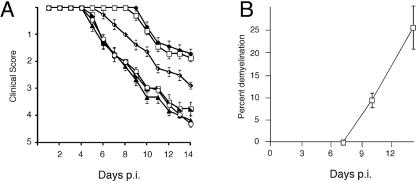
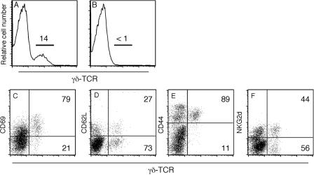
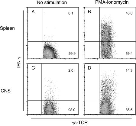
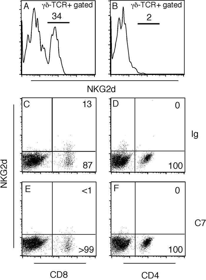
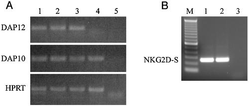
gammadelta T cells mediate demyelination in athymic (nude) mice infected with the neurotropic coronavirus mouse hepatitis virus strain JHM. Now, we show that these cells also mediate the same process in mice lacking alphabeta T cells (T-cell receptor beta-deficient [TCRbeta(-/-)] mice) and demyelination is gamma interferon (IFN-gamma) dependent. Most strikingly, our results also show a major role for NKG2D, expressed on gammadelta T cells, in the demyelinating process with in vivo blockade of NKG2D interactions resulting in a 60% reduction in demyelination. NKG2D may serve as a primary recognition receptor or as a costimulatory molecule. We show that NKG2D(+) gammadelta T cells in the JHM-infected central nervous system express the adaptor molecule DAP12 and an NKG2D isoform (NKG2D short), both required for NKG2D to serve as a primary receptor. These results are consistent with models in which gammadelta T cells mediate demyelination using the same effector cytokine, IFN-gamma, as CD8 T cells and do so without a requirement for signaling through the TCR.


Figures

FIG. 1.
FIG. 1.
Progression of clinical disease and demyelination in JHM-infected TCRβ−/− mice is dependent on γδ T cells and expression of IFN-γ and NKG2D. (A) TCRβ−/− mice were infected with JHM and treated with anti-γδ-TCR monoclonal antibody UC7-13D5 (n = 7, open squares), anti-IFN-γ monoclonal antibody XMG1.2 (n = 7, solid diamonds), anti-CD154 monoclonal antibody MR-1 (n = 4, hatched squares), or anti-NKG2D monoclonal antibody C7 (n = 8, hatched diamonds). Clinical signs were monitored as described in Materials and Methods. Mice that were not treated with antibody (n = 6, solid triangles) or were treated with control antibody (n = 20, open circles) developed the most severe disease. Each antibody was analyzed in at least two independent experiments. Samples were analyzed for statistical significance by comparing pairs of values at each day postinfection. Differences between mice receiving hamster Ig and those receiving anti-γδ TCR monoclonal antibody, anti-IFN-γ monoclonal antibody, or anti-NKG2D monoclonal antibody were statistically significant (day 5: P < 0.05; days 6 and 7: P < 0.01; days 8+: P < 0.001). The difference between treatment with anti-NKG2D monoclonal antibody and anti-IFN-γ monoclonal antibody or anti-γδ TCR monoclonal antibody was also statistically significant (day 7: P < 0.01; days 8+: P < 0.001). The clinical course after anti-IFN-γ monoclonal antibody versus anti-γδ TCR monoclonal antibody treatment was not significantly different (P > 0.05). (B) Demyelination was initially detectable between days 7 and 10 postinfection in JHM-infected TCRβ−/− mice.
FIG. 2.
FIG. 2.
Demyelination in the spinal cord of JHM-infected TCRβ−/− mice. Spinal cords from infected mice at 14 days postinfection were removed, fixed with zinc formalin, and embedded in paraffin. Serial 8-μm sections were stained for (A) demyelination, (B) viral antigen, (C) macrophages or microglia, and (D) axons, as described in Materials and Methods. Areas of demyelination (A) are accompanied by robust macrophage/microglia infiltration (C). Little viral antigen is detectable in these areas, although some can be found in adjacent, normal-appearing white matter (B). The loss of myelin is primary, as axonal staining is preserved through the area of demyelination (D). Scale bar: 100 μm.
FIG. 3.
FIG. 3.
Phenotype and depletion of γδ T cells. (A and B) JHM-infected mice were treated with hamster Ig (A) or anti-γδ-TCR monoclonal antibody UC7-13D5 (B) on days 0, 3, 6, 9, and 12 postinfection. At 14 days postinfection, γδ T cells constituted 10 to 20% of the lymphocyte infiltrate into the CNS (A). No γδ T cells were detectable in UC7-13D5-treated mice (B). (C to F) Brains were removed from JHM-infected TCRβ−/− mice and CNS-derived lymphocytes were stained with anti-γδ-TCR and antibodies against CD69 (C), CD62L (D), CD44 (E), and NKG2D (F). Most γδ T cells within the CNS had marker levels consistent with an activated phenotype: CD44+, CD62L, and CD69+. Approximately 40% of γδ T cells expressed the NK activating receptor NKG2D (F). Numbers indicate the percentage of γδ T cells in each group.
FIG. 4.
FIG. 4.
IFN-γ production by CNS-derived and splenic γδ T cells. Lymphocytes were harvested from the spleens (A and B) and CNS (C and D) of infected TCRβ−/− mice at 14 days postinfection. Cells were either treated with phorbol myristate acetate and ionomycin directly ex vivo (B and D) or not stimulated (A and C) and then analyzed for intracellular IFN-γ production as described in Materials and Methods.
FIG. 5.
FIG. 5.
Depletion of NKG2D+ cells. (A and B) JHM-infected TCRβ−/− mice were treated with hamster Ig (A) or the anti-NKG2D monoclonal antibody C7 (B) on days 0, 3, 6, 9, and 12 postinfection. There were no residual γδ T cells expressing NKG2D protein in C7-treated mice (B). (C to F) JHM-infected C57BL/6 mice were treated with anti-NKG2D monoclonal antibody C7 (E and F) or Ig (C and D). CD8 (C and E) and CD4 (D and F) cells were analyzed for expression of NKG2D. Only CD8 T cells expressed NKG2D (C) and treatment with monoclonal antibody C7 depleted this population (E).
FIG. 6.
FIG. 6.
Identification of DAP10, DAP12, and NKG2D-S mRNA in CNS-derived γδ T cells. NKG2D+ γδ T cells from the CNS of JHM-infected TCRβ−/− mice or NKG2D+ CD8 T cells from an uninfected C57BL/6 spleen were sorted as described in Materials and Methods. (A) mRNA was extracted from the sorted cells and subjected to RT-PCR for DAP10, DAP12, and hypoxanthine phosphoribosyltransferase (HPRT). Lanes 1 to 3, CNS-derived γδ T cells from three individual JHM-infected TCRβ−/− mice. Lane 4, splenic NKG2D+ CD8 T cells from an uninfected C57BL/6 mouse. Lane 5, negative control. (B) mRNA was subjected to RT-PCR for NKG2D-S. Lanes 1 and 2, CNS-derived γδ T cells from two individual JHM-infected mice. Lane 3, negative control.

Similar articles

Cited by

References

    1. Backstrom, E., B. J. Chambers, E. L. Ho, O. V. Naidenko, R. Mariotti, D. H. Fremont, W. M. Yokoyama, K. Kristensson, and H. G. Ljunggren. 2003. Natural killer cell-mediated lysis of dorsal root ganglia neurons via RAE1/NKG2D interactions. Eur. J. Immunol. 33:92-100. - PubMed
    1. Bauer, S., V. Groh, J. Wu, A. Steinle, J. H. Phillips, L. L. Lanier, and T. Spies. 1999. Activation of NK cells and T cells by NKG2D, a receptor for stress-inducible MICA. Science 285:727-729. - PubMed
    1. Becher, B., B. G. Durell, A. V. Miga, W. F. Hickey, and R. J. Noelle. 2001. The clinical course of experimental autoimmune encephalomyelitis and inflammation is controlled by the expression of CD40 within the central nervous system. J. Exp. Med. 193:967-974. - PMC - PubMed
    1. Benveniste, P., B. S. Chadwick, R. G. Miller, and J. Reimann. 1990. Characterization of cells with T-cell markers in athymic nude bone marrow and of their in vitro-derived clonal progeny. Comparison with euthymic bone marrow. J. Immunol. 144:411-419. - PubMed
    1. Bergmann, C. C., J. D. Altman, D. Hinton, and S. A. Stohlman. 1999. Inverted immunodominance and impaired cytolytic function of CD8+ T cells during viral persistence in the central nervous system. J. Immunol. 163:3379-3387. - PubMed

Publication types

MeSH terms

Substances

LinkOut - more resources