Comparative Study
. 2005 Aug;79(15):9458-69.
doi: 10.1128/JVI.79.15.9458-9469.2005.

Vaccinia virus A21 virion membrane protein is required for cell entry and fusion

Affiliations
Comparative Study

Vaccinia virus A21 virion membrane protein is required for cell entry and fusion

Alan C Townsley et al. J Virol. 2005 Aug.

Abstract

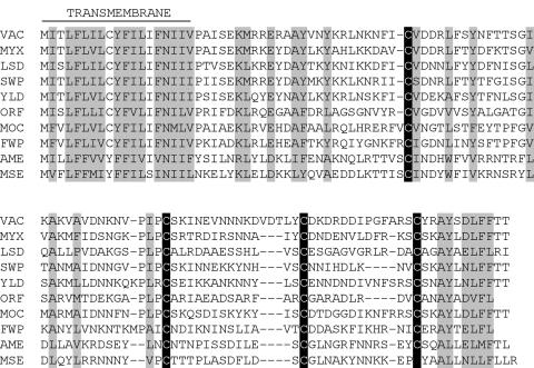
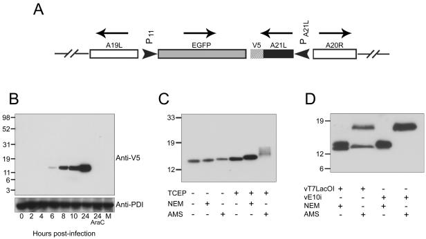
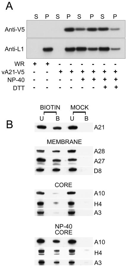
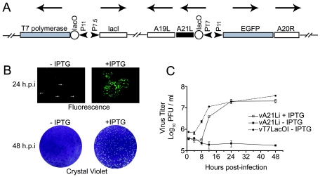
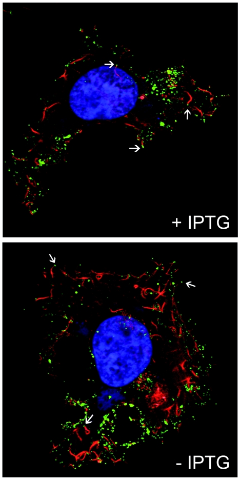
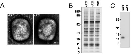
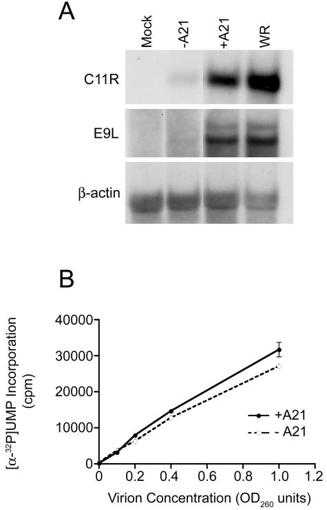
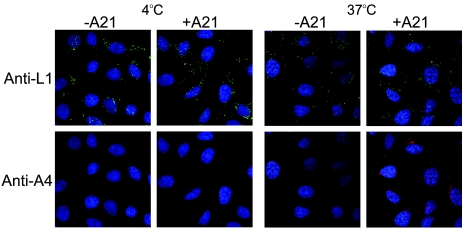
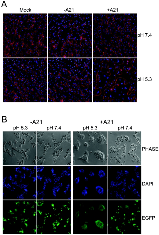
We provide the initial characterization of the product of the vaccinia virus A21L (VACWR140) gene and demonstrate that it is required for cell entry and low pH-triggered membrane fusion. The A21L open reading frame, which is conserved in all sequenced members of the poxvirus family, encodes a protein of 117 amino acids with an N-terminal hydrophobic domain and four invariant cysteines. Expression of the A21 protein occurred at late times of infection and was dependent on viral DNA replication. The A21 protein contained two intramolecular disulfide bonds, the formation of which required the vaccinia virus-encoded cytoplasmic redox pathway, and was localized on the surface of the lipoprotein membrane of intracellular mature virions. A conditional lethal mutant, in which A21L gene expression was regulated by isopropyl-beta-d-thiogalactopyranoside, was constructed. In the absence of inducer, cell-to-cell spread of virus did not occur, despite the formation of morphologically normal intracellular virions and extracellular virions with actin tails. Purified virions lacking A21 were able to bind to cells, but cores did not penetrate into the cytoplasm and synthesize viral RNA. In addition, virions lacking A21 were unable to mediate low pH-triggered cell-cell fusion. The A21 protein, like the A28 and H2 proteins, is an essential component of the poxvirus entry/fusion apparatus for both intracellular and extracellular virus particles.


Figures

FIG. 1.
FIG. 1.
Conservation of the A21L gene in Poxviridae. Multiple alignment of A21 orthologs, including a representative from each sequenced genus of the Chordopoxvirinae and Entomopoxvirinae subfamilies. Invariant cysteines are denoted by white text upon a black background. Other identical or similar residues are indicated by shading. VAC, vaccinia virus (Orthopoxvirus); MYX, myxoma virus (Leporipoxvirus); LSD, lumpy skin disease virus (Capripoxvirus); SWP, swinepox virus (Suipoxvirus); YLD, Yaba-like disease virus (Yatapoxvirus); ORF, ORF virus (Parapoxvirus); MOC, molluscum contagiosum virus (Molluscipoxvirus); FWP, fowlpox virus (Avipoxvirus); AME, Amsacta moorei entomopoxvirus (Entomopoxvirus B); MSE, Melanoplus sanguinipes entomopoxvirus (Entomopoxvirus B).
FIG. 2.
FIG. 2.
Expression of A21 and formation of intramolecular disulfide bonds. (A) Diagram of a portion of the genome of recombinant vA21-V5 containing a V5 epitope encoded at the end of the A21L gene. PA21L and P11 refer to the natural promoter of the A21L gene and the promoter for the gene encoding the 11K protein, respectively. Arrows over ORFs indicate the direction of transcription. (B) Western blot showing A21 expression. BS-C-1 cells were either mock-infected (M) or infected with 5 PFU of vA21-V5 per cell in the absence or presence of AraC and disrupted in SDS-PAGE loading buffer at indicated hours postinfection. Cell lysates were analyzed by SDS-PAGE and Western blotting with an anti-V5 MAb and anti-protein disulfide isomerase (anti-PDI) as a loading control. (C) Analysis of intramolecular disulfide bonds. BS-C-1 cells were infected with 5 PFU per cell of vA21-V5 for 18 h and then disrupted in SDS-PAGE loading buffer containing NEM or AMS. Some cells were treated with the reducing agent TCEP prior to alkylation with either NEM or AMS. SDS-PAGE and Western blotting were performed using an anti-V5 MAb. (D) Dependence of disulfide bond formation on expression of E10 protein. BS-C-1 cells were infected with vE10i or vT7LacOI in the absence of IPTG. Two hours later, the cells were transfected with a plasmid encoding A21 with a C-terminal V5-epitope tag under the control of the natural A21 promoter. At 18 h postinfection, cells were disrupted in SDS-PAGE loading buffer containing NEM or AMS. SDS-PAGE and Western blotting were carried out with an anti-V5 MAb. The positions of molecular mass standards in kDa are shown on the left axis of each panel.
FIG. 3.
FIG. 3.
A21 is a membrane protein exposed on the surface of IMV. (A) Extraction of A21 with a nonionic detergent. Purified VACV (WR) or vA21-V5 virions were suspended in Tris buffer, pH 7.4, and incubated at 37°C for 30 min with or without 1% NP-40 or 50 mM DTT. The virus suspensions were then centrifuged at 20,000 × g for 30 min at 4°C and separated into pellet (P) and supernatant (S) fractions. The fractions were analyzed by SDS-PAGE and Western blotting with anti-V5 and anti-L1 MAbs. (B) Biotinylation of IMV surface proteins. Untreated or NP-40-treated purified virions were reacted with 1 mg of sulfo-NHS-SS-biotin per ml for 30 min at 4°C (BIOTIN) or mock-labeled with PBS (MOCK). Excess reagent was quenched and virions were solubilized with SDS-PAGE sample buffer and incubated with neutravidin gel for 1 h at 4°C. The unbound (U) and the bound (B) proteins were treated with SDS-PAGE sample buffer containing 50 mM DTT and analyzed by SDS-PAGE and Western blotting with antibodies to membrane proteins A28, A27, and D8 and to core proteins A10, H4, and A3.
FIG. 4.
FIG. 4.
Construction of vA21i, a conditional lethal VACV with an inducible A21L gene. (A) Diagram of a portion of the genome of vA21i. Arrows over ORFs indicate the direction of transcription. Abbreviations: lacO, E. coli lac operator; P11, VACV late promoter; P7.5, VACV early/late promoter; lacI, E. coli lac repressor gene; PT7, bacteriophage T7 promoter. (B) Plaque formation by vA21i in the presence and absence of IPTG. BS-C-1 monolayers were infected with vA21i in the presence or absence of IPTG. At 24 h postinfection, cells were observed by fluorescence microscopy for the presence of EGFP. At 48 h postinfection, monolayers were fixed and stained with crystal violet. (C) Yields of vA21i in the presence or absence of IPTG. BS-C-1 monolayers were infected with vA21i or vT7LacOI at a multiplicity of 5 PFU per cell in the absence or presence of IPTG. The cells were harvested at indicated hours after infection, and virus titers were determined by plaque assay in the presence of 50 μM IPTG. Experiments were performed in duplicate, and data points represent the mean ± standard error.
FIG. 5.
FIG. 5.
Visualization of CEVs with actin tails. HeLa cell monolayers were infected at a multiplicity of 5 PFU per cell in the absence or presence of 50 μM IPTG for 20 h, fixed with 3% paraformaldehyde, and quenched with 2% glycine. Cell surface CEVs were immunolabeled with anti-B5R MAb and Cy5-conjugated goat anti-rat secondary antibody. Following this, cells were permeabilized by the addition of 0.1% Triton X-100 and stained with DAPI to visualize cellular and viral DNA and with Alexa Fluor 568 phalloidin to visualize filamentous actin. Arrows point to representative CEVs at the tips of actin tails.
FIG. 6.
FIG. 6.
Electron microscopy and SDS-PAGE of A21 and A21+ IMVs. (A) Electron micrographs of negatively stained sucrose gradient-purified A21 and A21+ IMVs. Virions were deposited on grids, washed with water, and stained with 7% uranyl acetate in 50% ethanol for 30 sec. (B) SDS-PAGE of sucrose gradient-purified A21, A21+, and VACV WR IMVs. The numbers of particles were determined from the optical density and equal amounts of the three types of virions were analyzed by SDS-PAGE and silver staining. The positions and molecular masses in kDa of marker proteins are shown on the left. (C) Western blot of A21 and A21+ virions. Following SDS-PAGE, Western blotting was performed with rabbit polyclonal antibody to the A21 peptide DKDRDDIPGFARSC.
FIG. 7.
FIG. 7.
In vivo and in vitro transcription mediated by A21 and A21+ virions. (A) Northern blot analysis. BS-C-1 cell monolayers were infected with 5 PFU per cell of wild type VACV WR, A21+ virions, or the corresponding OD260 of A21 virions. Total RNA was harvested at 3 h after infection and analyzed by gel electrophoresis and Northern blotting with [α-32P]dCTP-labeled double-stranded DNA probes to C11R, E9L, and β-actin transcripts. (B) In vitro transcription. Purified virions were incubated in reaction buffer containing NP-40, DTT, Mg++, ribonucleoside triphosphates, and [α-32P]UTP. Incorporation of [α-32P]UMP into trichloroacetic acid-insoluble material was determined by scintillation counting and is expressed as counts per minute (cpm).
FIG. 8.
FIG. 8.
Inability of A21 virions to release cores into the cytosol. HeLa cell monolayers were incubated with 20 PFU per cell of A21+ virions or the corresponding OD260 of A21 virions at 4°C for 1 h. Cells were washed extensively and fixed or incubated for a further 2 h at 37°C in the presence of cycloheximide before fixation. Autofluorescence was quenched with 2% glycine, and cells were permeabilized with 0.1% Triton X-100. Cells were incubated with mouse anti-L1 MAb to detect IMVs on the cell surface and rabbit anti-L4 antibody to detect cytoplasmic cores, followed by fluorescein isothiocyanate-conjugated goat anti-mouse (green) and rhodamine Red-X-conjugated goat anti-rabbit (red) antibody, respectively. DNA was visualized by staining with DAPI. Optical sections were obtained by confocal microscopy and are displayed as maximum intensity projections.
FIG. 9.
FIG. 9.
Inability of virions lacking A21 to mediate low pH-triggered cell-cell fusion. (A) Fusion from without. B-SC-1 monolayers were infected with either 200 PFU per cell of A21+ virions or the corresponding OD260 of A21 virions. Following this, a transient low pH treatment (pH 5.3) or control treatment (pH 7.4) was administered, and then the buffer was replaced with regular medium containing 300 μg of cycloheximide per ml. After 3 h, the cells were fixed and stained with Alexa Fluor 568 phalloidin and DAPI before visualization by confocal microscopy. (B) Fusion from within. BS-C-1 monolayers were infected with 5 PFU per cell of vA21i in the presence or absence of 50 μM IPTG for 18 h and transiently treated with either pH 5.3 or pH 7.4 buffer. The buffer was replaced with regular medium, and after 3 h the cells were stained with DAPI and visualized by phase microscopy and fluorescence microscopy for DNA and constitutively expressed EGFP.

Similar articles

Cited by

References

    1. Ahn, B.-Y., P. D. Gershon, and B. Moss. 1994. RNA-polymerase associated protein RAP94 confers promoter specificity for initiating transcription of vaccinia virus early stage genes. J. Biol. Chem. 269:7552-7557. - PubMed
    1. Alexander, W. A., B. Moss, and T. R. Fuerst. 1992. Regulated expression of foreign genes in vaccinia virus under the control of bacteriophage T7 RNA polymerase and the Escherichia coli lac repressor. J. Virol. 66:2934-2942. - PMC - PubMed
    1. Ansarah-Sobrinho, C., and B. Moss. 2004. Vaccinia virus G1 protein, a predicted metalloprotease, is essential for morphogenesis of infectious virions but not for cleavage of major core proteins. J. Virol. 78:6855-6863. - PMC - PubMed
    1. Antoine, G., F. Scheiflinger, F. Dorner, and F. G. Falkner. 1998. The complete genomic sequence of the modified vaccinia Ankara strain: comparison with other orthopoxviruses. Virology 244:365-396. - PubMed
    1. Bablanian, R., B. Baxt, J. A. Sonnabend, and M. Esteban. 1978. Studies on the mechanisms of vaccinia virus cytopathic effects. II. Early cell rounding is associated with virus polypeptide synthesis. J. Gen. Virol. 39:403-413. - PubMed

Publication types

MeSH terms

Associated data

LinkOut - more resources