. 2005 Aug;79(15):9540-55.
doi: 10.1128/JVI.79.15.9540-9555.2005.

The long terminal repeat-containing retrotransposon Tf1 possesses amino acids in gag that regulate nuclear localization and particle formation

Affiliations

The long terminal repeat-containing retrotransposon Tf1 possesses amino acids in gag that regulate nuclear localization and particle formation

Min-Kyung Kim et al. J Virol. 2005 Aug.

Abstract

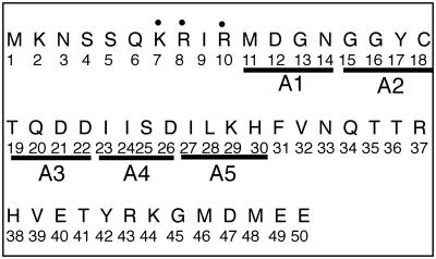
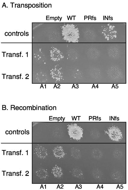
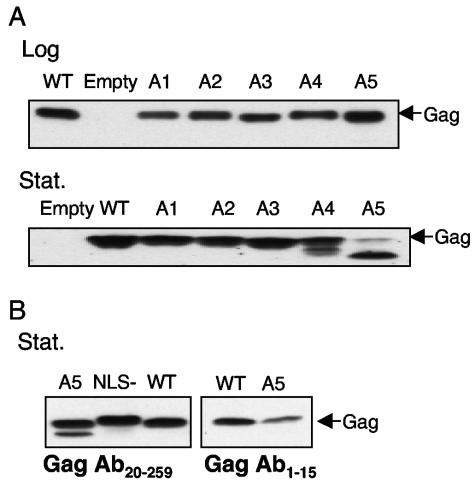
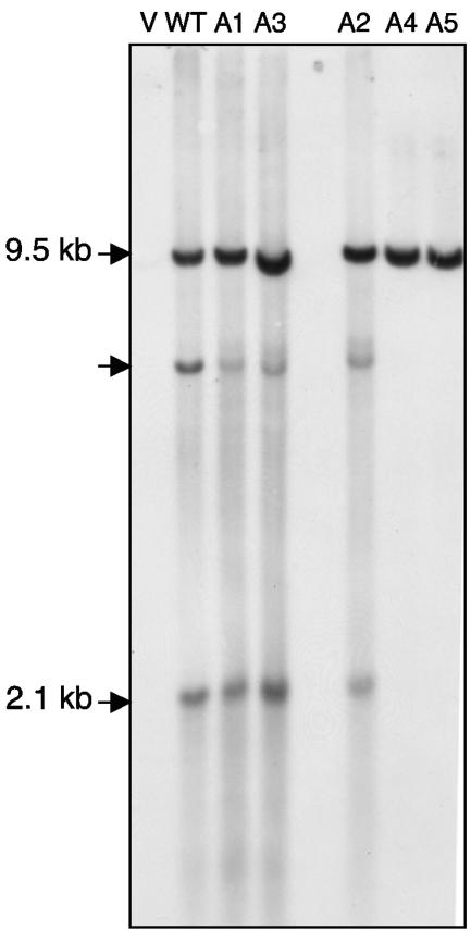
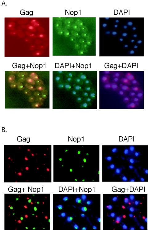
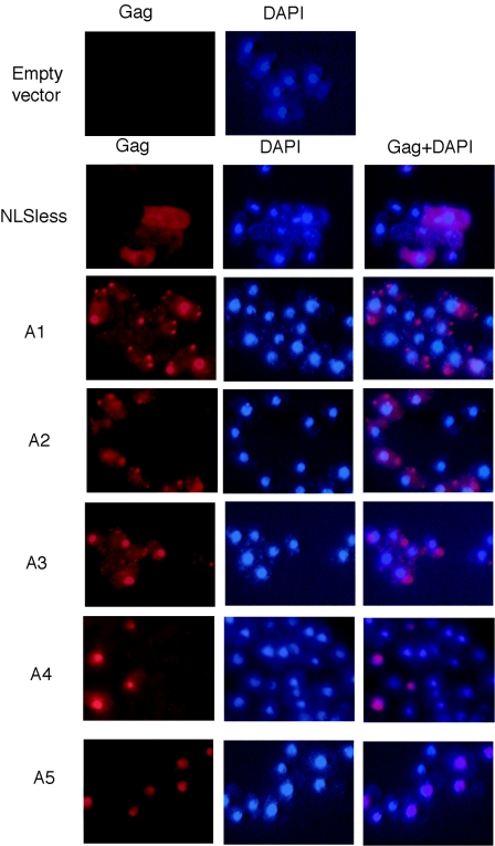
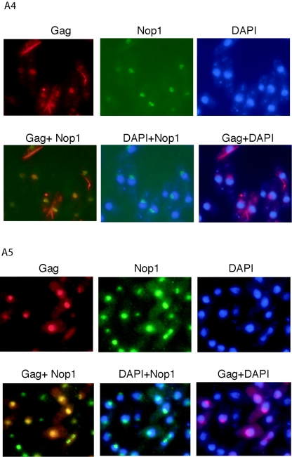
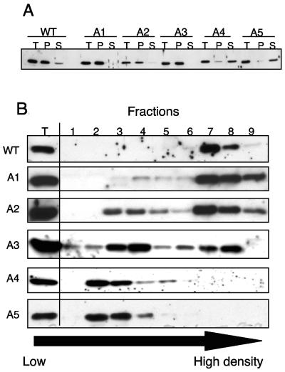
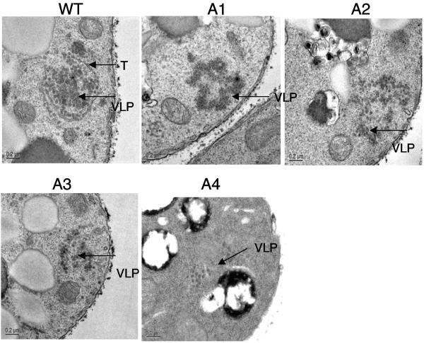
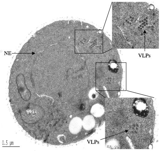
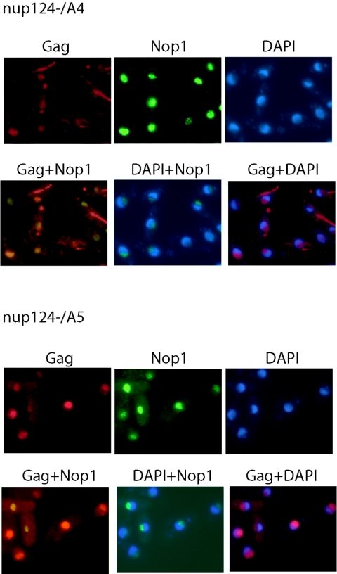
Tf1 is a long terminal repeat-containing retrotransposon of Schizosaccharomyces pombe that is studied to further our understanding of retrovirus propagation. One important application is to examine Tf1 as a model for how human immunodeficiency virus type 1 proteins enter the nucleus. The accumulation of Tf1 Gag in the nucleus requires an N-terminal nuclear localization signal (NLS) and the nuclear pore factor Nup124p. Here, we report that NLS activity is regulated by adjacent residues. Five mutant transposons were made, each with sequential tracts of four amino acids in Gag replaced by alanines. All five versions of Tf1 transposed with frequencies that were significantly lower than that of the wild type. Although all five made normal amounts of Gag, two of the mutations did not make cDNA, indicating that Gag contributed to reverse transcription. The localization of the Gag in the nucleus was significantly reduced by mutations A1, A2, and A3. These results identified residues in Gag that contribute to the function of the NLS. The Gags of A4 and A5 localized within the nucleus but exhibited severe defects in the formation of virus-like particles. Of particular interest was that the mutations in Gag-A4 and Gag-A5 caused their nuclear localization to become independent of Nup124p. These results suggested that Nup124p was only required for import of Tf1 Gag because of its extensive multimerization.


Figures

FIG. 1.
FIG. 1.
The five alanine mutations introduced in Gag. Five versions of Tf1 were generated each with a stretch of four amino acids in Gag changed to alanines. The mutations were made in the 20 residues after the N-terminal NLS of Gag. The key residues of the NLS are indicated by dots. The alanine mutations were named A1, A2, A3, A4, and A5. The positions in Gag of the residues changed to alanine are shown above the heavy lines labeled with the name of the mutant.
FIG. 2.
FIG. 2.
The transposition and homologous recombination activity of the alanine mutants. Two independently transformed strains (Transf. 1 and 2) with the mutated transposons, A1 (YHL8121 and YHL8122), A2 (YHL8123 and YHL8124), A3 (YHL8125 and YHL8126), A4 (YHL8127 and YHL8128), and A5 (YHL8129 and YHL8130), were tested for transposition and homologous recombination activity. The control strains included empty vector (YHL1032), wild-type Tf1 (YHL1282), Tf1 with a frameshift in protease (YHL1836), and Tf1 with a frameshift in integrase (YHL1554). (A) Transposition activity of Tf1 marked with neoAI was measured by inducing the expression of the element from the nmt1 promoter in the plasmid pHL449-1. After induction, the expression plasmid was removed with 5-FOA, and the frequency of transposition was measured on EMM supplemented with G418, as shown in panel A. (B) The accumulation of cDNA and its presence in the nucleus was detected by a homologous recombination assay. Once expression of Tf1-neoAI was induced from pHL449-1, patches of cells were replica printed to medium containing G418. Recombination between cDNA and pHL449-1 allowed growth on this medium.
FIG. 3.
FIG. 3.
The immunoblot of Gags expressed by the mutant transposons. (A) The levels of Gag in cells were measured by immunoblot analysis of whole-cell extracts. Both log-phase (OD600, 1.0) and stationary-phase (OD600, 10) cells were extracted from cultures containing WT (YHL1282), empty plasmid (YHL1032), A1 (YHL8121), A2 (YHL8123), A3 (YHL8125), A4 (YHL8127), and A5 (YHL8129). After electrophoresis on a 10% SDS-PAGE gel, the extracts were transferred to membranes and probed with an antibody specific for Gag residues 20 to 259 (Gag Ab20-259). (B) The levels of Gag produced in stationary-phase cells by wild-type Tf1 and A5 were analyzed with immunoblotting. The membranes were probed with Gag Ab20-259 and an antibody specific for the first 15 amino acids of Gag (Gag Ab1-15). NLSless (NLS−) was a version of Tf1 with a mutation in the NLS (10). This strain was included as a control to show that Gag can accumulate in the cytoplasm without degradation.
FIG. 4.
FIG. 4.
A blot of the cDNA produced by the five different alanine mutants. Total DNA was extracted from cells containing empty plasmid (V; YHL1032), WT (YHL1282), A1 (YHL8121), A2 (YHL8123), A3 (YHL8125), A4 (YHL8127), and A5 (YHL8129). Total DNA was digested with BstXI, subjected to electrophoresis on agaose, and probed with the neo gene. The 9.5-kb band was produced by the plasmid, and the 2.1-kb band was a fragment of the Tf1 cDNA. The band labeled with just an arrow was thought to be single LTR circles.
FIG. 5.
FIG. 5.
Indirect immunofluorescence of wild-type Gag in cells harvested at stationary phase and log phase. The immunolabeling of Gag was produced by a polyclonal antibody against Gag Ab20-259 (red). DNA in the cells was stained with DAPI (blue). Nop1 was detected by immunolabeling with a monoclonal antibody (green). Overlay images were created for Gag+Nop1, DAPI+Nop1, and Gag+DAPI. (A) The Gag, Nop1, and DNA of wild-type Tf1 (YHL1282) were visualized in stationary-phase cells. (B) The Gag, Nop1, and DNA of wild-type Tf1 were visualized in log-phase cells.
FIG. 6.
FIG. 6.
Indirect immunofluorescence of Gag in cells expressing the alanine mutants. The cellular localizations of Gag, Nop1, and DNA were determined in stationary-phase cells expressing A1 (YHL8121), A2 (YHL8123), A3 (YHL8125), A4 (YHL8127), and A5 (YHL8129). The immunolabeling of Gag was as described for Fig. 5.
FIG. 7.
FIG. 7.
Indirect immunofluorescence of Gag in log-phase cells expressing the alanine mutants A4 and A5. The localization of Gag, Nop1, and DNA was determined in cells expressing either A4 or A5. The immunolabeling of Gag was as described for Fig. 5.
FIG. 8.
FIG. 8.
Binding assays for interactions between Nup124p and Gag. The N-terminal half of Nup124p (N-nup124p) and versions of Gag proteins were fused to the C terminus of GST, expressed in bacteria, and purified. N-Nup124p with GST removed was incubated with each of the GST-Gags of the alanine mutants. The GST-Gags were precipitated with glutathione-Sepharose 4B beads, and the N-Nup124p associated with the Gag was visualized on SDS-PAGE gels stained with Coomassie blue. M, broad-range molecular-mass markers (Bio-Rad); N-nup, purified N-Nup124p. The plus and minus signs indicate the presence or absence of N-Nup124p in the binding reaction. (A) Each of the Gags with the alanine mutants was tested for binding with N-Nup124p. The gel was a 10% SDS-polyacrylamide gel. (B) GST-Gag without the first 30 amino acids of Gag (Δ30) was tested for binding to N-Nup124p. This gel was a 10 to 20% gradient SDS-PAGE gel. (C) Five 50-amino-acid segments of Gag were tested for binding to N-Nup124p. The segments of Gag tested were A (amino acids 1 to 50), B (amino acids 50 to 100), C (amino acids 100 to 150), D (amino acids 150 to 200), and E (amino acids 200 to 250). These gels were NuPAGE 4 to 12% bis-Tris gels. The asterisk marks the N-Nup124p that bound to the segment containing amino acids 200 to 250.
FIG. 9.
FIG. 9.
Differential sedimentation and gradient fractionation experiments of Gags with the alanine mutations. (A) Whole-cell extracts prepared from WT (YHL1282), A1 (YHL8121), A2 (YHL8123), A3 (YHL8125), A4 (YHL8127), and A5 (YHL8129) were centrifuged at 1,000 × g for 5 min. The supernatants were further centrifuged at 12,000 × g for 10 min. The samples loaded on an SDS-PAGE gel were the total cell extracts (T), the pellets from the 12,000 × g centrifugation (P), and the supernatant from the 12,000 × g centrifugation (S). (B) The supernatants from the 1,000 × g centrifugation described above were also sedimented on gradients of iodixanol centrifuged at 125,000 × g. The 0.2-ml fractions were collected from low density (fraction 1) to high density (fraction 9). The fractions were immunoblotted and probed with the Gag Ab20-259 antibody. Total cell extracts (T) from cells expressing the mutant transposons were centrifuged at 125,000 × g in OptiPrep gradients to isolate virus-like particles.
FIG. 10.
FIG. 10.
The EM analysis of VLPs produced by the alanine mutants. The cells expressing WT (YHL1282), A1 (YHL8121), A2 (YHL8123), A3 (YHL8125), A4 (YHL8127), and A5 (YHL8129) were harvested from stationary phase cultures and prepared for EM examination (see Materials and Methods). These EM images were representative of >1,000 sections of cells examined for each mutant. Although >2,000 sections of cells expressing Gag-A5 were examined, no particle structures were observed. VLPs are indicated by arrows labeled VLP, and the tube-like structures from cells expressing wild-type Tf1 were indicated by a T.
FIG. 11.
FIG. 11.
EM analysis of VLPs produced by alanine mutant A4. Images of A4 particles are shown at two different magnifications, 5,000× and 20,000× (boxed area). Four distinctive patches of VLPs were observed. NE, nuclear envelope.
FIG. 12.
FIG. 12.
The nuclear localization of Gag-A4 and Gag-A5 expressed in a strain with the nup124-1 mutation. The antibodies used for indirect immunofluorescence were the same as those used in the results shown in Fig. 5. The cultures expressing A4 (YHL8102) and A5 (YHL8104) were harvested in log phase.

Similar articles

Cited by

References

    1. Allen, N. P. C., L. Huang, A. Burlingame, and M. Rexach. 2001. Proteomic analysis of nucleoporin interacting proteins. J. Biol. Chem. 276:29268-29274. - PubMed
    1. Allen, N. P. C., S. S. Patel, L. Huang, R. F. Chalkley, A. Burlingame, M. Lutzmann, E. C. Hurt, and M. Rexach. 2002. Deciphering networks of protein interactions at the nuclear pore complex. Mol. Cell. Proteomics 1:930-946. - PubMed
    1. Aris, J. P., and G. Blobel. 1988. Identification and characterization of a yeast nucleolar protein that is similar to a rat liver nucleolar protein. J. Cell Biol. 107:17-31. - PMC - PubMed
    1. Atwood, A., J. Choi, and H. L. Levin. 1998. The application of a homologous recombination assay revealed amino acid residues in an LTR-retrotransposon that were critical for integration. J. Virol. 72:1324-1333. - PMC - PubMed
    1. Atwood, A., J. H. Lin, and H. L. Levin. 1996. The retrotransposon Tf1 assembles virus-like particles that contain excess Gag relative to integrase because of a regulated degradation process. Mol. Cell. Biol. 16:338-346. - PMC - PubMed

MeSH terms

LinkOut - more resources