. 2005 Aug;79(15):9556-65.
doi: 10.1128/JVI.79.15.9556-9565.2005.

Suppression of RNA interference by adenovirus virus-associated RNA

Affiliations

Suppression of RNA interference by adenovirus virus-associated RNA

M Gunnar Andersson et al. J Virol. 2005 Aug.

Abstract

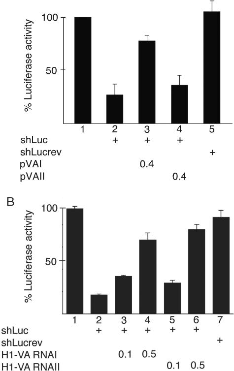
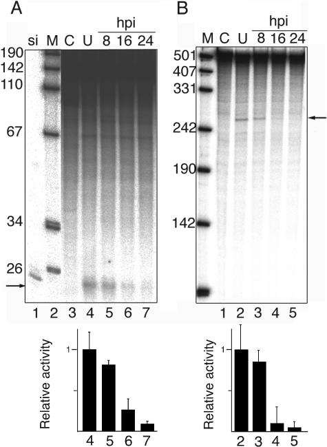
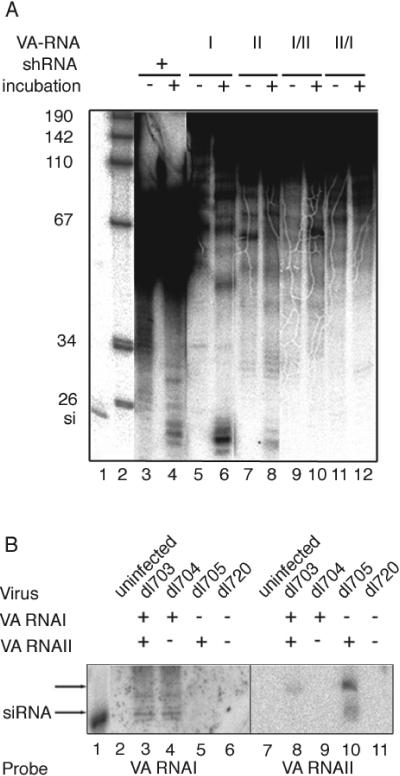
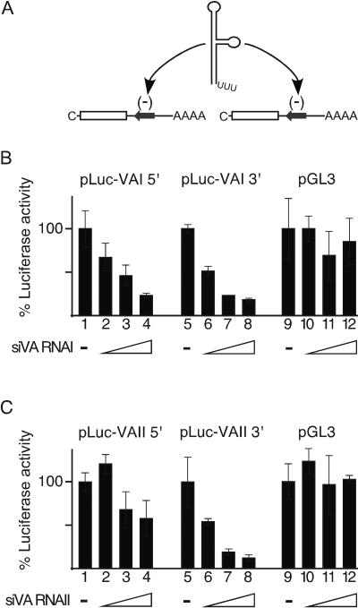
We show that human adenovirus inhibits RNA interference (RNAi) at late times of infection by suppressing the activity of two key enzyme systems involved, Dicer and RNA-induced silencing complex (RISC). To define the mechanisms by which adenovirus blocks RNAi, we used a panel of mutant adenoviruses defective in virus-associated (VA) RNA expression. The results show that the virus-associated RNAs, VA RNAI and VA RNAII, function as suppressors of RNAi by interfering with the activity of Dicer. The VA RNAs bind Dicer and function as competitive substrates squelching Dicer. Further, we show that VA RNAI and VA RNAII are processed by Dicer, both in vitro and during a lytic infection, and that the resulting short interfering RNAs (siRNAs) are incorporated into active RISC. Dicer cleaves the terminal stem of both VA RNAI and VA RNAII. However, whereas both strands of the VA RNAI-specific siRNA are incorporated into RISC, the 3' strand of the VA RNAII-specific siRNA is selectively incorporated during a lytic infection. In summary, our work shows that adenovirus suppresses RNAi during a lytic infection and gives insight into the mechanisms of RNAi suppression by VA RNA.


Figures

FIG. 1.
FIG. 1.
An adenovirus infection suppresses RNAi. 293 cells were cotransfected with a b3a2-GFP reporter plasmid and a vector expressing a 29-base-pair nonhomologous e1a2 or homologous b3a2 shRNA. At 16 h posttransfection the samples were infected with wild-type adenovirus (wt900; lanes 3 and 4) or mock infected (lanes 1 and 2). Fluorescent cells were counted under the microscope at 40 h posttransfection. The figure shows a typical result with mean and standard deviation from 10 counted fields. The effect of b3a2 shRNA in uninfected cells is variable but statistically significant (36 ± 18%; n = 6; P = 0.05). In contrast, no significant effect of b3a2 shRNA is seen in adenovirus-infected cells (8 ± 13%; n = 9; P = 0.05).
FIG. 2.
FIG. 2.
VA RNA restores expression of a silenced luciferase reporter. C33A cells were cotransfected with a luciferase reporter plasmid, a vector expressing an shRNA against luciferase (shLuc), or a control shRNA (shLucrev). The effect of VA RNA expression on the silencing of the luciferase reporter was measured by cotransfection of 0.4 μg of plasmid pVAI or pVAII (A) or 0.1 or 0.5 μg of plasmid H1-VA RNAI or H1-VA RNAII (B). The quantitative results are based on three independent experiments.
FIG. 3.
FIG. 3.
Time course of RNAi inactivation. Cytoplasmic extracts were prepared from uninfected 293 cells (U) or cells collected at 8, 16, or 24 h postinfection (hpi) with adenovirus (wt900) M, DNA size marker; C, uninfected extract. The reaction was stopped at time zero. (A) Dicer activity assayed against a 1,600-base-pair dsRNA. si, 32P-labeled 21-base-pair siRNA marker. The arrow indicates the position of 21-base-pair siRNA products. (B) RISC activity in the same extracts assayed against a 484-nucleotide, uniformly labeled mRNA in the presence of a synthetic siRNA. The arrow indicates the position of the 252-nucleotide 5′ cleavage product. Quantitative results based on three independent experiments are shown below respective autoradiograms.
FIG. 4.
FIG. 4.
The suppression of Dicer and RISC extracts at late times of adenovirus infection is specific. (A) An adenovirus infection does not inhibit protein kinase activity against a recombinant ASF/SF2 protein. Cytoplasmic extracts prepared from uninfected 293 cells (U) or cells infected with wild-type adenovirus for 8, 16, or 24 h were tested for their capacity to phosphorylate ASF/SF2 in vitro. Products were resolved on a 12% reducing SDS-polyacrylamide gel and subjected to autoradiography. (B) Restoration of Dicer activity in infected extracts by increasing the concentration of the dsRNA substrate. Dicer activity in cytoplasmic extracts prepared from uninfected or adenovirus-infected (16 h postinfection [hpi]) 293 cells was assayed against a 32P-labeled GL2 dsRNA. The figure shows the Dicer activity in infected extracts relative to uninfected extract at concentrations of 5, 20, and 100 nM dsRNA. The mean values and standard deviations from three independent experiments are shown. For further details see Fig. S1 in the supplemental material.
FIG. 5.
FIG. 5.
VA RNA expression is necessary for suppression of Dicer. (A) Cytoplasmic extracts prepared at 22 h postinfection from wild-type or VA RNA mutant virus-infected 293 cells were assayed for their activity of Dicer as described in Materials and Methods. (B) Quantitative results based on three independent experiments are shown.
FIG. 6.
FIG. 6.
The VA RNAs are processed into siRNA both in vitro and in vivo. (A) 32P-labeled VA RNAI, VA RNAII, or VA RNA hybrid molecules (VA RNAI/II and VA RNAII/I) or a 29-base-pair shRNA were incubated in S100 extracts. After purification the reaction products were separated on a 15% denaturing polyacrylamide gel and visualized by autoradiography. −, reactions stopped at time zero. The control RNAs, VA RNA I/II and II/I, are unable to form normal secondary structures. (B) Total RNA prepared 24 h postinfection from dl703 (wild type) or VA RNA mutant virus-infected 293 cells was separated on a 15% denaturing polyacrylamide gel and transferred to a Hybond membrane. VA RNAI- and VA RNAII-specific probes were used to detect small RNAs. si, 32P-labeled 21-base-pair siRNA marker. Note that the predominant product from VA RNAII is slightly larger than the siRNA marker.
FIG. 7.
FIG. 7.
The VA RNAs are cleaved by a recombinant Dicer into functional siRNAs. (A) Schematic diagram showing the structure of the luciferase reporter mRNAs expressed from pLuc-VAI 5′, pLuc-VAI 3′, pLuc-VAII 5′, and pLuc-VAII 3′, respectively. The VA RNA genes were separated within the terminal loop into two halves and cloned in the antiparallel orientation in the 3′ untranslated region of the luciferase reporter mRNA. (B and C) 293T cells were cotransfected with increasing amounts of siRNAs derived by Dicer cleavage of VA RNAI or VA RNAII (4, 20, and 100 ng) and 0.1 μg of reporter plasmids pLuc-VAI 5′ (lanes 1 to 4), pLuc-VAI 3′ (lanes 5 to 8), or pGL3 (lanes 9 to 12). Luciferase expression was measured 48 h posttransfection.
FIG. 8.
FIG. 8.
siRNAs derived from the terminal stem of VA RNAI and VA RNAII are incorporated into active RISC in adenovirus-infected cells. (A) Cytoplasmic extracts prepared from uninfected 293 cells or cells infected with dl703 (wild type) or VA RNA mutant viruses were assayed for RISC activity against synthetic Luc-VA transcripts with a target region complementary to the 5′ or 3′ half of VA RNAI (upper panel) or VA RNAII (lower panel). The arrows indicate the span of the VA RNA target regions. The predicted positions of the cleavage products generated by Dicer cleavage at the terminal stem of VA RNAI and VA RNAII are indicated by a dot. (B) Secondary structure of VA RNAI and VA RNAII (adapted from references and 37). Note that the VA RNAII structure is a computer model and has not been verified experimentally. The position where Dicer interacts with the VA RNAs, as deduced from the fragments generated in panel A, is indicated. The structure of VA RNAI contains an approximately 20-base-pair apical stem that could theoretically be cleaved by Dicer. Such a cleavage would have generated a 5′ fragment of 162 nucleotides and a 3′ fragment of 219 nucleotides. Cleavage of VA RNAII at the hypothetical apical stem would have generated fragments in the size range of 132 to 141 (5′ transcript) and 185 to 193 (3′ transcript) nucleotides, dependent on the exact position of Dicer interaction.

Similar articles

Cited by

References

    1. Akusjärvi, G., M. B. Mathews, P. Andersson, B. Vennström, and U. Pettersson. 1980. Structure of genes for virus-associated RNAI and RNAII of adenovirus type 2. Proc. Natl. Acad. Sci. USA 77:2424-2428. - PMC - PubMed
    1. Akusjärvi, G., and J. Stevenin. 2003. Remodelling of the host cell RNA splicing machinery during an adenovirus infection. Curr. Top. Microbiol. Immunol. 272:253-286. - PubMed
    1. Akusjärvi, G., C. Svensson, and O. Nygård. 1987. A mechanism by which adenovirus virus-associated RNAI controls translation in a transient expression assay. Mol. Cell. Biol. 7:549-551. - PMC - PubMed
    1. Ambros, V. 2004. The functions of animal microRNAs. Nature 431:350-355. - PubMed
    1. Ausubel, F. M., R. Brent, R. E. Kingston, D. D. Moore, J. G. Seidman, J. A. Smith, and K. Struhl (ed.). 1995. Current protocols in molecular biology. John Wiley & Sons, Inc., New York, N.Y.

Publication types