Comparative Study
. 2005 Aug;79(15):9651-64.
doi: 10.1128/JVI.79.15.9651-9664.2005.

mRNA decay during herpes simplex virus (HSV) infections: protein-protein interactions involving the HSV virion host shutoff protein and translation factors eIF4H and eIF4A

Affiliations
Comparative Study

mRNA decay during herpes simplex virus (HSV) infections: protein-protein interactions involving the HSV virion host shutoff protein and translation factors eIF4H and eIF4A

Pinghui Feng et al. J Virol. 2005 Aug.

Abstract

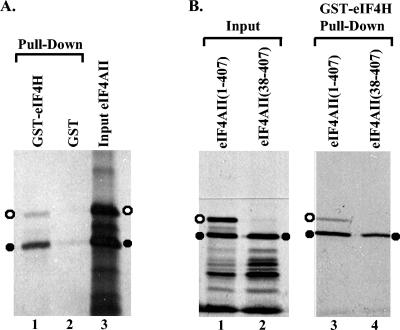
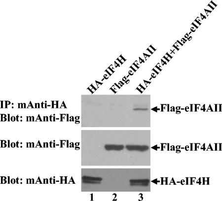
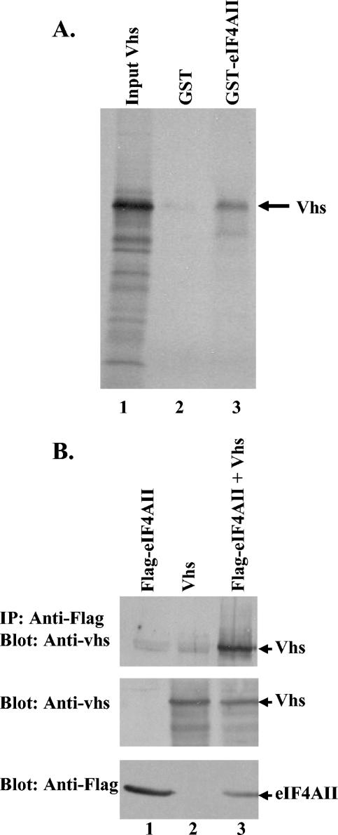
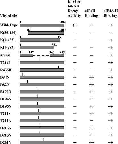
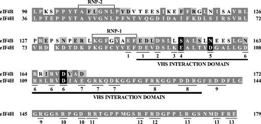
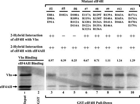
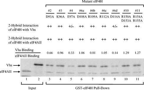
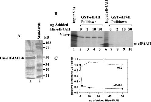
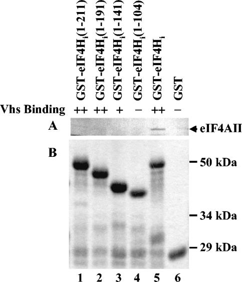
During lytic infections, the virion host shutoff (Vhs) protein of herpes simplex virus accelerates the degradation of both host and viral mRNAs. In so doing, it helps redirect the cell from host to viral protein synthesis and facilitates the sequential expression of different viral genes. Vhs interacts with the cellular translation initiation factor eIF4H, and several point mutations that abolish its mRNA degradative activity also abrogate its ability to bind eIF4H. In addition, a complex containing bacterially expressed Vhs and a glutathione S-transferase (GST)-eIF4H fusion protein has RNase activity. eIF4H shares a region of sequence homology with eIF4B, and it appears to be functionally similar in that both stimulate the RNA helicase activity of eIF4A, a component of the mRNA cap-binding complex eIF4F. We show that eIF4H interacts physically with eIF4A in the yeast two-hybrid system and in GST pull-down assays and that the two proteins can be coimmunoprecipitated from mammalian cells. Vhs also interacts with eIF4A in GST pull-down and coimmunoprecipitation assays. Site-directed mutagenesis of Vhs and eIF4H revealed residues of each that are important for their mutual interaction, but not for their interaction with eIF4A. Thus, Vhs, eIF4H, and eIF4A comprise a group of proteins, each of which is able to interact directly with the other two. Whether they interact simultaneously as a tripartite complex or sequentially is unclear. The data suggest a mechanism for linking the degradation of an mRNA to its translation and for targeting Vhs to mRNAs and to regions of translation initiation.


Figures

FIG. 1.
FIG. 1.
Binding of eIF4AII to a GST-eIF4H fusion protein. (A) GST or a GST-eIF4H fusion protein was produced in E. coli, bound to glutathione-Sepharose 4B, and incubated with rabbit reticulocyte lysates containing the [35S]methionine-labeled in vitro translation products of full-length eIF4AII mRNA. Bound proteins were eluted with 10 mM glutathione and analyzed by SDS-PAGE and autoradiography. Lane 1 contains proteins that bound to GST-eIF4H, lane 2 contains material that bound to GST, and lane 3 contains an aliquot of the starting material. The 45-kDa and 41-kDa products of in vitro translation of full-length eIF4AII mRNA are designated by the open and filled circles, respectively. (B) GST or GST-eIF4H was incubated with the in vitro translation products of full-length eIF4AII mRNA (lane 3) or an mRNA encoding only amino acids 38 through 407 of eIF4AII (lane 4). Bound proteins were eluted and analyzed as described in the legend to panel A. Lanes 1 and 2 contain aliquots of the starting material. The 45-kDa and 41-kDa eIF4AII polypeptides are designated by open and filled circles, respectively.
FIG. 2.
FIG. 2.
Coimmunoprecipitation of eIF4H and eIF4AII from mammalian cells. CV-1 cells were infected with 5 PFU/cell of the recombinant vaccinia virus vTF7. Thirty minutes later, they were transfected with 5 μg each of plasmids expressing HA-tagged eIF4H (lane 1), Flag-tagged eIF4AII (lane 2), or HA-eIF4H and Flag-eIF4AII (lane 3). Cell extracts were prepared 20 h after transfection and complexes containing HA-eIF4H were immunoprecipitated with an HA-specific monoclonal antibody (top). Immunoprecipitates were analyzed for coprecipitated Flag-eIF4AII by SDS-PAGE and Western blotting using a Flag-specific monoclonal antibody. To check for protein expression, aliquots of the cell lysates were analyzed directly by Western blotting using a Flag-specific monoclonal antibody (middle) or an HA-specific monoclonal antibody (bottom).
FIG. 3.
FIG. 3.
In vitro and in vivo binding of Vhs to eIF4AII. (A) Binding of Vhs to a GST-eIF4AII fusion protein. GST or a GST-eIF4AII fusion protein was produced in E. coli, bound to glutathione-Sepharose 4B, and incubated with rabbit reticulocyte lysates containing [35S]methionine-labeled in vitro-translated Vhs. Bound proteins were eluted with 10 mM glutathione and analyzed by SDS-PAGE and autoradiography. Lanes 2 and 3 contain proteins that bound to GST-eIF4AII and GST, respectively, while lane 1 contains an aliquot of the starting material. The Vhs protein is indicated by an arrow. (B) Coimmunoprecipitation of Vhs and eIF4AII from mammalian cells. CV-1 cells were infected with 5 PFU/cell of the recombinant vaccinia virus vTF7. Thirty minutes later, they were transfected with 5 μg (each) of plasmids expressing Flag-tagged eIF4AII (lane 1), the D194N allele of Vhs (lane 2), or Flag-eIF4AII and D194N Vhs (lane 3). Cell extracts were prepared 20 h after transfection, and complexes containing Flag-eIF4AII were immunoprecipitated using a Flag-specific monoclonal antibody (top). Immunoprecipitates were analyzed for coprecipitated Vhs by SDS-PAGE and Western blotting with a Vhs-specific rabbit antiserum. To check for protein expression, aliquots of the cell lysates were analyzed directly by Western blotting using a Vhs-specific antiserum (middle) or a Flag-specific monoclonal antibody (bottom).
FIG. 4.
FIG. 4.
eIF4AII binding by wild-type and mutant Vhs. [35S]methionine-labeled wild-type and mutant Vhs polypeptides were produced by in vitro transcription and translation and analyzed for the ability to bind a GST-eIF4AII fusion protein, as described in the legend for Fig. 1. Material that bound to GST-eIF4AII and was eluted with 10 mM glutathione is shown in the upper gels (GST-eIF4AII pull-down) of panels A and B. Aliquots of the input in vitro-translated material are shown in the lower gels (Input Vhs). The mutant and wild-type Vhs polypeptides are indicated at the top of each lane. Their structures are diagrammed in Fig. 5.
FIG. 5.
FIG. 5.
Summary of the in vivo mRNA-degradative activities of wild-type and mutant Vhs polypeptides and their abilities to bind eIF4H and eIF4AII. The structures of the wild-type and mutant Vhs polypeptides are diagrammed on the left. For deletion mutants, the Vhs residues included in the mutant proteins are indicated. For each point mutant, the location of the altered residue is indicated by the vertical line above the bar representing the protein. The in vivo mRNA-degradative activity of each Vhs protein is shown in the column immediately to the right of the diagram. This was assayed in transfected cells for all of the alleles and during virus infections for wild-type Vhs and the mutants K(1-382), Vhs ΔSma, and T214I. ++, wild-type activity; −, no detectable mRNA-degradative activity. The middle column indicates whether a Vhs protein binds (++) or does not bind (−) eIF4H in the yeast two-hybrid system and in GST pull-down assays. These data were reported previously (18). The right column indicates whether a Vhs protein binds (++) or does not bind (−) eIF4AII and summarizes the data shown in Fig. 4.
FIG. 6.
FIG. 6.
Residues of eIF4H whose alteration reduces Vhs binding. The region of sequence homology shared by eIF4H and eIF4B is shown, with the eIF4H amino acids and those eIF4B residues that are identical to amino acids in eIF4H indicated by white letters on a dark grey background. eIF4B residues that represent conservative changes relative to those in eIF4H are depicted by black letters on a light grey background. eIF4B residues that are nonconservative changes from eIF4H amino acids are indicated by black letters on a white background. eIF4H and eIF4B share an RNA recognition motif RNA-binding domain containing two RNP motifs (RNP-1 and RNP-2) that are boxed. The Vhs interaction domain of eIF4H, as defined by deletion mutagenesis (18), is indicated by the dark line under the eIF4H sequence. Thirteen eIF4H mutants were constructed in which clusters of charged amino acids were altered by site-directed mutagenesis. The amino acids that were altered in a particular mutant are underlined and indicated by the number of the mutant under the residue. For example, in mutant 1, E88, D90, and E91 were altered together. All of the indicated charged amino acids were changed to alanine except for mutant 8, in which R139 and K130 were both changed to threonine. The three charged eIF4H amino acids, whose modification to alanine significantly reduces Vhs binding, are indicated by white type on a black background, as are the corresponding residues of eIF4B.
FIG. 7.
FIG. 7.
Binding of mutant eIF4H polypeptides to Vhs and eIF4AII. Eight of the mutant eIF4H polypeptides diagrammed in Fig. 6 were tested for binding to Vhs and full-length eIF4AII using the recombination-based yeast two-hybrid system and GST pull-down assays. The number of each mutant is shown at the top of the figure, just above the amino acid substitutions that each mutant contains. The second and third lines contain the results of recombination based yeast two-hybrid assays to test the interaction of the mutant eIF4H polypeptides with Vhs and eIF4AII, respectively. ++ indicates that the mutant eIF4H allele yielded a number of colonies similar to that of the wild-type eIF4H in the recombination-based two-hybrid assay; − indicates that the number of colonies was reduced at least 10 fold relative to those of wild-type eIF4H. For GST pull-down assays, full-length mutant eIF4H polypeptides were expressed as GST-eIF4H fusion proteins in E. coli, bound to glutathione-Sepharose 4B, and incubated with rabbit reticulocyte lysates containing a mixture of [35S]methionine-labeled Vhs and eIF4AII produced by in vitro transcription and translation. Bound proteins were eluted with 10 mM glutathione and analyzed by SDS-PAGE and autoradiography as described in the legends to Fig. 1 and 3. The positions of Vhs and full-length eIF4AII are indicated by the arrows to the left of the gel. For each mutant, the relative amounts of bound Vhs and eIF4AII were determined by densitometric scanning of the autoradiogram and expressed as a ratio normalized to the ratio determined for wild-type eIF4H (data not shown). These ratios are indicated in line 4. Lane 1 contains an aliquot of the input mixture of Vhs and full-length eIF4AII; lane 2 contains material that bound to GST alone; lanes 3 through 10 contain material that bound to each of the mutant GST-eIF4H fusion proteins.
FIG. 8.
FIG. 8.
Binding of mutant eIF4H polypeptides to Vhs and eIF4AII. Five of the eIF4H mutants diagrammed in Fig. 6 and four additional mutants (mutants 6a, 6b, 6c, and 6d), were tested for binding to Vhs and full-length eIF4AII by using the recombination-based yeast two-hybrid system and GST pull-down assays. The number of each mutant is shown at the top of the figure, just above the amino acid substitutions that each mutant contains. The second and third lines contain the results of recombination based yeast two-hybrid assays to test the interaction of the mutant eIF4H polypeptides with Vhs and eIF4AII, respectively. ++, mutant eIF4H allele yielded a number of colonies similar to that of wild-type eIF4H by the recombination-based two-hybrid assay; +/−, the number of colonies was reduced at least fourfold but <10 fold relative to wild-type eIF4H. For GST pull-down assays, full-length mutant eIF4H polypeptides were expressed as GST-eIF4H fusion proteins in E. coli, bound to glutathione-Sepharose 4B, and incubated with rabbit reticulocyte lysates containing a mixture of [35S]methionine-labeled Vhs and eIF4AII produced by in vitro transcription and translation. Bound proteins were eluted with 10 mM glutathione and analyzed by SDS-PAGE and autoradiography as described in the legends to Fig. 1 and 3. The positions of Vhs and full-length eIF4AII are indicated by the arrows to the left of the gel. For each mutant, the relative amounts of bound Vhs and eIF4AII were determined by densitometric scanning of the autoradiogram and expressed as a ratio normalized to the ratio that was determined for wild-type eIF4H (data not shown). These ratios are indicated in line 4. Lane 1 contains an aliquot of the input full-length eIF4AII, lane 2 contains an aliquot of the input Vhs, and lanes 3 through 11 contain material that bound to each of the mutant GST-eIF4H fusion proteins.
FIG. 9.
FIG. 9.
eIF4AII does not compete with Vhs for binding to GST-eIF4H. (A) His-tagged eIF4AII was expressed in E. coli and isolated by metal chelate affinity chromatography. An aliquot was analyzed by SDS-PAGE and detected by staining the gel with Coomassie (lane 1). His-eIF4AII is indicated by the arrow. Lane 2 contains molecular mass standards. (B) GST-eIF4H was expressed in E. coli, bound to glutathione-Sepharose 4B, and incubated with rabbit reticulocyte lysates containing [35S]methionine-labeled in vitro-translated Vhs (lanes 2 to 5) or full-length eIF4AII (lanes 7 to 10). The incubations also contained 0 μg (lanes 2 and 7), 2 μg (lanes 3 and 8), 10 μg (lanes 4 and 9), or 50 μg (lanes 5 and 10) of the His-eIF4AII preparation shown in panel A. Material that bound GST-eIF4H and was eluted with 10 mM glutathione was analyzed by SDS-PAGE and autoradiography. Aliquots of the input Vhs and eIF4AII were run in lanes 1 and 6, respectively. The positions of the Vhs and full-length eIF4AII polypeptides are shown by arrows. (C) For each of the incubations, the amount of 35S-labeled Vhs or eIF4AII that bound GST-eIF4H was determined by densitometric scanning of the lanes shown in panel B. For each concentration of added His-eIF4AII, the amount of bound 35S-labeled Vhs or eIF4AII was plotted as a fraction of the amount that bound GST-eIF4H in the absence of added His-eIF4AII.
FIG. 10.
FIG. 10.
eIF4AII binding by deletion mutants of eIF4Hi. Deletion mutants of eIF4Hi were previously used to define the regions of eIF4Hi necessary to bind Vhs by the conventional yeast two-hybrid system and in GST pull-down experiments (18). These mutants were now used to examine the region of eIF4Hi required to bind full-length eIF4AII. Full-length wild-type [35S]methionine-labeled eIF4AII was produced by in vitro translation and analyzed for the ability to bind various fusion proteins containing GST fused to full-length eIF4Hi (lane 5) or deletion mutant forms of eIF4Hi (lanes 1 to 4). Full-length eIF4AII bound by the various GST-eIF4Hi polypeptides and eluted with 10 mM glutathione is shown in the autoradiogram in panel A. The material that bound to just GST is shown in lane 6. The eIF4Hi amino acids that were present in the wild-type and various mutant forms of eIF4Hi are shown in parentheses at the top of each lane of the gel. A Coomassie-stained gel of the mutant and wild-type GST-eIF4Hi proteins used in the GST pull-down reactions is shown in panel B. The ability of the eIF4Hi polypeptides to bind Vhs was reported previously (18) and is summarized above panel A. ++, the mutant eIF4Hi polypeptide binds Vhs as well as wild-type eIF4Hi by both the yeast two-hybrid and GST pull-down assays; +, the mutant eIF4Hi polypeptide binds Vhs as well as wild-type eIF4Hi by the yeast two-hybrid assay but binds threefold-less Vhs in GST pull-down experiments; −, there was no detectable interaction between the mutant eIF4Hi and Vhs by either the yeast two-hybrid or GST pull-down assay.

Similar articles

Cited by

References

    1. Allmang, C., E. Petfalski, A. Podtelejnikov, M. Mann, D. Tollervey, and P. Mitchell.c 1999. The yeast exosome and human PM-Scl are related complexes of 3′→5′ exonucleases. Genes Dev. 13:2148-2158. - PMC - PubMed
    1. Anderson, J. S. J., and R. Parker. 1998. The 3′ to 5′ degradation of yeast mRNAs is a general mechanism for mRNA turnover that requires the SKI2 DEVH box protein and 3′ to 5′ exonucleases of the exosome complex. EMBO J. 17:1497-1506. - PMC - PubMed
    1. Becker, Y., E. Tavor, Y. Asher, C. Berkowiltz, and M. Moyal. 1993. Effect of herpes simplex virus type-1 UL41 gene on the stability of mRNA from the cellular genes: beta-actin, fibronectin, glucose transporter-1, and docking protein, and on virus intraperitoneal pathogenicity of newborn mice. Virus Genes 7:133-143. - PubMed
    1. Callaghan, A. J., J. P. Aurikko, L. L. Ilag, G. J. Gunter, V. Chandran, K. Kuhnel, L. Poljak, A. J. Carpousis, C. V. Robinson, M. F. Symmons, and B. F. Luisi. 2004. Studies of the RNA degradosome-organizing domain of the Escherichia coli ribonuclease RNase E. J. Mol. Biol. 340:965-979. - PubMed
    1. Carpousis, A. J. 2002. The Escherichia coli RNA degradosome: structure, function and relationship in other ribonucleolytic multienzyme complexes. Biochem. Soc. Trans. 30:150-155. - PubMed

Publication types