. 2005 Aug;79(15):9725-34.
doi: 10.1128/JVI.79.15.9725-9734.2005.

Hepatitis C virus stimulates the expression of cyclooxygenase-2 via oxidative stress: role of prostaglandin E2 in RNA replication

Affiliations

Hepatitis C virus stimulates the expression of cyclooxygenase-2 via oxidative stress: role of prostaglandin E2 in RNA replication

Gulam Waris et al. J Virol. 2005 Aug.

Retraction in

Abstract

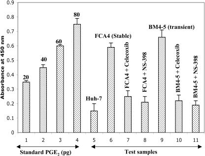
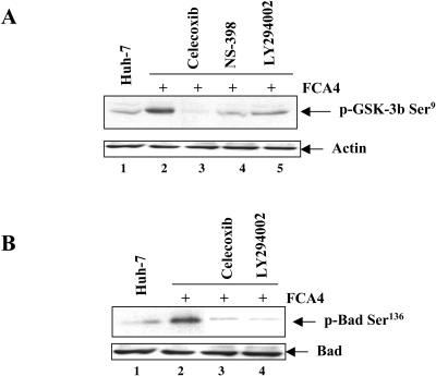
Hepatitis C virus (HCV) infection is a major cause of chronic liver disease, which can lead to the development of liver cirrhosis and hepatocellular carcinoma. Recently, the activation of cyclooxygenase-2 (Cox-2) has been implicated in the HCV-associated hepatocellular carcinoma. In this study, we focus on the signaling pathway leading to Cox-2 activation induced by HCV gene expression. Here, we demonstrate that the HCV-induced reactive oxygen species and subsequent activation of NF-kappaB mediate the activation of Cox-2. The HCV-induced Cox-2 was sensitive to antioxidant (pyrrolidine dithiocarbamate), Ca(2+) chelator (BAPTA-AM), and calpain inhibitor (N-acetyl-Leu-Leu-Met-H). The levels of prostaglandin E(2) (PGE(2)), the product of Cox-2 activity, are increased in HCV-expressing cells. Furthermore, HCV-expressing cells treated with the inhibitors of Cox-2 (celecoxib and NS-398) showed significant reduction in PGE(2) levels. We also observed the enhanced phosphorylation of Akt and its downstream substrates glycogen synthase kinase-3beta and proapoptotic Bad in the HCV replicon-expressing cells. These phosphorylation events were sensitive to inhibitors of Cox-2 (celecoxib and NS-398) and phosphatidylinositol 3-kinase (LY294002). Our results also suggest a potential role of Cox-2 and PGE(2) in HCV RNA replication. These studies provide insight into the mechanisms by which HCV induces intracellular events relevant to liver pathogenesis associated with viral infection.


Figures

FIG. 1.
FIG. 1.
HCV replicon induces expression of Cox-2. (A) Whole-cell lysates from cells stably expressing HCV replicons (FCA4) or Huh-7 cells transiently transfected with in vitro transcribed BM4-5 RNA or BM4-5 Pol RNA were fractionated by SDS-PAGE and immunoblotted with anti-Cox-2 monoclonal antibody. Lane 1, untransfected Huh-7 lysates; lane 2, FCA4 lysates; lanes 3 and 4, Huh-7 cells transfected with BM4-5 RNA; lane 5, Huh-7 cells transfected with BM4-5 RNA Pol. (B) Induction of Cox-2 mRNA in HCV replicon-containing cells. Total cellular mRNA was analyzed by using Cox-2-specific primers as described in Materials and Methods. Bar 1, Huh-7 cells; bars 2, 3, and 4, FCA4 cells and Huh-7 cells transfected with BM4-5 and BM4-5 Pol RNA. (C) Lane 1, untransfected Huh-7 lysates; lane 2, FCA4 lysates; lane 3, FCA4 lysates treated with PDTC (100 μM) for 6 h. (D) Luciferase reporter gene assay. Huh-7 and FCA4 cells were transfected with the Cox-2 luciferase reporter gene (containing NF-κB binding sites). At 36 h posttransfection, cells were treated with PDTC (100 μM) for 6 h, BAPTA-AM (50 μM) for 2 h, and ALLM (100 μM) for 24 h before the lysates were prepared for luciferase activity.
FIG. 1.
FIG. 1.
HCV replicon induces expression of Cox-2. (A) Whole-cell lysates from cells stably expressing HCV replicons (FCA4) or Huh-7 cells transiently transfected with in vitro transcribed BM4-5 RNA or BM4-5 Pol RNA were fractionated by SDS-PAGE and immunoblotted with anti-Cox-2 monoclonal antibody. Lane 1, untransfected Huh-7 lysates; lane 2, FCA4 lysates; lanes 3 and 4, Huh-7 cells transfected with BM4-5 RNA; lane 5, Huh-7 cells transfected with BM4-5 RNA Pol. (B) Induction of Cox-2 mRNA in HCV replicon-containing cells. Total cellular mRNA was analyzed by using Cox-2-specific primers as described in Materials and Methods. Bar 1, Huh-7 cells; bars 2, 3, and 4, FCA4 cells and Huh-7 cells transfected with BM4-5 and BM4-5 Pol RNA. (C) Lane 1, untransfected Huh-7 lysates; lane 2, FCA4 lysates; lane 3, FCA4 lysates treated with PDTC (100 μM) for 6 h. (D) Luciferase reporter gene assay. Huh-7 and FCA4 cells were transfected with the Cox-2 luciferase reporter gene (containing NF-κB binding sites). At 36 h posttransfection, cells were treated with PDTC (100 μM) for 6 h, BAPTA-AM (50 μM) for 2 h, and ALLM (100 μM) for 24 h before the lysates were prepared for luciferase activity.
FIG. 2.
FIG. 2.
HCV replicon induces production of PGE2 accumulation in HCV replicon-expressing cells. Huh-7 cells stably expressing the HCV subgenomic replicon (FCA4) or Huh-7 cells transiently transfected with BM4-5 RNA were treated with Cox-2 inhibitors celecoxib (50 μM) for 24 h and NS-398 (100 μM) for 24 h. After the drug treatment, culture cells were washed thoroughly with cold PBS, pH 7.4, and lysis reagent 1 (supplied by Amersham Biosciences) was added to the cells for 10 min. The PGE2 levels were then assayed by using the Biotrak Prostaglandin E2 Enzyme Immunoassay system (Amersham Pharmacia Biotech) according to the manufacturer's protocol.
FIG. 3.
FIG. 3.
HCV replicon induces Akt phosphorylation via PI3-kinase. Huh-7 and FCA4 cells were treated with antioxidant PDTC (100 μM) for 6 h, celecoxib (50 μM) for 24 h, and LY294002 (50 μM) for 12 h. Equal amounts of cellular lysates were immunoprecipitated with anti-Akt serum, fractionated by SDS-PAGE, and immunoblotted with anti-Akt Ser473 serum. Lanes 1, Huh-7 lysates; lanes 2, FCA4 lysates; lanes 3, FCA4 lysates treated with celecoxib (A), PDTC (B), or LY294002 (C). (Bottom) Total Akt in Huh-7 and HCV replicon-expressing cells.
FIG. 4.
FIG. 4.
Inhibitors of Cox-2 and PI3-kinase inhibit GSK3-β9 and Bad136 phosphorylation in HCV-expressing cells. (A) Equal amounts of cellular lysates from Huh-7 cells and FCA4 cells treated with inhibitors of Cox-2 celecoxib (5 μM) for 24 h, NS-398 (100 μM) for 24 h, and PI3-kinase (LY294002) (50 μM) for 12 h were subjected to SDS-PAGE and immunoblotted with anti-phospho-GSK-3βSer9. Lane 1, Huh-7 lysates; lane 2, FCA4 lysates; lanes 3 to 5, FCA4 lysates treated with celecoxib, NS-398, or LY294002, respectively. (B) Equal amounts of cellular lysates were immunoprecipitated with anti-Bad serum, fractionated by SDS-PAGE, and immunoblotted with anti-BadSer136 serum. Lane 1, Huh-7 lysates; lane 2, FCA4 lysates; lanes 3 and 4, FCA4 lysates treated with celecoxib and LY294002, respectively.
FIG. 5.
FIG. 5.
Effect of Cox-2 inhibitors and PGE2 on HCV RNA replication. (A and B) FCA4 cells and Huh-7 cells transiently transfected with in vitro-synthesized BM4-5 RNA were first incubated with the inhibitors of Cox-2 (50 μM celecoxib and 100μM NS-398) for 24 h. Total RNA was extracted and subjected to quantitative RT-PCR analysis. The data were expressed as relative HCV RNA levels related to the RNA level of the control cells. (C) Stably HCV replicon-expressing cells (FCA4) or Huh-7 cells transiently transfected with BM4-5 RNA were exogenously added with 0.1 μM and 0.5 μM of PGE2, respectively. Quantitative RT-PCR analyses were carried out as described above. (D) FCA4 cells were treated with the inhibitors of Cox-2 (50 μM celecoxib; 100 μM NS-398). Equal amounts of cellular lysates were subjected to SDS-PAGE and Western blotted with anti-NS5A serum. Lanes 1 and 2, equal amounts of Huh-7 and FCA4 lysates; lanes 3 and 4, FCA4 lysates treated with celecoxib and NS-398, respectively.
FIG. 6.
FIG. 6.
Model illustrating the mechanism(s) of HCV replicon-induced cell survival cascade via oxidative stress and activation of NF-κB and Cox-2. PGE2 generated by Cox-2 activity stimulates the phosphorylation of PI3-kinase-Akt, Bad136, and GSK-3β9 phosphorylation. By a mechanism not clearly understood, Cox-2 and PGE2 negatively regulate HCV RNA replication.

Similar articles

Cited by

References

    1. Bae, S. H., E. S. Jung, Y. M. Park, B. S. Kim, B. K. Kim, D. G. Kim, and W. S. Ryu. 2001. Expression of cyclooxygenase-2 (Cox-2) in hepatocellular carcinoma and growth inhibition of hepatoma cell lines by a Cox-2 inhibitor, NS-398. Clin. Cancer Res. 7:1410-1418. - PubMed
    1. Baldwin, A. S., Jr. 2001. Control of oncogenesis and cancer therapy resistance by the transcription factor NF-κB. J. Clin. Investig. 107:241-246. - PMC - PubMed
    1. Barbieri, S. B., S. Eligini, M. Brambilla, E. Tremoli, and S. Colli. 2003. Reactive oxygen species mediate cyclooxygenase-2 induction during monocyte to macrophage differentiation: critical role of NADPH oxidase. Cardiovasc. Res. 60:187-197. - PubMed
    1. Bartenschlager, R., and V. Lohmann. 2000. Replication of hepatitis C virus. J. Gen. Virol. 81:1631-1648. - PubMed
    1. Bartenschlager, R., and V. Lohmann. 2001. Novel cell culture systems for the hepatitis C virus. Antiviral Res. 52:1-17. - PubMed

Publication types

MeSH terms