

Deletion of the herpes simplex virus VP22-encoding gene (UL49) alters the expression, localization, and virion incorporation of ICP0
- PMID: 16014935
- PMCID: PMC1181569
- DOI: 10.1128/JVI.79.15.9735-9745.2005
Deletion of the herpes simplex virus VP22-encoding gene (UL49) alters the expression, localization, and virion incorporation of ICP0
Abstract
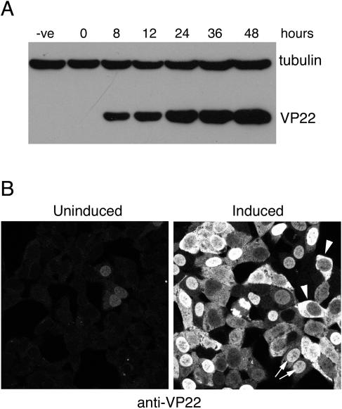
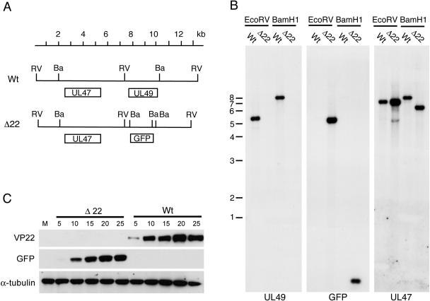
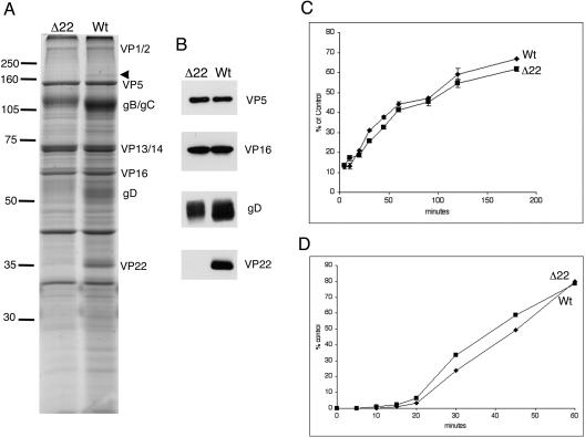
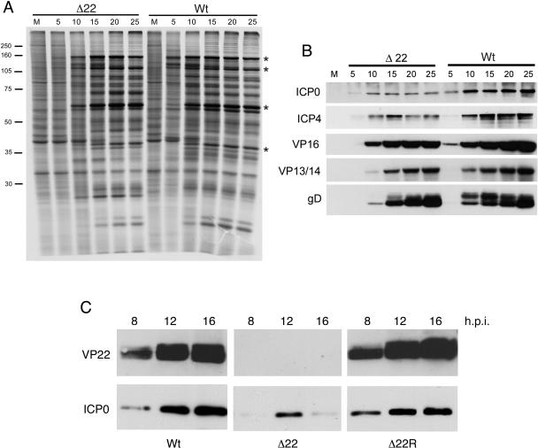
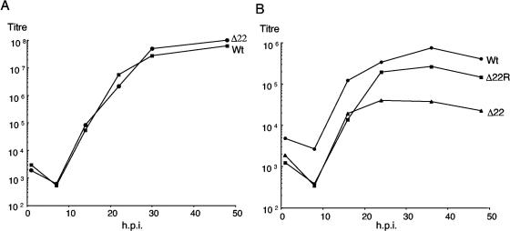
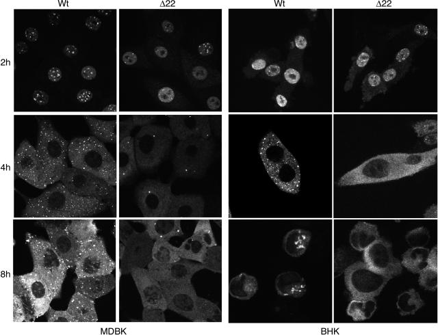
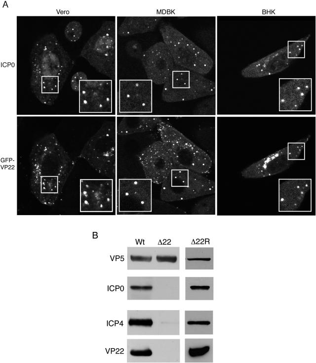
The role of the herpes simplex virus tegument protein VP22 is not yet known. Here we describe the characterization of a virus in which the entire VP22 open reading frame has been deleted. We show that VP22 is not essential for virus growth but that virus lacking VP22 (Delta22) displays a cell-specific replication defect in epithelial MDBK cells. Virus particles assembled in the absence of VP22 show few obvious differences to wild-type (WT) particles, except for a moderate reduction in glycoproteins gD and gB. In addition, the Delta22 virus exhibits a general delay in the initiation of virus protein synthesis, but this is not due to a glycoprotein-related defect in virus entry. Intriguingly, however, the absence of VP22 has an obvious effect on the intracellular level of the immediate-early (IE) protein ICP0. Moreover, following translocation from the nucleus to the cytoplasm, ICP0 is unable to localize to the characteristic cytoplasmic sites observed in a WT infection. We demonstrate that, in WT-infected cells, VP22 and ICP0 are concentrated in the same cytoplasmic sites. Furthermore, we show that, while ICP0 and ICP4 are components of WT extracellular virions, the altered localization of ICP0 in the cytoplasm of Delta22-infected cells correlates with an absence of both ICP0 and ICP4 from Delta22 virions. Hence, while a role has not yet been defined for virion IE proteins in virus infection, our results suggest that their incorporation is a specific event requiring the tegument protein VP22. This report provides the first direct evidence that VP22 influences virus assembly.
Figures
Similar articles
-
Overexpression of the herpes simplex virus type 1 tegument protein VP22 increases its incorporation into virus particles.Virology. 1996 Jun 1;220(1):60-8. doi: 10.1006/viro.1996.0286. Virology. 1996. PMID: 8659129
-
Phosphorylation of the herpes simplex virus tegument protein VP22 has no effect on incorporation of VP22 into the virus but is involved in optimal expression and virion packaging of ICP0.J Virol. 2005 Nov;79(22):14057-68. doi: 10.1128/JVI.79.22.14057-14068.2005. J Virol. 2005. PMID: 16254340 Free PMC article.
-
Herpes simplex virus tegument protein VP22 contains an internal VP16 interaction domain and a C-terminal domain that are both required for VP22 assembly into the virus particle.J Virol. 2005 Oct;79(20):13082-93. doi: 10.1128/JVI.79.20.13082-13093.2005. J Virol. 2005. PMID: 16189010 Free PMC article.
-
[Mechanisms of herpesvirus infection--virus entry into host cells and virus assembly].Uirusu. 2007 Dec;57(2):151-8. doi: 10.2222/jsv.57.151. Uirusu. 2007. PMID: 18357753 Review. Japanese.
-
Association of the herpes simplex virus major tegument structural protein VP22 with chromatin.Biochim Biophys Acta. 2010 Mar-Apr;1799(3-4):200-6. doi: 10.1016/j.bbagrm.2009.08.002. Epub 2009 Aug 12. Biochim Biophys Acta. 2010. PMID: 19682615 Review.
Cited by
-
Replication-competent herpes simplex virus 1 isolates selected from cells transfected with a bacterial artificial chromosome DNA lacking only the UL49 gene vary with respect to the defect in the UL41 gene encoding host shutoff RNase.J Virol. 2007 Oct;81(20):10924-32. doi: 10.1128/JVI.01239-07. Epub 2007 Aug 1. J Virol. 2007. PMID: 17670820 Free PMC article.
-
Direct and specific binding of the UL16 tegument protein of herpes simplex virus to the cytoplasmic tail of glycoprotein E.J Virol. 2011 Sep;85(18):9425-36. doi: 10.1128/JVI.05178-11. Epub 2011 Jul 6. J Virol. 2011. PMID: 21734044 Free PMC article.
-
Morphogenesis of a highly replicative EGFPVP22 recombinant Marek's disease virus in cell culture.J Virol. 2007 Nov;81(22):12348-59. doi: 10.1128/JVI.01177-07. Epub 2007 Sep 12. J Virol. 2007. PMID: 17855520 Free PMC article.
-
Functions of the ORF9-to-ORF12 gene cluster in varicella-zoster virus replication and in the pathogenesis of skin infection.J Virol. 2008 Jun;82(12):5825-34. doi: 10.1128/JVI.00303-08. Epub 2008 Apr 9. J Virol. 2008. PMID: 18400847 Free PMC article.
-
Cell cycle modulation by Marek's disease virus: the tegument protein VP22 triggers S-phase arrest and DNA damage in proliferating cells.PLoS One. 2014 Jun 19;9(6):e100004. doi: 10.1371/journal.pone.0100004. eCollection 2014. PLoS One. 2014. PMID: 24945933 Free PMC article.
References
-
- Chi, J. H., C. A. Harley, A. Mukhopadhyay, and D. W. Wilson. 2005. The cytoplasmic tail of herpes simplex virus envelope glycoprotein D binds to the tegument protein VP22 and to capsids. J. Gen. Virol. 86:253-261. - PubMed
-
- Dargin, D. 1986. The structure and assembly of herpes viruses, p. 359-437. In J. R. Harris and R. W. Horne (ed.), Electron microscopy of proteins, vol. 5. Virus structure. Academic Press, London, United Kingdom.
Publication types
MeSH terms
Substances
LinkOut - more resources
Full Text Sources
