Comparative Study
. 2005 Aug;79(15):9756-64.
doi: 10.1128/JVI.79.15.9756-9764.2005.

The capsid protein of satellite Panicum mosaic virus contributes to systemic invasion and interacts with its helper virus

Affiliations
Comparative Study

The capsid protein of satellite Panicum mosaic virus contributes to systemic invasion and interacts with its helper virus

Rustem T Omarov et al. J Virol. 2005 Aug.

Abstract

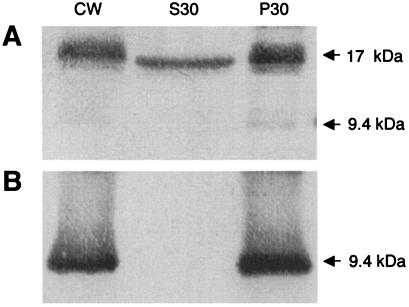
Satellite panicum mosaic virus (SPMV) depends on its helper Panicum mosaic virus (PMV) for replication and spread in host plants. The SPMV RNA encodes a 17-kDa capsid protein (CP) that is essential for formation of its 16-nm virions. The results of this study indicate that in addition to the expression of the full-length SPMV CP from the 5'-proximal AUG start codon, SPMV RNA also expresses a 9.4-kDa C-terminal protein from the third in-frame start codon. Differences in solubility between the full-length protein and its C-terminal product were observed. Subcellular fractionation of infected plant tissues showed that SPMV CP accumulates in the cytosol, cell wall-, and membrane-enriched fractions. However, the 9.4-kDa protein exclusively cofractionated with cell wall- and membrane-enriched fractions. Earlier studies revealed that the 5'-untranslated region (5'-UTR) from nucleotides 63 to 104 was associated with systemic infection in a host-specific manner in millet plants. This study shows that nucleotide deletions and insertions in the 5'-UTR plus simultaneous truncation of the N-terminal part of the CP impaired SPMV spread in foxtail millet, but not in proso millet plants. In contrast, the expression of the full-length version of SPMV CP efficiently compensated the negative effect of the 5'-UTR deletions in foxtail millet. Finally, immunoprecipitation assays revealed the presence of a specific interaction between the capsid proteins of SPMV and its helper virus (PMV). Our findings show that the SPMV CP has several biological functions, including facilitating efficient satellite virus infection and movement in millet plants.


Figures

FIG. 1.
FIG. 1.
SPMV genome and expression of CP. (A) Schematic representation of the 824-nt SPMV RNA (solid line) and the full-length 17-kDa CP. The restriction enzyme sites used to assemble the cDNA constructs and the positions of the predicted in-frame start codons (arrows) are indicated. The 17-kDa CP (AUG1) and N-terminal-truncated open reading frames (AUG2 to AUG4) are indicated by white rectangles. (B) In vitro translation products labeled with [35S]methionine were generated from infectious transcripts of SPMV wild type and SPMV/U-91. (C) Western blot analyses of SPMV CP products in millet plants coinfected with the helper virus and the wild type (SPMV) or its mutants (UAA-234 and U-91) probed with anti-SPMV CP polyclonal antiserum. Arrows indicate the SPMV CP-specific proteins and their molecular masses.
FIG. 2.
FIG. 2.
Differences in solubility of the 17-kDa SPMV CP and the 9.4-kDa N-terminally truncated SPMV/U-91 CP isolated from millet plants coinoculated with Panicum mosaic virus. (A) Extraction and immunoblot detection of the capsid protein from SPMV and SPMV/U-91 under native (-SDS) and denaturing (+SDS) conditions. The lower panel represents a gel stained with Coomassie brilliant blue R to verify equal protein loading. (B) Extractions of the 9.4-kDa CP from SPMV/U-91-infected tissue were made in buffer (100 mM Tris-HCl, pH 7.5) (lane 1), buffer plus 1 M NaCl (lane 2), buffer plus 2% Triton X-100 (lane 3), or buffer plus 1% SDS (lane 4). In lane 4, the asterisk indicates an SPMV CP antibody-specific protein that was consistently observed.
FIG. 3.
FIG. 3.
Serological detection of SPMV CP in proso millet plants collected 14 days after inoculation of PMV plus SPMV or PMV plus SPMV/U-91 transcripts. The fractionation of cellular proteins by differential centrifugation is represented by CW (cell wall proteins), S30 (cytosolic proteins), and P30 (membranes). The proteins (indicated by arrows) were separated by SDS-PAGE and analyzed by Western blotting using SPMV CP-specific antiserum. (A) Plants infected with PMV and SPMV. (B) Plants infected with PMV and SPMV/U-91.
FIG. 4.
FIG. 4.
Host-dependent movement of SPMV/ΔBsmI-MscI in millet following coinfection with PMV. (A) RNA was isolated from upper noninoculated leaves of infected foxtail and proso millet plants. The upper panel represents an RNA blot probed with 32P-labeled SPMV. The lower panel represents an ethidium bromide-stained gel to verify equal loading of RNA, represented by rRNA, for each sample. (B and C) Western blots of SPMV CP (B) or PMV CP (C) isolated from upper leaves of infected millet plants. The expressed proteins are indicated by arrows.
FIG. 5.
FIG. 5.
SPMV RNA accumulation in upper noninoculated leaves with PMV plus SPMV/U-301. (A) Immunodetection of the 26-kDa PMV CP (upper panel) and the 17-kDa SPMV CP (lower panel) in upper uninoculated leaves of proso and foxtail millet plants 14 dpi with PMV plus wild-type SPMV or SPMV/U-301 (a double frameshift mutant to abolish CP expression). Molecular mass markers (in kilodaltons) are indicated on the rightmost side of the blot. (B) SPMV RNA accumulation as detected by Northern blotting using an SPMV-specific probe (upper panel). The lower panel is rRNA from an ethidium bromide-stained agarose gel, indicating equal loading for each sample.
FIG. 6.
FIG. 6.
SPMV RNA and CP accumulation from 5′-UTR mutants of SPMV and SPMV/AUC. The upper noninoculated leaves of proso and foxtail millet plants coinfected with PMV and the respective mutants were used for Western and Northern blotting. Equal loading of RNA was determined by visualizing rRNA levels on ethidium bromide-stained agarose gels, prior to transfer to membranes. For panels A and B, lanes 1, 2, and 3 represent mock, PMV, and PMV plus SPMV inoculations, respectively. (A) SPMV 5′-UTR derivatives that retain the expression of the full-length CP (lanes 4 to 6), representing mixed infections of PMV plus SPMV/BamHI, SPMV/ΔSpeI-BamHI, or SPMV/insertClaI, respectively. (B) SPMV/AUC-derived constructs that express the 9.4-kDa C-terminal CP. Lanes 4 to 6 represent mixed infections of PMV plus SPMV/AUC, SPMV/AUC/ΔSpeI-BamHI, or SPMV/AUC/insertClaI, respectively.
FIG. 7.
FIG. 7.
Biomolecular interactions between PMV and SPMV capsid proteins. (A) Healthy (mock) and PMV- and SPMV-infected (P+S) proso millet plants were labeled in planta with [35S]methionine. The left panel shows total protein extracts, and the right panel is an autoradiograph after a pull-down assay using SPMV CP-specific antiserum for immunoprecipitation (IP). The asterisk indicates the position of a host protein (in healthy and infected plants). The PMV (26-kDa) and SPMV (17-kDa) capsid proteins are indicated with arrows. (B) Immunoprecipitation and detection of PMV CP from extracts of infected proso millet leaves. PMV-inoculated plants were subjected to immunoprecipitation, as separate assays, with SPMV CP or PMV CP antiserum. The resultant precipitate was eluted, subjected to Western blotting, and analyzed for the presence of PMV CP using PMV CP antibody as a probe. (C and D) Coimmunoprecipitation of SPMV and PMV capsid proteins from extracts of infected proso millet leaves. PMV- and SPMV-infected plants were subjected to immunoprecipitation using antiserum specific for either SPMV CP or PMV CP. The immunoprecipitates were eluted and separately subjected to Western blotting and probed with polyclonal antiserum specific for PMV CP (C) or SPMV CP (D). The asterisk indicates the position of the dimeric form of SPMV CP (∼34 kDa). The arrowheads show positions and molecular masses of precipitated proteins.

Similar articles

Cited by

References

    1. Allison, R., C. Thompson, and P. Ahlquist. 1990. Regeneration of a functional RNA virus genome by recombination between deletion mutants and requirement for cowpea chlorotic mottle virus 3a and coat genes for systemic infection. Proc. Natl. Acad. Sci. USA 87:1820-1824. - PMC - PubMed
    1. Batten, J. S. 2002. Replication and translation of panicum mosaic virus. Ph.D. dissertation. Texas A&M University, College Station.
    1. Buzen, F. G., C. L. Niblett, G. R. Hooper, J. Hubbard, and M. A. Newman. 1984. Further characterization of panicum mosaic virus and its associated satellite virus. Phytopathology 74:313-318.
    1. Callaway, A., D. Giesman-Cookmeyer, E. T. Gillock, T. L. Sit, and S. A. Lommel. 2001. The multifunctional capsid proteins of plant RNA viruses. Annu. Rev. Phytopathol. 39:419-460. - PubMed
    1. Chapman, S., T. Kavanagh, and D. Baulcombe. 1992. Potato virus X as a vector for gene expression in plants. Plant J. 2:549-557. - PubMed

Publication types

MeSH terms

LinkOut - more resources