Comparative Study
. 2005 Aug;79(15):9765-76.
doi: 10.1128/JVI.79.15.9765-9776.2005.

A virus essential for insect host-parasite interactions encodes cystatins

Affiliations
Comparative Study

A virus essential for insect host-parasite interactions encodes cystatins

E Espagne et al. J Virol. 2005 Aug.

Abstract

Cotesia congregata is a parasitoid wasp that injects its eggs in the host caterpillar Manduca sexta. In this host-parasite interaction, successful parasitism is ensured by a third partner: a bracovirus. The relationship between parasitic wasps and bracoviruses constitutes one of the few known mutualisms between viruses and eukaryotes. The C. congregata bracovirus (CcBV) is injected at the same time as the wasp eggs in the host hemolymph. Expression of viral genes alters the caterpillar's immune defense responses and developmental program, resulting in the creation of a favorable environment for the survival and emergence of adult parasitoid wasps. Here, we describe the characterization of a CcBV multigene family which is highly expressed during parasitism and which encodes three proteins with homology to members of the cystatin superfamily. Cystatins are tightly binding, reversible inhibitors of cysteine proteases. Other cysteine protease inhibitors have been described for lepidopteran viruses; however, this is the first description of the presence of cystatins in a viral genome. The expression and purification of a recombinant form of one of the CcBV cystatins, cystatin 1, revealed that this viral cystatin is functional having potent inhibitory activity towards the cysteine proteases papain, human cathepsins L and B and Sarcophaga cathepsin B in assays in vitro. CcBV cystatins are, therefore, likely to play a role in host caterpillar physiological deregulation by inhibiting host target proteases in the course of the host-parasite interaction.


Figures

FIG. 1.
FIG. 1.
Nucleotide and amino acid sequence alignment of the CcBV cystatins. (A) Nucleotide sequence alignment of the three CcBV cystatin ORFs. Differences in nucleotide sequences are boxed. (B) Amino acid sequence alignment of the three CcBV preprotein cystatins. The potential cleavage site for cystatin 2 and cystatin 3 (same site) and cystatin 1 are indicated by white triangles. The three active sites which are the hallmarks of cystatins are indicated by stars: G (residue 28), Qx(V)xG (starting residue 72), and W (residue 119). The four cysteine residues potentially forming two disulfide bonds are indicated by black diamonds. Differing amino acids are boxed (same family, light grey boxing; different family, dark grey boxing).
FIG. 2.
FIG. 2.
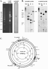
Mapping of Cotesia congregata bracovirus cystatins in the viral genome. (A) Field inversion gel electrophoresis of 250 ng of viral DNA, followed by hybridization using cystatin 1 cDNA as a probe. The unique hybridization signal observed corresponds to the circle (indicated by a white asterisk) of the estimated linear size (19 kb). The 2.5-kb ladder from Bio-Rad was used as linear molecular weight marker in this experiment. (B) Southern blot analysis of viral DNA (200 ng per lane), nondigested (ND) or after restriction digestion with XbaI-XhoI (Xa, Xo), BamHI (B), or SacI (S), using either cystatin 1 cDNA or cystatin 2 cDNA as a probe. Hybridizing fragments corresponding to cystatin 1 are indicated by a black square. Hybridizing fragments corresponding to cystatin 2 are indicated by a black star. Hybridizing fragments corresponding to cystatin 3 are indicated by a black asterisk. (C) Restriction map of CcBV circle19, harboring the three cystatin genes. Lengths of expected fragments after restriction digestion are indicated. Hybridizing fragments are highlighted with corresponding symbols (box, star, or asterisk). Note the presence of a BamHI site (grey circle) in cystatin 3, which gives rise to a partial digest.
FIG. 3.
FIG. 3.
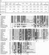
Pairwise BLAST and multiple sequence alignment of CcBV cystatins with insect and noninsect cystatins. (A) Pairwise BLAST of CcBV cystatins with insect and noninsect cystatins. Numbers indicate percentage identity/percentage similarity. Preprotein sequences were compared. (B) Multiple sequence alignment of CcBV cystatins with related cystatins using the Clustalw program (http://clustalw.genome.jp/) combined with the BoxShade Server (http://www.ch.embnet.org/software/BOX_form.html). Preprotein sequences were compared in this alignment. The numbering refers to CcBV Cystatins labeled with the number one have the first amino acid of the signal peptides. The three conserved active sites of cystatins are indicated by stars beneath the concensus sequence: G residue, Qx(V)xG motif and (P)W residues. The cysteine residues in conserved positions are indicated by black diamonds. Agam, Anopheles gambiae (mosquito, Diptera; GenBank accession number EST AJ284933); Amel, Apis mellifera (honeybee, Hymenoptera; Honeybee Genome Project accession number Amel_1.1contig2738), Bm: Brugia malayi (filarial nematode; GenBank accession number AAC47623); Bt, Bos taurus (bovine; GenBank accession number P01045); Gg, Gallus gallus (chicken; GenBank accession number NP_990831); Hs. Homo sapiens (GenBank accession number NP_000090); Mm, Mus musculus (mouse; GenBank accession number NP_067380); Sp, Sarcophaga peregrina (flesh fly, Diptera; SwissProt accession number P31727).
FIG. 4.
FIG. 4.
Expression profile of CcBV cystatin genes in parasitized Manduca sexta by Northern blot analysis (A) Time course experiment using RNA extracted from nonparasitized (NP) or parasitized Manduca sexta fat body at different time points postparasitization as indicated. (B) Tissue-specific expression using RNA extracted from nonparasitized (NP) or parasitized Manduca sexta at different time points postparasitization as indicated. A total of 20 μg of RNA was loaded in each lane. Cystatin 1 cDNA was used as a probe. As a control, blots were hybridized with Manduca sexta actin. Levels of cystatin expression are indicated relative to that of the control actin. The values correspond to the ratios obtained after Instant Imager scanning of the Northern blots presented.
FIG. 5.
FIG. 5.
Enzymatic assays for evaluation of stably transformed cell lines expressing recombinant cystatin 1 protein. The assays were performed using 10% (vol/vol) cell culture supernatant in each reaction mixture as described in Materials and Methods. The following conditions are shown: standard, standard curve without inhibition; transformed Hi5 supernatants (10:1 and 100:1), supernatants from the relevant stably transformed cell lines (HI5-C10 and HI5-C100) expressing cystatin 1 protein; Hi5 supernatant, control supernatant from nontransfected HighFive cell cultures of the same cell density as transfected Hi5 cells; negative, assay without papain to examine substrate self-hydrolysis. Note that addition of 10% control supernatant had a very minor impact on the assay kinetics, while supernatants from cystatin 1 expressing cultures inhibited papain very strongly. The effect of self-hydrolysis was negligible.
FIG. 6.
FIG. 6.
Silver-stained SDS-polyacrylamide gel electrophoresis (18%) gel of 20-μl samples from several steps of the cystatin 1 protein purification procedure. Lanes: 1, concentrated, desalted cell culture supernatant before application to CM-papain matrix; 2, postbinding flowthrough fraction; 3 and 4, wash fractions (5 bed volumes each); 5 to 8, eluate fractions (1 bed volume each); 9, CM-papain matrix beads after elution; 10, molecular mass standard. The arrow points to the prominent protein band that migrates at the expected molecular weight for cystatin 1 (see text).
FIG. 7.
FIG. 7.
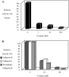
Cysteine protease inhibitory activity of CcBV cystatin 1. Various concentrations of cystatin 1 were incubated with papain (1 nM) (A) or human cathepsin B (1.8 nM), cathepsin L (0.6 nM), or Sarcophaga cathepsin B (approximately 1 nM) (B). The relative activity of each cysteine protease was determined in presence of the recombinant cystatin 1. Incubation of each protease with the substrate and without addition of cystatin 1 corresponded to 100% activity of the enzymes.

Similar articles

Cited by

References

    1. Abe, K., Y. Emori, H. Kondo, K. Suzuki, and S. Arai.c 1987. Molecular cloning of a cysteine proteinase inhibitor of rice (oryzacystatin). Homology with animal cystatins and transient expression in the ripening process of rice seeds. J. Biol. Chem. 262:16793-16797. - PubMed
    1. Abrahamson, M., M. Alvarez-Fernandez, and C. M. Nathanson. 2003. Cystatins. Biochem. Soc. Symp. 2003:179-199. - PubMed
    1. Albrecht, U., T. Wyler, R. Pfister-Wilhelm, A. Gruber, P. Stettler, P. Heiniger, F. Kurt, D. Schümperli, and B. Lanzrein. 1994. Polydnavirus of the parasitic wasp Chelonus inanitus (Braconidae): characterization, genome organization and time point of replication. J. Gen. Virol. 75:3353-3363. - PubMed
    1. Altschul, S. F., W. Gish, W. Miller, E. W. Myers, and D. J. Lipman. 1990. Basic local alignment search tool. J. Mol. Biol. 215:403-410. - PubMed
    1. Anastasi, A., M. A. Brown, A. A. Kembhavi, M. J. Nicklin, C. A. Sayers, D. C. Sunter, and A. J. Barrett. 1983. Cystatin, a protein inhibitor of cysteine proteinases. Improved purification from egg white, characterization, and detection in chicken serum. Biochem. J. 211:129-138. - PMC - PubMed

Publication types

MeSH terms

Associated data

LinkOut - more resources