Comparative Study
. 2005 Aug;79(15):9786-98.
doi: 10.1128/JVI.79.15.9786-9798.2005.

Mapping of the hepatitis B virus pre-S1 domain involved in receptor recognition

Affiliations
Comparative Study

Mapping of the hepatitis B virus pre-S1 domain involved in receptor recognition

Azeneth Barrera et al. J Virol. 2005 Aug.

Abstract

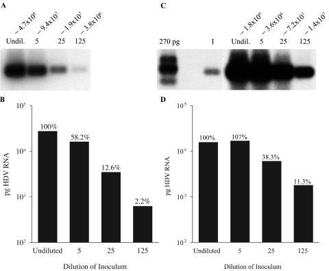
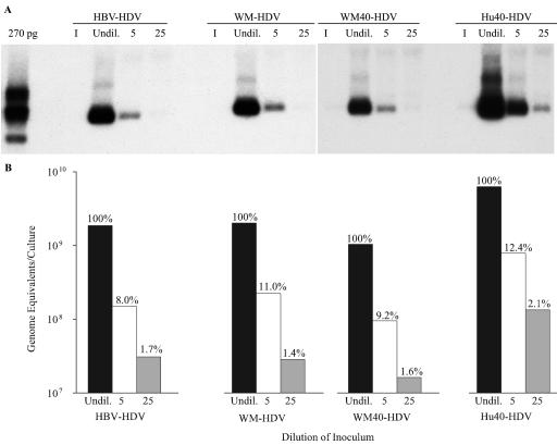
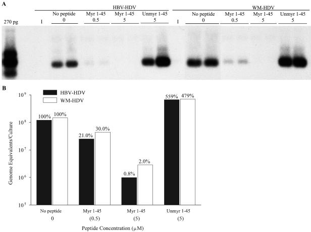
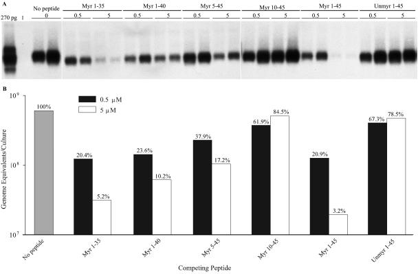
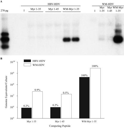
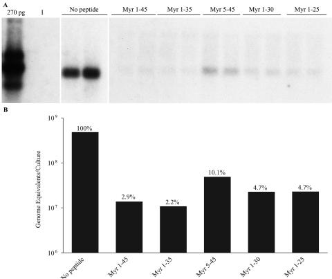
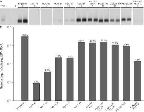
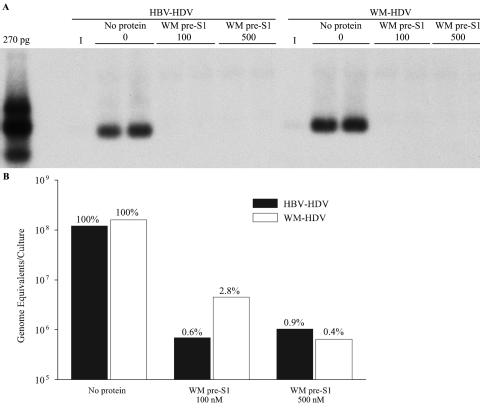
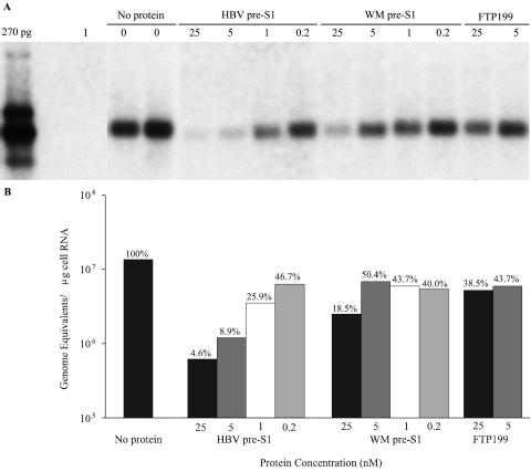
Hepatitis B virus (HBV) and woolly monkey hepatitis B virus (WMHBV) are primate hepadnaviruses that display restricted tissue and host tropisms. Hepatitis D virus (HDV) particles pseudotyped with HBV and WMHBV envelopes (HBV-HDV and WM-HDV) preferentially infect human and spider monkey hepatocytes, respectively, thereby confirming host range bias in vitro. The analysis of chimeric HBV and WMHBV large (L) envelope proteins suggests that the pre-S1 domain may comprise two regions that affect infectivity: one within the amino-terminal 40 amino acids of pre-S1 and one downstream of this region. In the present study, we further characterized the role of the amino terminus of pre-S1 in infectivity by examining the ability of synthetic peptides to competitively block HDV infection of primary human and spider monkey hepatocytes. A synthetic peptide representing the first 45 residues of the pre-S1 domain of the HBV L protein blocked infectivity of HBV-HDV and WM-HDV, with a requirement for myristylation of the amino terminal residue. Competition studies with truncated peptides suggested that pre-S1 residues 5 to 20 represent the minimal domain for inhibition of HDV infection and, thus, presumably represent the residues involved in virus-host receptor interaction. Recombinant pre-S1 proteins expressed in insect cells blocked infection with HBV-HDV and WM-HDV at a concentration of 1 nanomolar. The ability of short pre-S1 peptides to efficiently inhibit HDV infection suggests that they represent suitable ligands for identification of the HBV receptor and that a pre-S1 mimetic may represent a rational therapy for the treatment of HBV infection.


Figures

FIG. 1.
FIG. 1.
Titration and saturation of HDV infectivity in human hepatocytes. (A) Human hepatocytes were inoculated with doses of HBV-HDV that were undiluted (4.7 × 10⁸ ge) or diluted 5-fold (9.4 × 10⁷ ge), 25-fold (1.9 × 10⁷ ge), and 125-fold (3.8 × 10⁶ ge). Cultures were harvested on day 12 postinoculation, and 5 μg of total cell RNA (approximately 15% of RNA from a 35-mm dish) was analyzed by Northern blot hybridization by using a riboprobe for HDV genomic RNA. (B) The hybridized membrane from panel A was digitally scanned with a phosphorimager. The amount of HDV RNA is expressed in picograms per culture and was derived by linear regression analysis of HDV RNA standards loaded on the same gel. Percent HDV RNA was derived from undiluted HBV-HDV-infected cultures as 100%. (C) Human hepatocytes were inoculated with doses of HBV-HDV that were undiluted (1.8 × 10⁹ ge) or diluted 5-fold (3.6 × 10⁸ ge), 25-fold (7.2 × 10⁷ ge), and 125-fold (1.4 × 10⁷ ge). Cultures were harvested on day 11 postinoculation, and total cellular RNA was analyzed as described for panel A. A 270-pg HDV-RNA standard was run on the gel for comparison. HDV RNA extracted from the equivalent of 25% of the inoculum was analyzed under the same conditions (lane I). (D) The hybridized membrane from panel C was analyzed by a phosphorimager as described for panel B.
FIG. 2.
FIG. 2.
Titration of HDV psuedotypes of HBV, WMHBV, a chimeric HBV, and a chimeric WMHBV in spider monkey hepatocytes. (A) Spider monkey hepatocytes were inoculated with doses of HBV-HDV, WM-HDV, WM40-HDV, or Hu40-HDV that were undiluted (6.4 × 10⁷ ge) or diluted 5-fold (1.3 × 10⁷ ge) and 25-fold (2.6 × 10⁶ ge). Cultures were harvested on day 12 postinoculation, and total cellular RNA was analyzed as described in the legend for Fig. 1A. A 270-pg HDV-RNA standard was run on the gel for comparison. RNA extracted from the equivalent of 10% of the inocula was analyzed under the same conditions (lanes I) but was undetectable in the exposure shown. (B) Levels of HDV RNA from the same cultures analyzed in panel A were quantified by TaqMan RT-PCR and expressed as genomic equivalents per culture. Percent genome equivalents of HDV were derived from undiluted-virus-infected cultures as 100%.
FIG. 3.
FIG. 3.
Inhibition of HDV infection with pre-S1 peptides. (A) Spider monkey hepatocytes were inoculated in duplicate with HBV-HDV or WM-HDV and competed with myristylated or unmyristylated HBV 1-45 peptides at 0.5 μM and 5 μM concentrations. Viruses were competed with peptides for 2 h at 4°C, and then cultures were washed to remove excess peptide and virus. Cultures were harvested on day 12 postinoculation, and total cellular RNA was analyzed as described in the legend for Fig. 1A. Lanes I, RNA extracted from the equivalent of 10% of the inocula. (B) Levels of HDV RNA from the same cultures analyzed in panel A were quantified by TaqMan RT-PCR and expressed as genomic equivalents per culture. Percent genome equivalents of HDV were derived from no-peptide control cultures as 100%.
FIG. 4.
FIG. 4.
Mapping of the pre-S1 domain for inhibition of HDV infectivity. (A) Human hepatocytes were inoculated in duplicate with HBV-HDV and competed with myristylated peptides consisting of HBV 1-35, 1-40, 5-45, 10-45, and 1-45 or an unmyristylated HBV 1-45 peptide at 0.5 μM and 5 μM concentrations. Virus was competed with peptides for 16 h at 37°C, and then cultures were washed to remove excess peptide and virus. Cultures were harvested on day 9 postinoculation, and total cellular RNA was analyzed as described in the legend for Fig. 1A. Lane I, RNA extracted from the equivalent of 20% of the inocula. (B) Levels of HDV RNA from the same cultures analyzed in panel A were quantified by TaqMan RT-PCR and expressed as genomic equivalents per culture. Percent genome equivalents of HDV were derived from no-peptide control cultures as 100%.
FIG. 5.
FIG. 5.
Peptide inhibition of HDV infection in spider monkey hepatocytes. (A) Spider monkey hepatocytes were inoculated in duplicate for HBV-HDV or in individual wells for WM-HDV and competed with myristylated peptides consisting of HBV 1-35 and 1-45 and WMHBV 1-35 at a 5 μM concentration. Viruses were competed with peptides for 16 h at 37°C, and then cultures were washed to remove excess peptide and virus. Cultures were harvested on day 7 postinoculation, and total cellular RNA was analyzed as described in the legend for Fig. 1A. Lanes I, RNA extracted from the equivalent of 20% of the inocula. (B) Levels of HDV RNA from the same cultures analyzed in panel A were quantified by TaqMan RT-PCR and expressed as genomic equivalents per culture. Percent genome equivalents of HDV were derived from WM-Myr 1-35-competed cultures as 100%.
FIG. 6.
FIG. 6.
Residues 1 to 25 of the pre-S1 domain inhibit HDV infectivity. (A) Human hepatocytes were inoculated in duplicate with HBV-HDV particles and competed with myristylated peptides consisting of HBV 1-45, 1-35, 5-45, 1-30, and 1-25 at a 5 μM concentration. Virus was competed with peptides for 16 h at 37°C, and then cultures were washed to remove excess peptide and virus. Cultures were harvested on day 9 postinoculation, and total cellular RNA was analyzed as described in the legend for Fig. 1A. Lane I, RNA extracted from the equivalent of 20% of the inocula. (B) Levels of HDV RNA from the same cultures analyzed in panel A were quantified by TaqMan RT-PCR and expressed as genomic equivalents per culture. Percent genome equivalents of HDV were derived from no-peptide control cultures as 100%.
FIG. 7.
FIG. 7.
Mapping of the minimal pre-S1 region capable of inhibiting HDV infection. (A) Spider monkey hepatocytes were inoculated in duplicate with Hu40-HDV particles and competed with myristylated peptides consisting of HBV 2-16, 2-20, 1-25, 2-35, 2-45 (Δ5-9), and 1-45 and WM-Myr 2-35 at a 5 μM concentration and HBV Unmyr 1-45 at 25 μM and 75 μM concentrations. Virus was competed with peptide for 16 h at 37°C (except for Pre-Bound), and then cultures were washed to remove excess peptide and virus. Pre-Bound indicates that cultures were incubated with 5 μM Myr 1-45 for 2 h at 4°C, washed to remove excess peptide, and then inoculated with virus for 16 h at 37°C. Cultures were harvested on day 9 postinoculation, and total cellular RNA was analyzed as described in the legend for Fig. 1A. (B) Levels of HDV RNA from the same cultures analyzed in panel A were quantified by TaqMan RT-PCR and expressed as genomic equivalents per culture. Percent genome equivalents of HDV were derived from no peptide control cultures as 100%.
FIG. 8.
FIG. 8.
Inhibition of HDV infection with WM pre-S1 protein that was purified from insect cells. (A) Spider monkey hepatocytes were inoculated in duplicate with HBV-HDV or WM-HDV and competed with WM pre-S1 at 100 nM or 500 nM concentrations or no competing protein. Viruses were competed with WM pre-S1 for 2 h at 4°C, and then cultures were washed to remove excess protein and virus. Cultures were harvested on day 12 postinoculation, and total cellular RNA was analyzed as described in the legend for Fig. 1A. Lanes I, RNA extracted from the equivalent of 10% of the inocula. (B) Levels of HDV RNA from the same cultures analyzed in panel A were quantified by TaqMan RT-PCR and expressed as genomic equivalents per culture. Percent genome equivalents of HDV were derived from no-competing-protein control cultures as 100%.
FIG. 9.
FIG. 9.
Inhibition of HDV infection with proteins purified from insect cells. (A) Human hepatocytes were inoculated with HBV-HDV and competed with HBV pre-S1, WM pre-S1, or FTP199 at 25 nM, 5 nM, 1 nM, and 0.2 nM concentrations. Virus was competed for 16 h at 37°C, and then cultures were washed to remove excess protein and virus. Cultures were harvested on day 9 postinoculation, and total cellular RNA was analyzed as described in the legend for Fig. 1A. Lane I, RNA extracted from the equivalent of 20% of the inocula. (B) Levels of HDV RNA from the same cultures analyzed in panel A were quantified by TaqMan RT-PCR and expressed as genomic equivalents per μg. Percent genome equivalents of HDV were derived from no-competing-protein control cultures as 100%.

References

    1. Barrera, A., B. Guerra, H. Lee, and R. E. Lanford. 2004. Analysis of host range phenotypes of primate hepadnaviruses by in vitro infections of hepatitis D virus pseudotypes. J. Virol. 78:5233-5243. - PMC - PubMed
    1. Barrera, A., and R. E. Lanford. 2004. Infection of primary chimpanzee hepatocytes with recombinant hepatitis D virus particles: a surrogate model for hepatitis B virus. Methods Mol. Med. 95:131-142. - PubMed
    1. Breiner, K. M., S. Urban, and H. Schaller. 1998. Carboxypeptidase D (gp180), a Golgi-resident protein, functions in the attachment and entry of avian hepatitis B viruses. J. Virol. 72:8098-8104. - PMC - PubMed
    1. Bruss, V., J. Hagelsten, E. Gerhardt, and P. R. Galle. 1996. Myristylation of the large surface protein is required for hepatitis B virus in vitro infectivity. Virology 218:396-399. - PubMed
    1. Chouteau, P., J. Le Seyec, I. Cannie, M. Nassal, C. Guguen-Guillouzo, and P. Gripon. 2001. A short N-proximal region in the large envelope protein harbors a determinant that contributes to the species specificity of human hepatitis B virus. J. Virol. 75:11565-11572. - PMC - PubMed

Publication types

MeSH terms

LinkOut - more resources