Comparative Study
. 2005 Aug;79(15):9799-809.
doi: 10.1128/JVI.79.15.9799-9809.2005.

Diversity, divergence, and evolution of cell-free human immunodeficiency virus type 1 in vaginal secretions and blood of chronically infected women: associations with immune status

Affiliations
Comparative Study

Diversity, divergence, and evolution of cell-free human immunodeficiency virus type 1 in vaginal secretions and blood of chronically infected women: associations with immune status

Sharon T Sullivan et al. J Virol. 2005 Aug.

Abstract

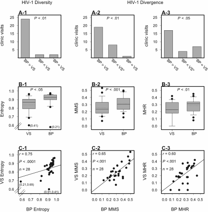
Most human immunodeficiency virus type 1 (HIV-1) infections are believed to be the result of exposure to the virus in genital secretions. However, prevention and therapeutic strategies are usually based on characterizations of HIV-1 in blood. To understand better the dynamics between HIV-1 quasispecies in the genital tract and blood, we performed heteroduplex assays on amplified env products from cell-free viral RNA in paired vaginal secretion (VS) and blood plasma (BP) samples of 14 women followed for 1.5 to 3.5 years. Diversity and divergence were less in VS than in BP (P = 0.03 and P < 0.01, respectively), and divergence at both sites was correlated with blood CD4(+) cell levels (VS, P = 0.05; BP, P = 0.01). Evolution of quasispecies was observed in 58% of the women; the loss or gain of quasispecies in VS or BP was always accompanied by such changes at the other site. In addition, sustained compartmentalization of quasispecies in VS was found for four women, even as CD4(+) cell levels decreased to low levels (<50 cells/microl). Quasispecies changes over time were associated with fluctuations in CD4(+) cell levels; concordant increases or decreases in VS and BP divergence had greater CD4(+) cell level changes than intervals with discordant changes (P = 0.05), and women with evolving quasispecies had greater decreases in CD4(+) cell levels compared to that for women who maintained the same quasispecies (P < 0.05). Thus, diversity, divergence, and evolution of cell-free HIV-1 in VS can be different from that in BP, and dynamics between their respective quasispecies are associated with changes in CD4(+) cell levels.


Figures

FIG. 1.
FIG. 1.
HMA profiles of HIV-1 env (C2V4 region) quasispecies amplified from viral RNA in matched VS and BP collected at three clinic visits for each woman. HMA profiles are grouped according to a woman's mean peripheral blood CD4+ cell level: (A) <200 cells/μl, (B) 200 to 400 cells/μl, and (C) >400 cells/μl. Lane 1 in each gel is a DNA ladder of 500, 700, 1,000, 1,500, and 2,000 base pairs. The bands between 1,500 and 2,000 base pairs in each HMA lane are single-stranded species that are not included in the HMA analysis. *, VS samples with <10 amplifiable copies of C2V4 RNA as determined by limiting-dilution analysis. a, CD4+-cell counts were available for only one clinic visit for this woman.
FIG. 2.
FIG. 2.
Analyses of HIV-1 RNA (env C2V4) quasispecies diversity and divergence in matched VS and BP by HMA. (A) Number of clinic visits at which diversity (A-1), median divergence (A-2), and maximum divergence (A-3) were greater in BP (BP > VS) or in VS (BP < VS) or equal (BP = VS). *, Vaginal infections were diagnosed at two of these clinic exams. (B) Entropy (B-1), MMS (B-2), and MHR (B-3) values of VS and BP. Values within the 25% to 75% statistical quartiles are represented by grey boxes, and the solid and dashed lines through the boxes are the medians and means, respectively. Values within the 10% to 90% statistical intervals are represented by error bars, and outlier data are shown as solid circles. (C) Spearman rank correlations of entropy (C-1), MMS (C-2), and MHR (C-3) values between matched samples of VS and BP. In B-1 and C-1, entropy values in parentheses are for two outliers that lie beyond plot scales.
FIG. 3.
FIG. 3.
Analyses of HIV-1 RNA (env C2V4) quasispecies maximum divergence in matched VS and BP with blood CD4+ cell levels. Comparison of MHR values in VS (A) and BP (B) with low (<200 cells/μl), medium (200 to 400 cells/μl), and high (>400 cells/μl) CD4+ cell levels. MHR values within the 25% to 75% statistical quartiles are represented by grey boxes, and the solid lines through the boxes are the medians. MHR values within the 10% to 90% statistical intervals are represented by error bars, and outlier data are shown as solid circles.
FIG. 4.
FIG. 4.
Changes in the maximum divergence of HIV-1 RNA (env C2V4) quasispecies and CD4+ cell levels. (A) Spearman rank correlation of changes in MHR values of matched VS and BP during a time interval. (B) Comparison of CD4+ cell level changes during time intervals with concordant or discordant changes in MHR values between matched samples of VS and BP. CD4+ cell level changes within the 25% to 75% statistical quartiles are represented by grey boxes, and the median changes are indicated by solid lines through boxes. CD4+ cell level changes were unavailable for one interval in each group. *, A vaginal infection was diagnosed at a clinic visit included in two of these intervals during which discordant changes in divergence were observed.
FIG. 5.
FIG. 5.
HTA analysis of HIV-1 RNA (env C2V4) quasispecies evolution in VS and BP using a person-specific fluorescently labeled probe from BP. (A) Validation of the HTA using a cloned, person-specific probe from BP. The fluorescently labeled clone generated from env C2V4 RT-nPCR products of BP (from first clinic visit) was hybridized against itself (lane 1) or against other BP clones from clinic visits 1, 2, and 3 (clones from clinic visits 1, 2, and 3 are in lanes 2, 3, and 4, respectively). Clones from clinic visits 1, 2, and 3 were combined in various concentrations and hybridized with the fluorescent probe (lanes 5 to 10). Hybridized products were electrophoresed on a 6% polyacrylamide gel, and gel images were produced using a fluorescent scanner. Relative concentrations of each clone in a hybridization mixture are indicated for each lane. (B to D) HTA profiles of HIV-1 RNA (env C2V4) quasispecies of VS and BP collected at three clinic visits (1, 2, and 3) for each woman. A person-specific HTA probe was generated for each woman using a fluorescently labeled C2V4 PCR product from a single clone of the BP quasispecies at the first clinic visit. The first lane in all gel scans is the patient-specific probe alone, and the highest bands in all lanes are single-stranded species of the fluorescent probe. *, VS samples with <10 amplifiable copies of C2V4 RNA as determined by limiting-dilution analysis.
FIG. 6.
FIG. 6.
Comparison of the linear regression profiles of CD4+ cell levels over time of women with and without detectable evolution of the major HIV-1 RNA (env C2V4) quasispecies in VS and BP. (A) Women without detectable C2V4 evolution in matched VS and BP, (B) women with similar C2V4 evolution in VS and BP, and (C) women with different C2V4 evolution in VS and BP. Calculated CD4+ cell declines/year are in parentheses following patient numbers.

Similar articles

Cited by

References

    1. Albert, J., B. Abrahamsson, K. Nagy, E. Aurelius, H. Gaines, G. Nystrom, and E. M. Fenyo. 1990. Rapid development of isolate-specific neutralizing antibodies after primary HIV-1 infection and consequent emergence of virus variants which resist neutralization by autologous sera. AIDS 4:107-112. - PubMed
    1. Altfeld, M., E. S. Rosenberg, R. Shankarappa, J. S. Mukherjee, F. M. Hecht, R. L. Eldridge, M. M. Addo, S. H. Poon, M. N. Phillips, G. K. Robbins, P. E. Sax, S. Boswell, J. O. Kahn, C. Brander, P. J. R. Goulder, J. A. Levy, J. I. Mullins, and B. D. Walker. 2001. Cellular immune responses and viral diversity in individuals treated during acute and early HIV-1 infection. J. Exp. Med. 193:169-180. - PMC - PubMed
    1. Arendrup, M., C. Nielsen, J.-E. S. Hansen, C. Pedersen, L. Mathiesen, and J. O. Nielsen. 1992. Autologous HIV-1 neutralizing antibodies: emergence of neutralization-resistant escape virus and subsequent development of escape virus neutralizing antibodies. J. Acquir. Immune Defic. Syndr. 5:303-307. - PubMed
    1. Artenstein, A. W., T. C. VanCott, K. V. Sitz, M. L. Robb, K. F. Wagner, S. C. D. Veit, A. F. Rogers, R. P. Garner, J. W. Byron, P. R. Burnett, and D. L. Birx. 1997. Mucosal immune responses in four distinct compartments of women infected with human immunodeficiency virus type 1: a comparison by site and correlation with clinical information. J. Infect. Dis. 175:265-271. - PubMed
    1. Belec, L., T. Dupre, T. Prazuck, C. Tevi-Benissan, J.-M. Kanga, O. Pathey, X.-S. Lu, and J. Pillot. 1995. Cervicovaginal overproduction of specific IgG to human immunodeficiency virus (HIV) contrasts with normal or impaired IgA local response in HIV infection. J. Infect. Dis. 172:691-697. - PubMed

Publication types