. 2005 Aug;79(15):9831-41.
doi: 10.1128/JVI.79.15.9831-9841.2005.

Age-dependent role for CCR5 in antiviral host defense against herpes simplex virus type 2

Affiliations

Age-dependent role for CCR5 in antiviral host defense against herpes simplex virus type 2

Nina Ank et al. J Virol. 2005 Aug.

Abstract

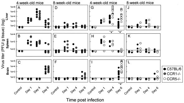
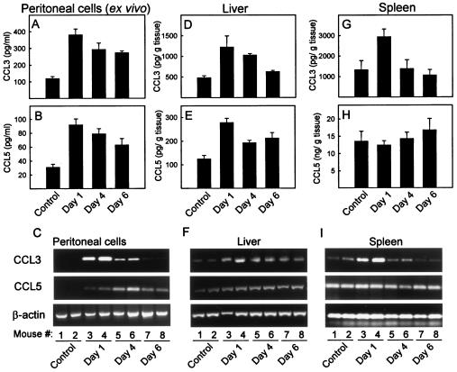
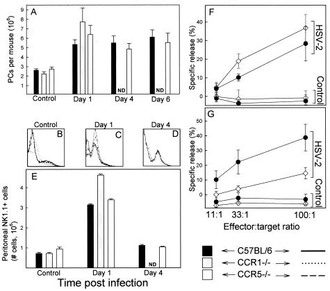
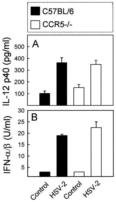
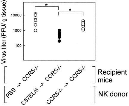
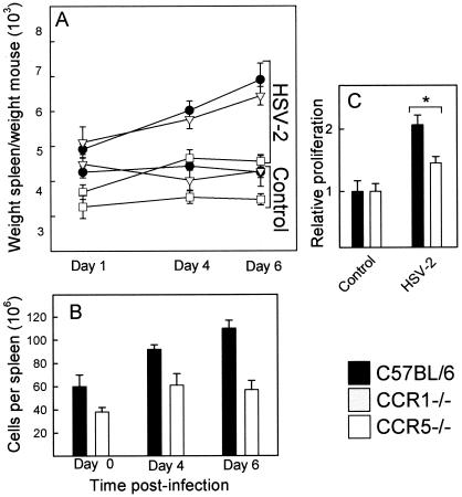
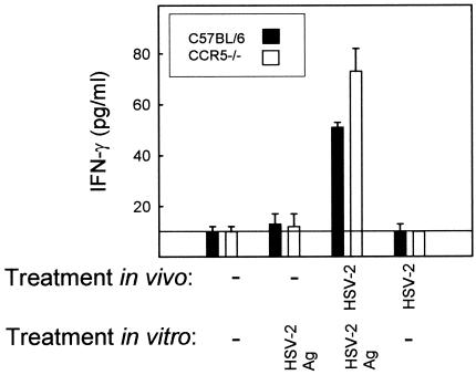
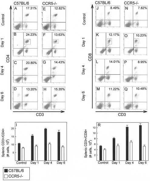
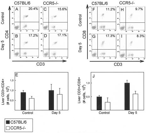
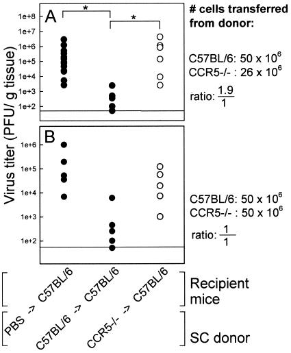
Elimination of viral infections is dependent on rapid recruitment and activation of leukocytes with antiviral activities to infected areas. Chemokines constitute a class of cytokines that have regulatory effects on leukocyte migration and activity. In this study we have studied the role of CC chemokine receptor 1 (CCR1) and CCR5 in host defense during a generalized herpes simplex virus type 2 (HSV-2) infection. Whereas both 4- and 8-week-old CCR1(-/-) mice resembled wild-type mice (C57BL/6) with respect to defense against the infection, significantly higher virus titers were seen in the livers and brains of 4-week-old CCR5(-/-) mice. At the age of 8 weeks, CCR5(-/-) were indistinguishable from wild-type mice and cleared the infection from liver and spleen. Although 4-week-old CCR5(-/-) mice were able to recruit natural killer (NK) cells to the site of infection, these cells had reduced cytotoxic activity compared to NK cells from wild-type mice. This was not due to lower production of alpha/beta interferon or interleukin-12, two well-described activators of cytotoxic activity in NK cells. We also noted that the spleens of young CCR5(-/-) mice did not increase in size during infection as did the spleens of wild-type and CCR1(-/-) mice. This observation was accompanied by impaired proliferation of CCR5(-/-) splenocytes (SCs) ex vivo. Moreover, migration of CD8(+) T cells to the liver in response to infection was impaired in CCR5(-/-) mice, and adoptive transfer of SCs from CCR5(-/-) mice infected for 6 days into newly infected wild-type mice did not improve antiviral activity in the liver, in contrast to what was seen in mice receiving immune SCs from wild-type mice. Altogether, this study shows that CCR5 plays an age-dependent role in host defense against HSV-2 by supporting both the innate and adaptive immune response.


Figures

FIG. 1.
FIG. 1.
Virus titers during a generalized HSV-2 infection in mice lacking CCR1 and CCR5. Four- and 8-week-old C57BL/6, CCR1−/−, and CCR5−/− mice were infected i.p. with 106 PFU of HSV-2. At the indicated time points p.i. livers, spleens, and brains were harvested and HSV-2 titers were determined by plaque assay. The results are shown as dots represent individual mice. Statistically significant differences (P < 0.05) are marked by asterisks (n = 4 to 12).
FIG. 2.
FIG. 2.
Expression of CCL3 and CCL5 during a generalized virus infection in 4-week-old mice. Four-week-old female C57BL/6 mice were infected i.p. with 106 PFU of HSV-2, and livers, spleens, and PCs were harvested at indicated time points p.i. (A and B) The PCs were cultured for 24 h, and supernatants were harvested for measurement of CCL3 and CCL5 by ELISA. (D, E, G, and H) Livers and spleens were homogenized, and CCL3 and CCL5 were measured in the supernatants. The results are shown as mean ± the SEM (n = 3 to 9). (C, F, and I) RNA was isolated from PCs, livers, and spleens from mice treated as indicated. CCL3, CCL5, and β-actin mRNA were detected by RT-PCR.
FIG. 3.
FIG. 3.
Accumulation and activation of leukocytes and NK1.1 positive cells in the peritoneal cavity of C57BL/6, CCR1−/−, and CCR5−/− mice during HSV-2 infection. (A) PCs from 4-week-old infected and control mice were harvested and counted. The cell numbers are shown as mean ± the SEM. (B to E) Accumulation of NK cells in the peritoneal cavity. NK1.1-positive cells in the PC population were identified by flow cytometry. Representative diagrams are shown in panels B to D, and panel E shows a summary of an analysis of three mice per group. Values are presented as mean ± the SEM. ND, not done. (F and G) NK activity of PCs after HSV-2 infection. PCs were harvested from WT, CCR1−/−, and CCR5−/− mice infected for 24 h with HSV-2 and assayed for cytotoxic activity toward YAC-1 cells in different ratios of effector to target cells. The results are shown as mean ± the SEM.
FIG. 4.
FIG. 4.
HSV-induced expression of IL-12 p40 and IFN-α/β in 4-week-old WT and CCR5−/− mice. PCs were harvested from WT and CCR5−/− mice and sat in culture for 24 h in the presence or absence of HSV-2 (3 × 10⁶ PFU/ml). The supernatants were harvested, and the content of IL-12 p40 (A) and IFN-α/β (B) was measured by ELISA and bioassay, respectively. The results are shown as mean ± the SEM.
FIG. 5.
FIG. 5.
Adoptive transfer of NK cell activity from C57BL/6 and CCR5−/− mice to HSV-2-infected CCR5−/− mice. PCs were harvested from mice infected for 24 h with HSV-2, and 4 × 10⁶ of the nonadherent cells were injected i.p. into each CCR5−/− mice infected with 106 PFU of HSV-2 2 h previously. Two days later the mice were sacrificed, and virus titers in the livers were determined by plaque assay on Vero cells. Statistically significant differences (P < 0.05) are marked by asterisks.
FIG. 6.
FIG. 6.
Expansion of SCs during a generalized virus infection. (A) Spleens were harvested from 4-week-old C57BL/6, CCR1−/−, and CCR5 −/− mice at the indicated time points after infection with 106 PFU of HSV-2. The weights of the spleens were measured and plotted as a function of time of infection. The results are shown as mean ± the SEM (n = 10 to 50). (B) Spleens were harvested from C57BL/6 and CCR5−/− mice at the indicated times after HSV-2 infection, and SCs were isolated and counted. The results are shown as mean ± the SEM (n = 3 to 6). (C) SCs from C57BL/6 and CCR5−/− mice, either uninfected or infected for 5 days with HSV-2, were harvested and cultured. The proliferation of SCs ex vivo was measured and is shown as the mean ± the SEM (n = 5; P = 0.008).
FIG. 7.
FIG. 7.
Production of IFN-γ by T cells from 4-week-old mice infected in vivo and cultured in vitro in the presence or absence of viral restimulation. Splenic T cells were isolated from control and day-6-infected mice and sat in culture for 72 h with irradiated syngeneic SCs in the presence or absence of 3 × 10⁷ PFU of heat-inactivated HSV-2. IFN-γ levels in the supernatants were measured by ELISA. The results are shown as means ± the SEM (n = 5).
FIG. 8.
FIG. 8.
T-cell subsets in the spleen of C57BL/6 and CCR5−/− mice during HSV-2 infection. CD3/CD4-double-positive (A to I) and CD3/CD8-double-positive (J to R) cells in the spleens of 4-week-old control and virus-infected mice were identified by flow cytometry. Representative dot blots are shown in panels A to H (CD3/CD4) and J to Q (CD3/CD8), whereas panels I and R show summaries of analyses of three mice per group, with the results shown as means ± the SEM.
FIG. 9.
FIG. 9.
T-cell subsets in the liver of C57BL/6 and CCR5−/− mice during HSV-2 infection. CD3/CD4-double-positive (A to E) and CD3/CD8-double-positive (F to J) cells in the liver of untreated and virus-infected 4-week-old mice were identified by flow cytometry. Representative dot blots are shown in panels A to D (CD3/CD4) and F to I (CD3/CD8), whereas panels E and J show summaries of analyses of three mice per group, with the results shown as means ± the SEM.
FIG. 10.
FIG. 10.
Adoptive transfer of SCs from C57BL/6 and CCR5−/− mice to HSV-2-infected C57BL/6 mice. Four-week-old C57BL/6 mice were infected i.p. with 106 PFU of HSV-2. Six hours later the mice received SCs harvested from 4-week-old C57BL/6 and CCR5−/− mice infected for 6 days with HSV-2. Two days later livers were harvested, and the viral loads in the organs were determined by plaque titration. (A) SCs were given to the recipient mice in the same ratios as they were recovered from the C57BL/6 and CCR5−/− mice in the experiment depicted in Fig. 6 (5 × 10⁷ and 2.6 × 10⁷ cells, respectively). (B) The same number of SCs (5 × 10⁷ cells) were given to the recipient mice irrespective of the number of SCs harvested from the infected C57BL/6 and CCR5−/− mice. The results are shown as dots representing individual mice (n = 5 to 13). Statistically significant differences (P < 0.05) are marked by asterisks.

Similar articles

Cited by

References

    1. Aliberti, J., C. Sousa, M. Schito, S. Hieny, T. Wells, G. B. Huffnagle, and A. Sher. 2000. CCR5 provides a signal for microbial induced production of IL-12 by CD8 α+ dendritic cells. Nat. Immunol. 1:83-87. - PubMed
    1. Biron, C. A., K. B. Nguyen, G. C. Pien, L. P. Cousens, and T. P. Salazar-Mather. 1999. Natural killer cells in antiviral defense: function and regulation by innate cytokines. Annu. Rev. Immunol. 17:189-220. - PubMed
    1. Cook, D. N., M. A. Beck, T. M. Coffman, S. L. Kirby, J. F. Sheridan, I. B. Pragnell, and O. Smithies. 1995. Requirement of MIP-1 α for an inflammatory response to viral infection. Science 269:1583-1585. - PubMed
    1. Croft, M. 2003. Co-stimulatory members of the TNFR family: keys to effective T-cell immunity? Nat. Rev. Immunol. 3:609-620. - PubMed
    1. Dawson, T. C., M. A. Beck, W. A. Kuziel, F. Henderson, and N. Maeda. 2000. Contrasting effects of CCR5 and CCR2 deficiency in the pulmonary inflammatory response to influenza A virus. Am. J. Pathol. 156:1951-1959. - PMC - PubMed

Publication types

Substances