. 2005 Aug;79(15):9842-53.
doi: 10.1128/JVI.79.15.9842-9853.2005.

A peptide derived from RNA recognition motif 2 of human la protein binds to hepatitis C virus internal ribosome entry site, prevents ribosomal assembly, and inhibits internal initiation of translation

Affiliations

A peptide derived from RNA recognition motif 2 of human la protein binds to hepatitis C virus internal ribosome entry site, prevents ribosomal assembly, and inhibits internal initiation of translation

Renuka Pudi et al. J Virol. 2005 Aug.

Abstract

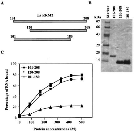
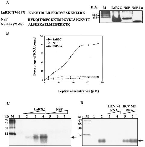
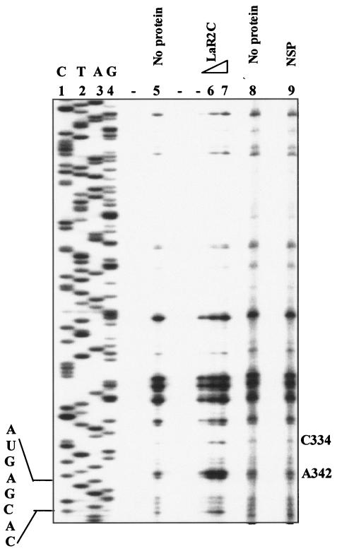
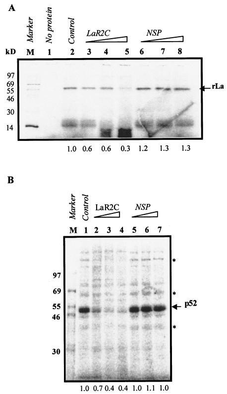
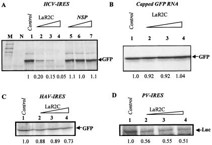
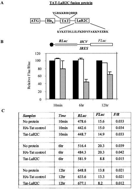
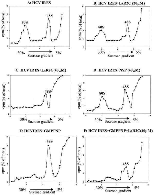
Human La protein is known to interact with hepatitis C virus (HCV) internal ribosome entry site (IRES) and stimulate translation. Previously, we demonstrated that mutations within HCV SL IV lead to reduced binding to La-RNA recognition motif 2 (RRM2) and drastically affect HCV IRES-mediated translation. Also, the binding of La protein to SL IV of HCV IRES was shown to impart conformational alterations within the RNA so as to facilitate the formation of functional initiation complex. Here, we report that a synthetic peptide, LaR2C, derived from the C terminus of La-RRM2 competes with the binding of cellular La protein to the HCV IRES and acts as a dominant negative inhibitor of internal initiation of translation of HCV RNA. The peptide binds to the HCV IRES and inhibits the functional initiation complex formation. An Huh7 cell line constitutively expressing a bicistronic RNA in which both cap-dependent and HCV IRES-mediated translation can be easily assayed has been developed. The addition of purified TAT-LaR2C recombinant polypeptide that allows direct delivery of the peptide into the cells showed reduced expression of HCV IRES activity in this cell line. The study reveals valuable insights into the role of La protein in ribosome assembly at the HCV IRES and also provides the basis for targeting ribosome-HCV IRES interaction to design potent antiviral therapy.


Figures

FIG. 1.
FIG. 1.
Effect of deletions on the binding of La-RRM2 to HCV IRES RNA. (A) Schematic representation of N- and C-terminal deletions within La-RRM2. The corresponding amino acid numbers of the truncated proteins are indicated. (B) The purified protein samples (as indicated on top of the lanes) were analyzed by SDS-10% Tris-Tricine gel followed by silver staining. (C) Filter-binding assay to study the binding of La101-208, La101-180, and La120-208 to HCV IRES. 32P-labeled HCV IRES RNA was bound to increasing concentrations of La RRM2 or truncated proteins (as indicated by the symbols within the panel). The amount of bound RNA was determined by binding to the nitrocellulose filters. The percentage of bound RNA was plotted against the protein concentration (nanomolars).
FIG. 2.
FIG. 2.
Ability of the peptide LaR2C to bind to HCV IRES RNA. (A) The sequences of the peptides LaR2C, NSP, and NSP-La are indicated. The LaR2C, NSP, and NSP-La peptides were analyzed by resolving by SDS-12% Tris-Tricine gel electrophoresis followed by silver staining. (B) Filter-binding assay to study the binding of the peptide LaR2C to HCV IRES. 32P-labeled HCV IRES RNA was bound to increasing concentrations of the peptide LaR2C, NSP, or NSP-La (as indicated by the symbols within the panel). The amount of bound RNA was determined by binding to the nitrocellulose filters. The percentage of bound RNA was plotted against the peptide concentration (micromolars). (C) UV cross-linking of LaR2C and NSP to HCV IRES. 32P-labeled HCV IRES RNA was UV cross-linked with increasing concentrations (20, 40, and 60 μM) of either LaR2C or NSP (as indicated above the panel), digested with RNase A, and resolved by SDS-15% PAGE followed by phosphorimaging. Lane 1 represents the no-protein control. Lane M represents the 14C protein molecular weight marker. The corresponding molecular masses are indicated to the left of the panel. (D) Competition assay to determine specificity of the binding of LaR2C to HCV IRES RNA. LaR2C preincubated with 100- and 200-fold excesses of unlabeled HCV wild-type (wt) RNA or HCV M2 RNA (where the SL IV region was mutated), as indicated above the lanes, was bound to 32P-labeled HCV IRES RNA and UV cross-linked. The complexes were treated with RNase A and resolved by SDS-15% PAGE followed by phosphorimaging. The band corresponding to LaR2C is indicated to the right of the panels by arrows. Lane 1 represents the no-protein control. Lane M represents the 14C protein molecular weight marker. The corresponding molecular masses are indicated to the left of the panel.
FIG. 3.
FIG. 3.
Primer extension inhibition (toe-printing) analysis. Increasing concentrations of the LaR2C peptide (20 and 40 μM [lanes 6 and 7]) or 40 μM NSP (lane 9) was incubated with HCV IRES RNA (18 to 383 nucleotides) as described above and analyzed for the toe prints. Lanes 5 and 8 represent the no-protein control. Lanes 1 to 4 show the DNA sequencing ladder corresponding to the HCV RNA (18 to 383 nucleotides) obtained by using the same end-labeled primer. The nucleotide indicated on the right of the panel signifies the corresponding positions on the HCV IRES RNA. The nucleotide positions corresponding to GCAC and iAUG are indicated on the left.
FIG. 4.
FIG. 4.
Effect of LaR2C on the binding of recombinant La protein and other cellular proteins to HCV IRES RNA. (A) 32P-labeled HCV IRES RNA was preincubated with increasing concentrations (20, 40, and 60 μM) of LaR2C or NSP as indicated above the lanes and then bound to recombinant purified La protein. The UV cross-linked complexes were treated with RNase A and resolved by SDS-10% PAGE followed by phosphorimaging. The band corresponding to recombinant La (rLa) is indicated to the right of the panel. Lane M represents the 14C-protein molecular weight marker. The corresponding molecular masses are indicated to the left of the panel. The intensity of the band corresponding to La protein in each lane was quantitated using densitometry and represented in numbers below the lanes. (B) 32P-labeled HCV IRES RNA was preincubated with increasing concentrations (20, 40, and 60 μM) of LaR2C and then bound to HeLa cytoplasmic extract (2.5 μg). The UV cross-linked RNA-protein complexes were treated with RNase A and resolved by SDS-5 to 15% gradient PAGE followed by phosphorimaging. The band corresponding to p52 is indicated to the right of the panel. The protein bands whose intensities were reduced are indicated by asterisks. The intensity of the band corresponding to p52 in each lane was quantitated using densitometry and represented in numbers below the respective lanes. Lane M represents the 14C-protein molecular weight marker. The corresponding molecular masses are indicated to the left of the panel.
FIG. 5.
FIG. 5.
Effect of LaR2C on HCV IRES-mediated translation in vitro. (A) One microgram of uncapped HCV IRES-GFP RNA was translated in RRL in the absence (lane 1) or presence of increasing concentrations (20, 40, and 60 μM) of either LaR2C (lanes 2 to 4) or NSP (lanes 5 to 7). The translation of GFP was analyzed on an SDS-12.5% polyacrylamide gel followed by phosphorimaging. The band corresponding to GFP is indicated to the right of the panel. (B) One microgram of capped GFP RNA was translated in RRL in the absence (lane 1) or presence of increasing concentrations (20, 40, and 60 μM) of LaR2C (lanes 2 to 4). The translation of GFP was analyzed on an SDS-12.5% polyacrylamide gel followed by phosphorimaging. The band corresponding to GFP is indicated to the right of the panel. (C and D) Two micrograms of either capped HAV-bicistronic RNA (containing FLuc-HAV-GFP in order) or PV bicistronic RNA (containing RLuc-PV-FLuc in order) was translated in the absence (lane 1) and presence of increasing concentrations (20, 40, and 60 μM) of LaR2C (lanes 2 to 4). The translation of the reporter genes was analyzed on an SDS-12.5% polyacrylamide gel followed by phosphorimaging. The bands corresponding to GFP (representing HAV IRES function [C]) and FLuc (representing PV-IRES function [D]) are shown in the picture for clarity only. The intensities of the GFP band and the FLuc band in each lane were quantitated using densitometry and represented in numbers below the respective lanes.
FIG. 6.
FIG. 6.
Effect of TAT-LaR2C fusion protein on HCV IRES-mediated translation in vivo. (A) Schematic representation of the TAT-LaR2C fusion protein. The amino acid sequences of TAT and LaR2C are highlighted. (B) Huh7 monolayer cells expressing HCV bicistronic RNA were overlaid with 100 nM of either HA-TAT or TAT-LaR2C fusion protein for 10 min. The cells were then harvested after different time points (10 min, 6 h, and 12 h) and lysed, and the RLuc and FLuc levels were measured using a Dual Luciferase assay system. The relative ratio of FLuc to RLuc was plotted at each time point. Black bars represent control cells, white bars represent cells overlaid with HA-TAT, and gray bars represent cells treated with TAT-LaR2C fusion protein. The HCV bicistronic construct used in the cell line is indicated on top of panel B. Data from the transfection experiments are expressed as means ± standard deviations of three independent replicates. (C) Absolute levels of RLuc and FLuc activities (in relative light units) of a representative experiment are presented in the table.
FIG. 7.
FIG. 7.
Effect of LaR2C peptide on ribosomal assembly on the HCV IRES RNA. Sucrose gradient sedimentation profiles of 32P-labeled HCV IRES RNA in the absence (A) or presence of increasing concentrations of the LaR2C peptide (B and C) or the NSP (D) after incubation in RRL and separated on a 5 to 30% sucrose gradient are shown. The fractions (200 μl) were manually collected from the bottom of the tube, and scintillation counts were measured. The counts per minute of each fraction were shown as the percentage of the total counts added to the reaction (∼2 × 10⁵ cpm) and were plotted against the volume of the gradient solution (0 to 8 ml). The ribosomal peaks corresponding to 48S and 80S are indicated. Panel E represents the sedimentation profile of HCV IRES RNA incubated in RRL in the presence of 10 mM GMP-PNP alone, and panel F is that obtained in the presence of both LaR2C (40 μM) and GMP-PNP (10 mM).

Similar articles

Cited by

References

    1. Ali, N., and A. Siddiqui. 1995. Interaction of polypyrimidine tract-binding protein with the 5′ noncoding region of the hepatitis C virus RNA genome and its functional requirement in internal initiation of translation. J. Virol. 69:6367-6375. - PMC - PubMed
    1. Ali, N., and A. Siddiqui. 1997. The La antigen binds 5′ NCR of HCV RNA and in the context of the initiator AUG and stimulates IRES-mediated translation. Proc. Natl. Acad. Sci. USA 94:2249-2254. - PMC - PubMed
    1. Ali, N., G. J. M. Pruijn, D. J. Kenan, J. D. Keene, and A. Siddiqui. 2000. Human La antigen is required for the hepatitis C virus internal ribosome entry site-mediated translation. J. Biol. Chem. 275:27531-27540. - PubMed
    1. Bartenschlager, R. 1999. The NS3/4A proteinase of the hepatitis C virus: unravelling structure and function of an unusual enzyme and a prime target for antiviral therapy. J. Viral. Hepat. 6:165-181. - PubMed
    1. Becker-Hapak, M., S. S. McAllister, and S. F. Dowdy. 2001. TAT-mediated protein transduction into mammalian cells. Methods 24:247-256. - PubMed

Publication types

LinkOut - more resources