Comparative Study
. 2005 Aug;79(15):9862-71.
doi: 10.1128/JVI.79.15.9862-9871.2005.

Murine coronavirus requires lipid rafts for virus entry and cell-cell fusion but not for virus release

Affiliations
Comparative Study

Murine coronavirus requires lipid rafts for virus entry and cell-cell fusion but not for virus release

Keum S Choi et al. J Virol. 2005 Aug.

Abstract

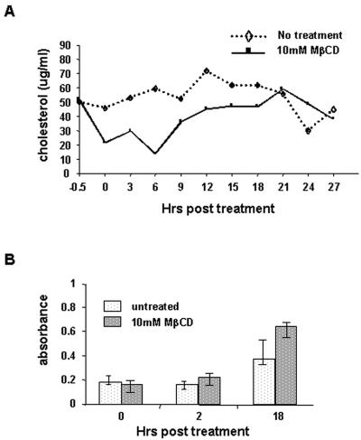
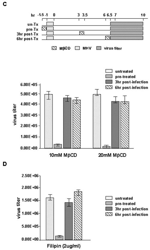
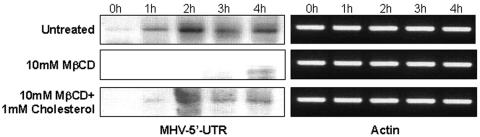
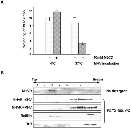
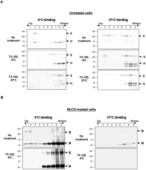
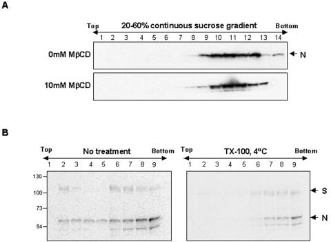
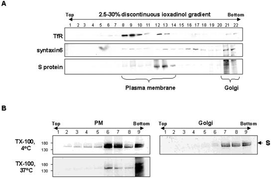
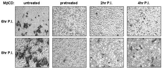
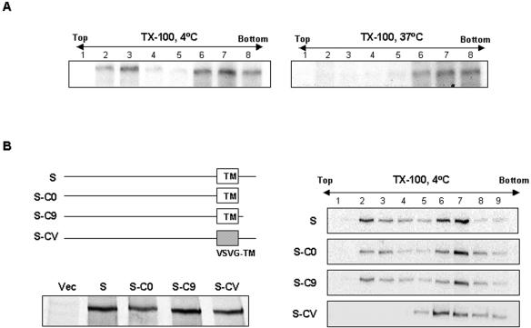
Thorp and Gallagher first reported that depletion of cholesterol inhibited virus entry and cell-cell fusion of mouse hepatitis virus (MHV), suggesting the importance of lipid rafts in MHV replication (E. B. Thorp and T. M. Gallagher, J. Virol. 78:2682-2692, 2004). However, the MHV receptor is not present in lipid rafts, and anchoring of the MHV receptor to lipid rafts did not enhance MHV infection; thus, the mechanism of lipid rafts involvement is not clear. In this study, we defined the mechanism and extent of lipid raft involvement in MHV replication. We showed that cholesterol depletion by methyl beta-cyclodextrin or filipin did not affect virus binding but reduced virus entry. Furthermore, MHV spike protein bound to nonraftraft membrane at 4 degrees C but shifted to lipid rafts at 37 degrees C, indicating a redistribution of membrane following virus binding. Thus, the lipid raft involvement in MHV entry occurs at a step following virus binding. We also found that the viral spike protein in the plasma membrane of the infected cells was associated with lipid rafts, whereas that in the Golgi membrane, where MHV matures, was not. Moreover, the buoyant density of the virion was not changed when MHV was produced from the cholesterol-depleted cells, suggesting that MHV does not incorporate lipid rafts into the virion. These results indicate that MHV release does not involve lipid rafts. However, MHV spike protein has an inherent ability to associate with lipid rafts. Correspondingly, cell-cell fusion induced by MHV was retarded by cholesterol depletion, consistent with the association of the spike protein with lipid rafts in the plasma membrane. These findings suggest that MHV entry requires specific interactions between the spike protein and lipid rafts, probably during the virus internalization step.


Figures

FIG. 1.
FIG. 1.
Cholesterol depletion by MβCD and its effects on MHV replication in DBT cells. (A) Reduction of cholesterol level in cells. After treatment with 10 mM MβCD for 30 min, cells were collected every 3 h up to 27 h after treatment. The amount of cholesterol in the lysates was assayed using the Amplex Red Cholesterol Assay kit. (B) Cytotoxicity of MβCD to the cells. Cell viability was assayed at 0, 2, and 18 h p.i. using WST-1 reagent. (C and D) Virus titers released from either MβCD- or filipin-treated cells. (C, top) Time frames of the experiment. Cells were either pretreated before virus infection or treated at 3 or 6 h p.i. with 10 mM or 20 mM MβCD for 30 min and washed. Filipin treatment was done at a concentration of 2 μg/ml for 1 h. Culture supernatant was collected from 6.5 h to 10 h p.i., and a plaque assay was performed. Samples were duplicated and experiments repeated three times. Arrow bars indicate the standard deviations of three independent experiments.
FIG. 1.
FIG. 1.
Cholesterol depletion by MβCD and its effects on MHV replication in DBT cells. (A) Reduction of cholesterol level in cells. After treatment with 10 mM MβCD for 30 min, cells were collected every 3 h up to 27 h after treatment. The amount of cholesterol in the lysates was assayed using the Amplex Red Cholesterol Assay kit. (B) Cytotoxicity of MβCD to the cells. Cell viability was assayed at 0, 2, and 18 h p.i. using WST-1 reagent. (C and D) Virus titers released from either MβCD- or filipin-treated cells. (C, top) Time frames of the experiment. Cells were either pretreated before virus infection or treated at 3 or 6 h p.i. with 10 mM or 20 mM MβCD for 30 min and washed. Filipin treatment was done at a concentration of 2 μg/ml for 1 h. Culture supernatant was collected from 6.5 h to 10 h p.i., and a plaque assay was performed. Samples were duplicated and experiments repeated three times. Arrow bars indicate the standard deviations of three independent experiments.
FIG. 2.
FIG. 2.
The effect of MβCD on viral RNA uptake. Cells were either untreated or treated with 10 mM MβCD for 30 min and then infected with MHV-A59. In the cholesterol replenishment experiment, MβCD was removed after the 30-min treatment, and cells were incubated with MEM containing 1 mM cholesterol for 1 h before viral infection. Total cellular RNAs were isolated at 0, 1, 2, 3, and 4 h p.i. MHV RNA was detected by RT-PCR using 5′ UTR-specific primers. RT-PCR of actin mRNA was used as a control.
FIG. 3.
FIG. 3.
Effects of cholesterol depletion on virus binding and association of MHV receptor with lipid rafts. (A) Binding of radiolabeled MHV virion. 35S-labeled MHV virion (106 cpm) was incubated with either untreated or MβCD-treated cells for 1 h at 4°C or 37°C. Unbound virion was removed by being washed three times with PBS. Cells were harvested and resuspended in hypotonic buffer, and the radioactivity was determined with a scintillation counter. Each sample was done in triplicate, and bars indicate standard deviation. (B) Association of MHVR with lipid rafts. MHVR-overexpressing 293A cells were either uninfected or infected with MHV-A59, and membrane flotation analysis was done after treatment for 1 h with 1% TX-100 at 4°C. MHVR was detected by immunoblotting. Flotillin and transferrin receptor were used as positive and negative control, respectively, for lipid raft association.
FIG. 4.
FIG. 4.
Redistribution of MHV virion during virus entry. (A) Raft-association of viral proteins during virus entry. 35S-labeled MHV virion (106 cpm) was incubated with MHVR-overexpressing 293A cells for 1 h at either 4°C (left) or 37°C (right). Lysates were treated with 1% TX-100 for 1 h at 4°C or 37°C, and membrane flotation analysis was subsequently done. Viral S and N proteins were detected by immunoprecipitation with polyclonal anti-MHV antibody. (B) Effect of cholesterol depletion on the association of viral protein with lipid rafts. MHVR-overexpressing 293A cells were pretreated with 10 mM MβCD for 30 min, and then radiolabeled virus was added and incubated at either 4°C (left) or 37°C (right) for 1 h. Lysates were treated with 1% TX-100 for 1 h at 4°C, followed by membrane flotation analysis. The exposure time for the both panels was the same.
FIG. 5.
FIG. 5.
Lack of incorporation of lipid rafts into MHV virion. (A) Buoyant density of virion produced from cells untreated or treated with 10 mM MβCD. MHV from the culture supernatant was purified by two-step sucrose centrifugation. Samples were collected into 1-ml fractions, and viral N proteins were detected by immunoblotting. (B) Lack of association of viral proteins with lipid rafts in the virion. 35S-labeled MHV virion was treated with 1% TX-100 for 1 h at 4°C, and then membrane flotation analysis was done. The viral S and N proteins were detected by immunoprecipitation with polyclonal anti-MHV antibody.
FIG. 6.
FIG. 6.
Raft-association of the viral S proteins in plasma and Golgi membranes. (A) Fractionation of total membranes into plasma and Golgi membranes. Membranes prepared from 35S-translabeled MHV-infected cells were fractionated in a 2.5%, 10%, 17.5%, and 25% iodixanol step gradient. Samples were collected into 0.8-ml fractions. Transferrin receptor (TfR) and syntaxin 6 were used as plasma and Golgi membrane markers, respectively. S proteins were detected by immunoprecipitation with polyclonal anti-MHV antibody. (B) Association of viral proteins from plasma and Golgi membranes with lipid rafts. Fractions from lanes 8 to 14 or 21 and 22 of panel A were pooled and analyzed by membrane flotation gradients. Viral proteins were detected by immunoprecipitation with polyclonal anti-MHV antibody.
FIG. 7.
FIG. 7.
Inhibition of cell-cell fusion by MβCD. Cells were treated with 10 mM MβCD for 30 min at several different time points during virus infection: pretreatment and after being treated at 2 and 4 h p.i. Cells were fixed at 6 and 8 h p.i. and immunostained with anti-N monoclonal antibody, followed by incubation with β-galactosidase-conjugated secondary antibody. Syncytium formation was visualized by X-Gal (5-bromo-4-chloro-3-indolyl-β-galactoside) staining (shown as dark areas).
FIG. 8.
FIG. 8.
Determination of raft association domains of S proteins. (A) Raft association of S protein overexpressed in 293A cells. (B) Domains required for lipid raft association of S protein. (Left, top) Diagram of S protein mutants. (Bottom) The expression level of mutants was determined by immunoprecipitation with anti-MHV polyclonal antibody from 35S-translabeled cellular lysates. Membrane flotation analysis was done for the truncated mutants (right).

Similar articles

Cited by

References

    1. Ahn, A., D. L. Gibbons, and M. Kielian. 2002. The fusion peptide of Semliki Forest virus associates with sterol-rich membrane domains. J. Virol. 76:3267-3275. - PMC - PubMed
    1. Ali, A., R. T. Avalos, E. Ponimaskin, and D. P. Nayak. 2000. Influenza virus assembly: effect of influenza virus glycoproteins on the membrane association of M1 protein. J. Virol. 74:8709-8719. - PMC - PubMed
    1. Bavari, S., C. M. Bosio, E. Wiegand, G. Ruthel, A. B. Will, T. W. Geisbert, M. Hevey, C. Schmaljohn, A. Schmaljohn, and M. J. Aman. 2002. Lipid raft microdomains: a gateway for compartmentalized trafficking of Ebola and Marburg viruses. J. Exp. Med. 195:593-602. - PMC - PubMed
    1. Bender, F. C., J. C. Whitbeck, M. Ponce de Leon, H. Lou, R. J. Eisenberg, and G. H. Cohen. 2003. Specific association of glycoprotein B with lipid rafts during herpes simplex virus entry. J. Virol. 77:9542-9552. - PMC - PubMed
    1. Bock, J. B., J. Klumperman, S. Davanger, and R. H. Scheller. 1997. Syntaxin 6 functions in trans-Golgi network vesicle trafficking. Mol. Biol. Cell 8:1261-1271. - PMC - PubMed

Publication types

Substances

LinkOut - more resources