. 2005 Aug;79(15):9982-90.
doi: 10.1128/JVI.79.15.9982-9990.2005.

Genetic analysis of the polyomavirus DnaJ domain

Affiliations

Genetic analysis of the polyomavirus DnaJ domain

Kerry A Whalen et al. J Virol. 2005 Aug.

Abstract

Polyomavirus T antigens share a common N-terminal sequence that comprises a DnaJ domain. DnaJ domains activate DnaK molecular chaperones. The functions of J domains have primarily been tested by mutation of their conserved HPD residues. Here, we report detailed mutagenesis of the polyomavirus J domain in both large T (63 mutants) and middle T (51 mutants) backgrounds. As expected, some J mutants were defective in binding DnaK (Hsc70); other mutants retained the ability to bind Hsc70 but were defective in stimulating its ATPase activity. Moreover, the J domain behaves differently in large T and middle T. A given mutation was twice as likely to render large T unstable as it was to affect middle T stability. This apparently arose from middle T's ability to bind stabilizing proteins such as protein phosphatase 2A (PP2A), since introduction of a second mutation preventing PP2A binding rendered some middle T J-domain mutants unstable. In large T, the HPD residues are critical for Rb-dependent effects on the host cell. Residues Q32, A33, Y34, H49, M52, and N56 within helix 2 and helix 3 of the large T J domain were also found to be required for Rb-dependent transactivation. Cyclin A promoter assays showed that J domain function also contributes to large T transactivation that is independent of Rb. Single point mutations in middle T were generally without effect. However, residue Q37 is critical for middle T's ability to form active signaling complexes. The Q37A middle T mutant was defective in association with pp60(c-src) and in transformation.


Figures

FIG. 1.
FIG. 1.
Mutations in the polyomavirus DnaJ domain. The amino acid sequence encoded by the first exon (residues 1 to 79) of the polyomavirus T antigens is shown for wild type. Mutations were created in expression vectors for either middle T or large T. The amino acid changes are shown above the wild-type sequence for the large T mutants and below the wild-type sequence for middle T mutants. The dashes represent residues that were not mutated for these studies. Unstable mutants are indicated by an asterisk. Mutants defective in function are indicated by a dagger (†).
FIG. 2.
FIG. 2.
Effects of DnaJ mutants on large T and middle T stability. (A) Examples of expression of large T DnaJ mutants. NIH 3T3 cells in 100-mm dishes at 20% confluence were transfected with 3 μg of empty vector (lane 1) or vectors expressing wild-type (lane 2), P43S (lane 3), Q36A (lane 4), W59F (lane 5), F62Y (lane 6), and T64S (lane 7) large T antigens. At ca. 40 h posttransfection, cells were extracted. After SDS-PAGE of the cell extracts, large T was blotted with anti-T antibody. (B) Comparison of large T and middle T DnaJ mutant stability. Extracts from K10R (lane 1), E11D (lane 2), R12K (lane 3), or L13V (lane 4) mutants large T or middle T, as well as control (lane 5) and wild-type (lane 6), transfected cells were blotted with anti-T antibody as in panel A. (C) Cells transfected with wild-type middle T (lane l), NG59 middle T (lane 2), G25A (lane 3), G25A/NG59 double mutant (lane 4), L55V (lane 5), and L55V/NG59 double mutant (lane 6) were extracted at ca. 40 h posttransfection. After SDS-PAGE, blotting was done with anti-T antibody.
FIG. 3.
FIG. 3.
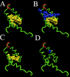
Locations in the DnaJ structure of large T and middle T mutations that affect stability or function. (A) Mutated residues that result in protein instability in the large T background are highlighted in yellow: K10R, L13V, L14V, L17V, P20A, G25A, F27Y, M30V, L55V, and W59F. The structure was originally reported by Berjanskii et al. (2) and is displayed using Visual Molecular Dynamics. (B) The large T mutations that cause the protein to be inactive in E2F or cyclin A promoter assays are shown in blue: Q32A, A33G, Y34F, H49R, M52V, and N56T. The unstable mutants are shown in yellow. (C) Mutated residues that cause middle T instability are highlighted in yellow: R12K, L13V, L14V, and M30V. (D) The defective mutant at position Q37A is highlighted in blue. In all panels the HPD residues are red.
FIG. 4.
FIG. 4.
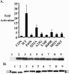
Effects of non-HPD large T J mutants on the Rb-family members. (A) The upper panel depicts E2F-containing promoter activation. NIH 3T3 cells were cotransfected with the 6XE2F-luciferase construct, and the indicated cytomegalovirus (CMV) expression plasmids (wild type, P43S, Q32A, A33G, Y34F, H49R, M52V, and N56T). Error bars indicate the standard error of the mean. The P value of wild type compared to each of the mutants was <0.05 as determined by one-way analysis of variance. Cells were extracted ca. 40 h posttransfection and assayed for luciferase activity. The lower panel shows results for extracts from the promoter assays separated by SDS-PAGE and blotted for large T antigens with anti-T antibody: control (lane 1), wild-type large T (lane 2), P43S (lane 3), Q32A (lane 4), A33G (lane 5), Y34F (lane 6), H49R (lane 7), M52V (lane 8), and N56 T (lane 9). (B) DnaJ mutant large T-induced p130 mobility shift in C33A cells. C33A cells on 60-mm dishes at 80% confluence were transfected with 3 μg of HA-p130 and 1 μg of the CMV expression vectors wild-type large T (lane 1), wild type (1.5 μg) (lane 2), Q32A (lane 3), A33G (lane 4), Y34F (lane 5), H49R(lane 6), M52V(lane 7), N56T (lane 8), and P43S (lane 9). Cell extracts made approximately 40 h posttransfection were subjected to SDS-PAGE and blotted for HA-p130 using HA11. The arrows indicate differentially phosphorylated species of p130.
FIG. 5.
FIG. 5.
Interactions of large T J domain mutants with Hsc70. (A) Binding to Hsc70: NIH 3T3 cells at 20% confluence in 100-mm dishes cotransfected with 2 μg of vector expressing wild type (lanes 1 and 2), P43S (lanes 3 and 4), CMV empty vector (lanes 5 and 6), Q32A (lanes 7 and 8), A33G (lanes 9 and 10), Y34F (lanes 11 and 12), H49R (lanes 13 and 14), and M52V (lanes 15 and 16), N56T (lanes 17 and 18), and 5 μg of myc-tagged Hsc70 were extracted ca. 40 h posttransfection. After extraction, immunoprecipitation was carried out with anti-myc 9E10 antibody (even lanes) or control monoclonal (odd lanes). After SDS-PAGE, Hsc70 was blotted with 9E10 (anti-myc Hsc70) or large T was blotted with PN116 (anti-T). (B) Western blots of cell extracts used for the immunoprecipitations in panel A. (C) H49R and Y34F are defective in the stimulation of Hsc70 ATPase activity. ATP hydrolysis measurements were carried out as described in Materials and Methods. ATP hydrolysis catalyzed by Hsc70, Hsc70 plus 0.8 μg of wild-type NT, Hsc70 plus 0.8 μg of Y34F NT, and Hsc70 of 0.8 μg of H49R NT was measured. The data represent the means from experiments done in triplicate, and error bars indicate the standard error.
FIG. 6.
FIG. 6.
Decreased activity of J domain mutants on the cyclin A promoter parallels that seen on E2F-containing promoters. The upper panel shows results for NIH 3T3 cells cotransfected with cyclin A −37/−33 luciferase construct along with the indicated CMV expression vectors (wild type, P43S, Q32A, A33G, Y34F, H49R, M52V, and N56T). The cells were placed in 0.2% calf serum at 24 h posttransfection. Cells were extracted approximately 40 h posttransfection and assayed for luciferase activity. Error bars represent the standard error of the mean. The P value of wild type compared to each of the mutants was <0.05 as determined by one-way analysis of variance. The lower panel shows results extracts from the promoter assays separated by SDS-PAGE and blotted for large T antigens with anti-T antibody: control (lane 1), wild-type large T (lane 2), P43S (lane 3), Q32A (lane 4), A33G (lane 5), Y34F (lane 6), H49R (lane 7), M52V (lane 8), and N56T (lane 9).
FIG. 7.
FIG. 7.
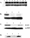
Q37A middle T is defective in association with c-src. (A) NIH cells were transfected with the indicated CMV expression vectors control (lane 1), wild-type middle T (lane 2), K10R (lane 3), A33G (lane 4), G46A (lane 5), M52V (lane 6), N56T (lane 7), F62Y (lane 8), and G74A (lane 9). The cells were extracted ca. 40 h posttransfection, and extracts were immunoprecipitated with anti-T antibody bound to protein A-Sepharose beads. Immunoprecipitated middle T was labeled with [γ]ATP in an in vitro kinase reaction. After SDS-PAGE, 32P-labeled middle T was analyzed by using a PhosphorImager (results shown in the upper panel). A fraction of the immunoprecipitates loaded in the same order as described above were separated by SDS-PAGE and blotted with anti-middle T antibody (results shown in the lower panel). (B) Pools of NIH 3T3 cells were made that express wild type, INS107A, or Q37A middle T antigens. Middle T immunoprecipitates from those or control cells were labeled in an in vitro kinase reaction and analyzed as described above (upper panel). A fraction of the immunoprecipitates were separated by SDS-PAGE and blotted with anti-middle T antibody (lower panel). (C) Middle T was immunoprecipitated from extracts of the cell pools. After separation on SDS-PAGE, Western blots were probed with GD11 anti-src antibody (upper panel). A fraction of the immunoprecipitates were separated by SDS-PAGE and blotted with anti-middle T antibody (lower panel). (D) Aliquots of the same immunoprecipitate shown in panel C analyzing src were used to test for association with PP2A by probing with antibody specific for PP2A subunit. Lanes (B to D): 1, wild type; 2, INS107A; 3, Q37A.
FIG. 8.
FIG. 8.
Q37A middle T is defective in transformation assays. NIH 3T3 cells were transfected with vector expressing wild-type middle T (A), INS107AL (B), or Q37A middle T (C) and grown for 14 days under reduced serum conditions. The cells were fixed and stained, and foci were counted.

Similar articles

Cited by

References

    1. Berger, H., and E. Wintersberger. 1986. Polyomavirus small T antigen enhances replication of viral genomes in 3T6 mouse fibroblasts. J. Virol. 60:768-770. - PMC - PubMed
    1. Berjanskii, M. V., M. I. Riley, A. Xie, V. Semenchenko, W. R. Folk, and S. R. Van Doren. 2000. NMR structure of the N-terminal J. domain of murine polyomavirus T antigens: implications for DnaJ-like domains and for mutations of T antigens. J. Biol. Chem. 275:36094-36103. - PubMed
    1. Brewster, C. E., H. R. Glover, and S. M. Dilworth. 1997. pp60c−src binding to polyomavirus middle T-antigen (MT) requires residues 185 to 210 of the MT sequence. J. Virol. 71:5512-5520. - PMC - PubMed
    1. Campbell, K., K. Auger, B. Hemmings, T. Roberts, and P. D. 1995. Identification of regions in polyomavirus middle T and small t antigens important for association with protein phosphatase 2A. J. Virol. 69:3721-3728. - PMC - PubMed
    1. Campbell, K. S., K. P. Mullane, I. A. Aksoy, H. Stubdal, J. Zalvide, J. M. Pipas, P. A. Silver, T. M. Roberts, B. S. Schaffhausen, and J. A. DeCaprio. 1997. DnaJ/hsp40 chaperone domain of SV40 large T antigen promotes efficient viral DNA replication. Genes Dev. 11:1098-1110. - PubMed

Publication types

MeSH terms