. 2005 Aug;79(15):9991-10002.
doi: 10.1128/JVI.79.15.9991-10002.2005.

Molecular basis for mitochondrial localization of viral particles during beet necrotic yellow vein virus infection

Affiliations

Molecular basis for mitochondrial localization of viral particles during beet necrotic yellow vein virus infection

Clarisse Valentin et al. J Virol. 2005 Aug.

Abstract

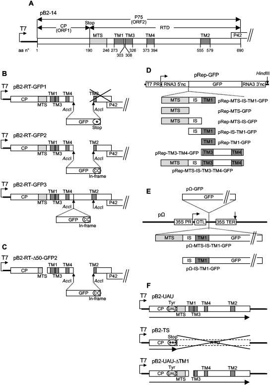
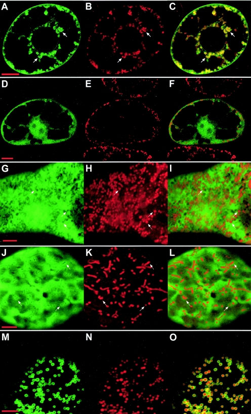
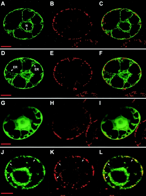
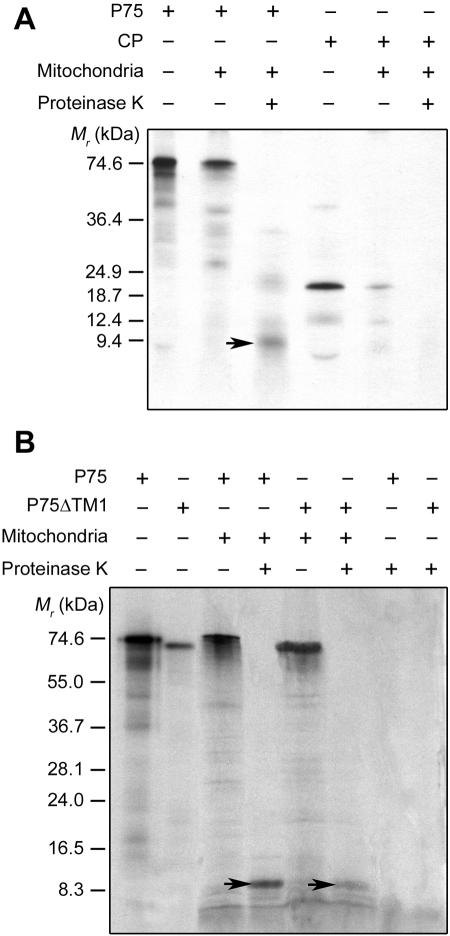
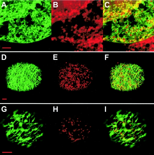
During infection, Beet necrotic yellow vein virus (BNYVV) particles localize transiently to the cytosolic surfaces of mitochondria. To understand the molecular basis and significance of this localization, we analyzed the targeting and membrane insertion properties of the viral proteins. ORF1 of BNYVV RNA-2 encodes the 21-kDa major coat protein, while ORF2 codes for a 75-kDa minor coat protein (P75) by readthrough of the ORF1 stop codon. Bioinformatic analysis highlighted a putative mitochondrial targeting sequence (MTS) as well as a major (TM1) and two minor (TM3 and TM4) transmembrane regions in the N-terminal part of the P75 readthrough domain. Deletion and gain-of-function analyses based on the localization of green fluorescent protein (GFP) fusions showed that the MTS was able to direct a reporter protein to mitochondria but that the protein was not persistently anchored to the organelles. GFP fused either to MTS and TM1 or to MTS and TM3-TM4 efficiently and specifically associated with mitochondria in vivo. The actual role of the individual domains in the interaction with the mitochondria seemed to be determined by the folding of P75. Anchoring assays to the outer membranes of isolated mitochondria, together with in vivo data, suggest that the TM3-TM4 domain is the membrane anchor in the context of full-length P75. All of the domains involved in mitochondrial targeting and anchoring were also indispensable for encapsidation, suggesting that the assembly of BNYVV particles occurs on mitochondria. Further data show that virions are subsequently released from mitochondria and accumulate in the cytosol.


Figures

FIG. 1.
FIG. 1.
Organization of the gene constructs used for the present study. (A) Plasmid pB2-14 contains the complete cDNA for BNYVV RNA-2 in pBluescribe (4, 53). Only ORF1 and ORF2 are represented. Amino acid (aa) numbers are indicated. (B) In plasmids pB2-RT-GFP1 and pB2-RT-GFP2 (15), the GFP gene replaces the AccI1415-AccI1828 sequence of pB2-14, truncating the TM2 domain; in pB2-RT-GFP3, the GFP gene is inserted in frame into the AccI1415 site of pB2-14. (C) pB2-RT-Δ50-GFP2 derives from pB2-RT-GFP2 and carries a deletion of nucleotides 793 to 894, truncating the MTS domain. (D) The MTS, IS, TM1, and TM3-TM4 domains, individually or in combination, were inserted in frame with the GFP gene between the upstream and downstream noncoding sequences of the cDNA for BNYVV RNA-3 in the pRep replicon (16). (E) The GFP gene alone or the MTS-IS-TM1 or IS-TM1 domain fused to the GFP gene was inserted into the expression cassette of the pΩ plasmid (41). (F) In pB2-UAU, the CP sequence ends with a tyrosine codon, allowing full readthrough, whereas in pB2-TS the CP sequence ends with three stop codons, preventing readthrough; pB2-UAU-ΔTM1 carries a deletion of nucleotides 961 to 1053, eliminating the TM1 domain. *, stop codons; “(CC)” indicates that two extra C residues were added at the end of the GFP sequence to ensure the in-frame insertion; UAU, the stop codon of ORF1 was mutated to a UAU tyrosine codon. For more details, see Materials and Methods.
FIG. 2.
FIG. 2.
Expression of GFP-fused P75 deletion mutants in plant protoplasts. N. tabacum BY2 protoplasts were infected with BNYVV RNA-1 and the in vitro transcripts derived from constructs pB2-RT-GFP1 (panels A to C), pB2-RT-Δ50-GFP2 (panels D to F), pRep-GFP (panels G to I), pRep-MTS-GFP (panels J to L), and pRep-MTS-IS-TM1-GFP (panels M to O). Expression was analyzed by confocal microscopy 48 h after infection. Panels A, D, G, J, and M display the green fluorescence of the GFP; panels B, E, H, K, and N display for the same cells the red fluorescence of the mitochondrion-specific dye MitoTracker; and panels C, F, I, L, and O display merged images. Arrows point to areas which clearly illustrate the localization of the fusion proteins as discussed in the text. Bars, 10 μm for A to F and 5 μm for G to O.
FIG. 3.
FIG. 3.
Expression of GFP fusions with different P75 domains in plant protoplasts. N. tabacum BY2 protoplasts were infected with BNYVV RNA-1 and the in vitro transcripts derived from constructs pRep-TM1-GFP (panels A to F), pRep-TM3-TM4-GFP (panels G to I), and pRep-MTS-IS-TM3-TM4-GFP (panels J to L). Expression was analyzed by confocal microscopy 48 h after infection. Panels A, D, G, and J display the green fluorescence of the GFP; panels B, E, H, and K display for the same cells the red fluorescence of the mitochondrion-specific dye MitoTracker; and panels C, F, I, and L display merged images. Arrows point to the nuclear envelope (N) or the endoplasmic reticulum (ER) in panels A to F or to mitochondria in panels J to L. Bars, 10 μm.
FIG. 4.
FIG. 4.
In vitro anchoring of full-length P75 to isolated plant mitochondria. (A) Labeled P75 and CP were obtained by in vitro transcription/translation of pB2-UAU and pB2-TS, respectively, in the presence of [3H]leucine. (B) Labeled P75 and P75ΔTM1 were obtained by in vitro transcription/translation of pB2-UAU and pB2-UAU-ΔTM1, respectively, in the presence of [35S]methionine. Translation products were incubated with isolated S. tuberosum mitochondria under standard import conditions. Mitochondria were subsequently mock treated or treated with proteinase K. Translation products were also treated with proteinase K in the absence of mitochondria. Final samples were analyzed by conventional sodium dodecyl sulfate-polyacrylamide gel electrophoresis and fluorography, along with in vitro-translated or stained marker polypeptides of known molecular masses (left of the panels). The P75-specific 9-kDa polypeptide protected against proteinase K in the mitochondrial fractions is indicated by arrows.
FIG. 5.
FIG. 5.
Expression of a BNYVV RNA-2 GFP fusion construct in plant protoplasts. N. tabacum BY2 protoplasts were infected with BNYVV RNA-1 and the in vitro transcript derived from construct pB2-RT-GFP3. The time course of expression and localization was analyzed by confocal microscopy. The images illustrate the three successive localizations of the fluorescence, as rings around mitochondria (panels A to C, taken 24 h after infection), associated with a filamentous network (panels D to F, taken at 30 h), and clustered in the cytosol (panels G to I, taken at 48 h). Panels A, D, and G display the green fluorescence of the GFP; panels B, E, and H display for the same cells the red fluorescence of the mitochondrion-specific dye MitoTracker; and panels C, F, and I display merged images. Bars, 5 μm.

Similar articles

Cited by

References

    1. Adams, M. J., J. F. Antoniw, and J. G. Mullins. 2001. Plant virus transmission by plasmodiophorid fungi is associated with distinctive transmembrane regions of virus-encoded proteins. Arch. Virol. 146:1139-1153. - PubMed
    1. Ahting, U., T. Waizenegger, W. Neupert, and D. Rapaport. 2005. Signal-anchored proteins follow a unique insertion pathway into the outer membrane of mitochondria. J. Biol. Chem. 280:48-53. - PubMed
    1. Beatch, M. D., and T. C. Hobman. 2000. Rubella virus capsid associates with host cell protein p32 and localizes to mitochondria. J. Virol. 74:5569-5576. - PMC - PubMed
    1. Bleykasten, C., D. Gilmer, H. Guilley, K. E. Richards, and G. Jonard. 1996. Beet necrotic yellow vein virus 42 kDa triple gene block protein binds nucleic acid in vitro. J. Gen. Virol. 77:889-897. - PubMed
    1. Bouzoubaa, S., L. Quillet, H. Guilley, G. Jonard, and K. Richards. 1987. Nucleotide sequence of Beet necrotic yellow vein virus RNA-1. J. Gen. Virol. 68:615-626.

Publication types

Substances