. 2005 Aug 3;24(15):2753-67.
doi: 10.1038/sj.emboj.7600748. Epub 2005 Jul 14.

TAF4 inactivation in embryonic fibroblasts activates TGF beta signalling and autocrine growth

Affiliations

TAF4 inactivation in embryonic fibroblasts activates TGF beta signalling and autocrine growth

Gabrielle Mengus et al. EMBO J. .

Abstract

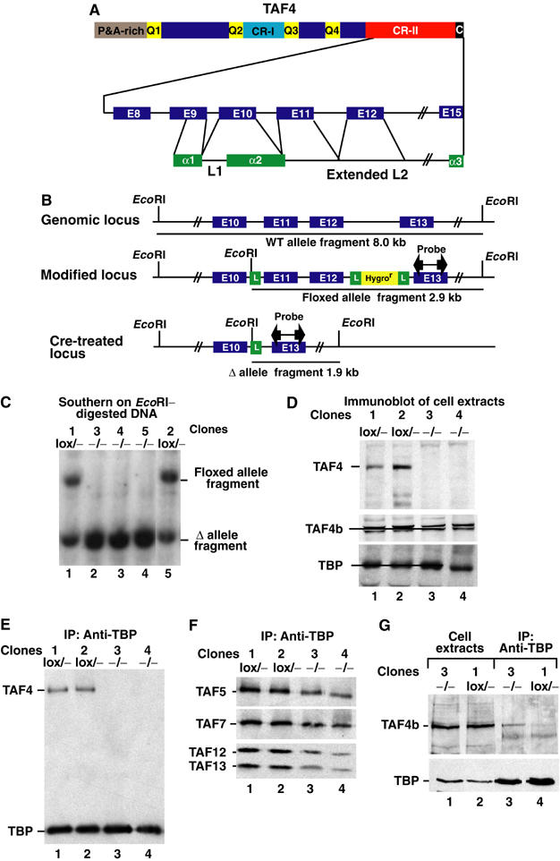
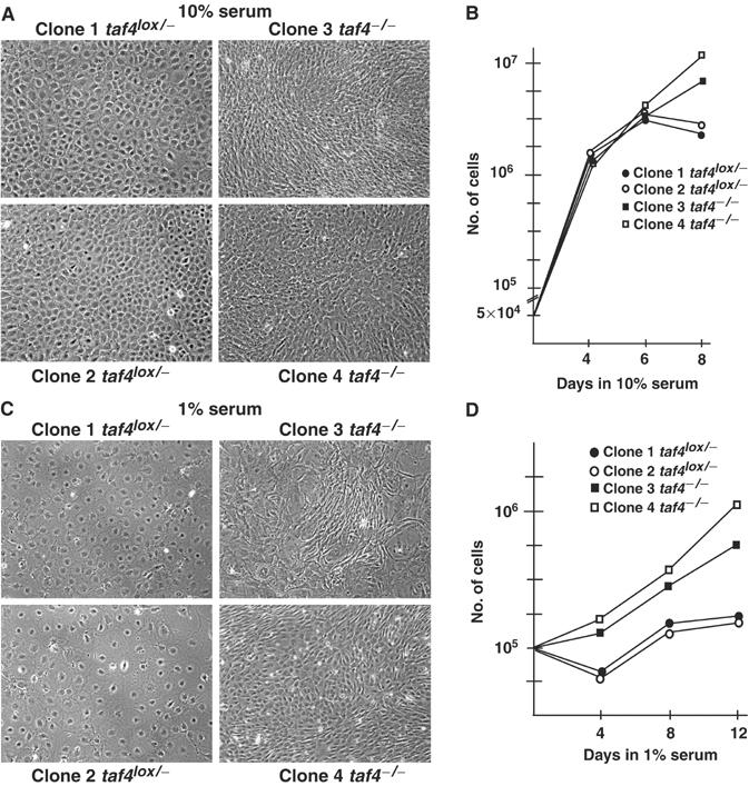
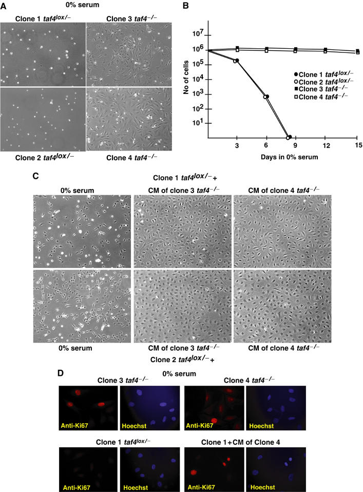
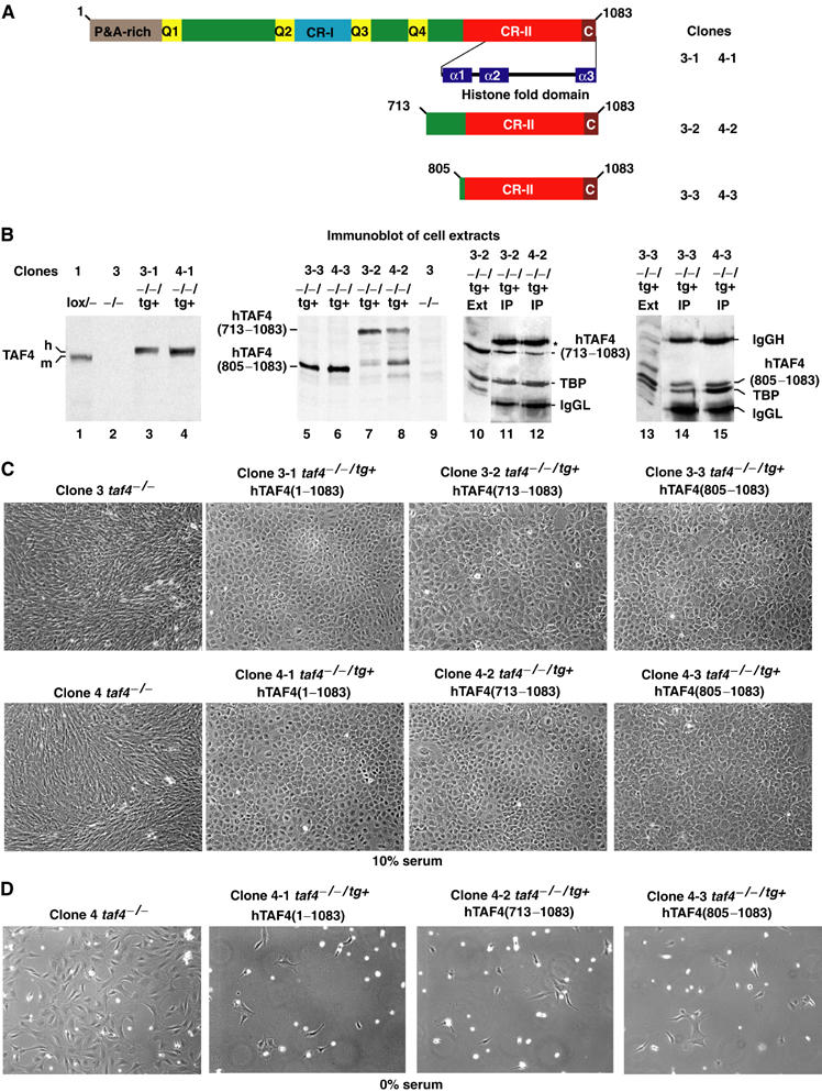
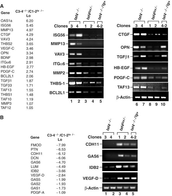
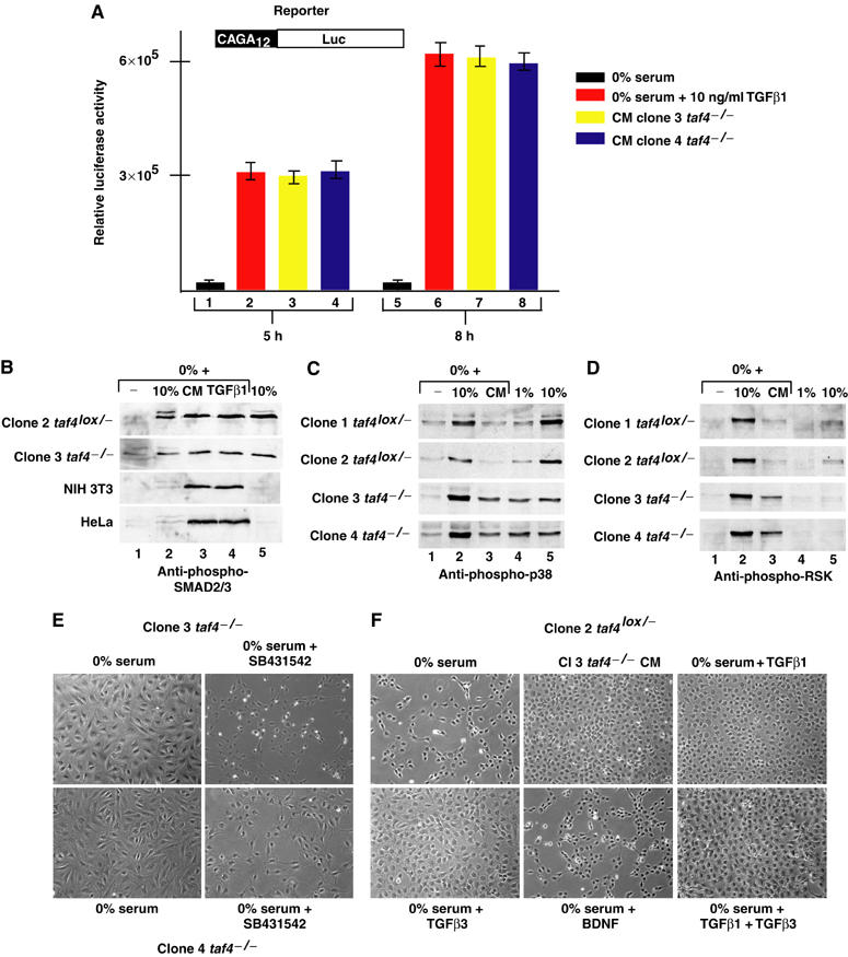
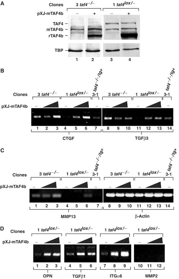
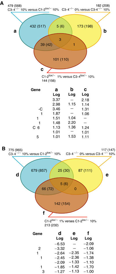
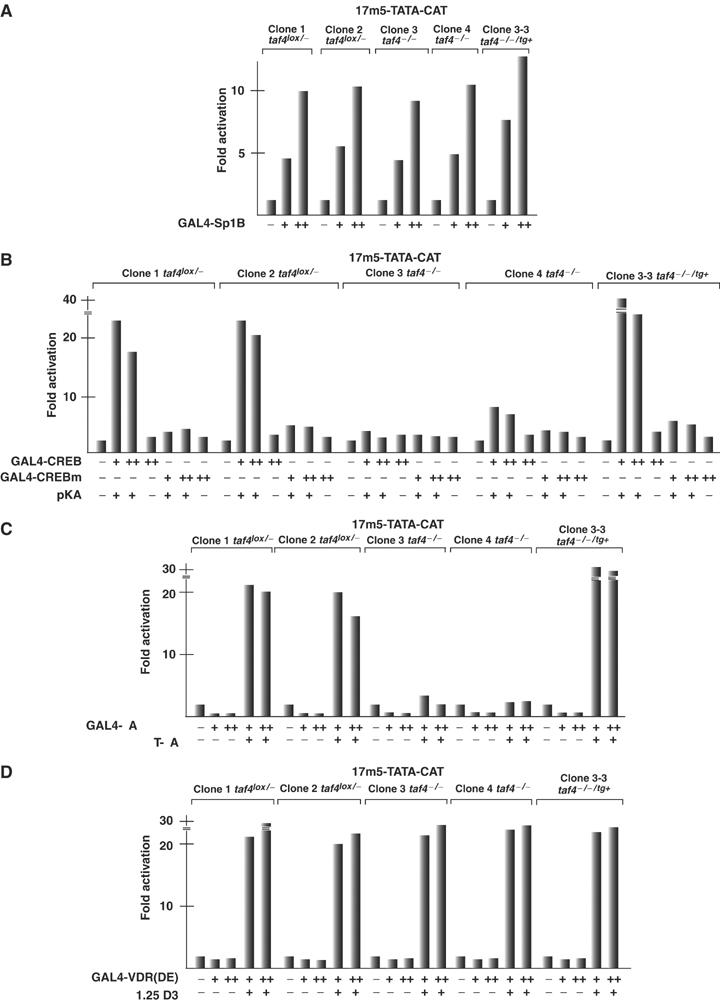
We have inactivated transcription factor TFIID subunit TBP-associated factor 4 (TAF4) in mouse embryonic fibroblasts. Mutant taf4(-/-) cells are viable and contain intact TFIID comprising the related TAF4b showing that TAF4 is not an essential protein. TAF4 inactivation deregulates more than 1000 genes indicating that TFIID complexes containing TAF4 and TAF4b have distinct target gene specificities. However, taf4(-/-) cell lines have altered morphology and exhibit serum-independent autocrine growth correlated with the induced expression of several secreted mitotic factors and activators of the transforming growth factor beta signalling pathway. In addition to TAF4 inactivation, many of these genes can also be induced by overexpression of TAF4b. A competitive equilibrium between TAF4 and TAF4b therefore regulates expression of genes controlling cell proliferation. We have further identified a set of genes that are regulated both by TAF4 and upon adaptation to serum starvation and which may be important downstream mediators of serum-independent growth. Our study also shows that TAF4 is an essential cofactor for activation by the retinoic acid receptor and CREB, but not for Sp1 and the vitamin D3 receptor.


Figures

Figure 1
Figure 1
Structure of TAF4. (A) The TAF4 protein is schematised showing the proline–alanine-rich (P&A) and glutamine-rich (Q1–Q4) regions and conserved regions I and II (CR-I and CR-II). Lower part: The intron/exon (E) structure of the CR-II region is schematised in relation to the HFD. (B) The native and modified taf4 alleles are schematised. The exons are indicated along with EcoRI sites, loxP sites (L) and the hygromycin resistance cassette. The location of the probe used in Southern blots is shown along with the EcoRI fragments generated from each allele. (C) Southern blots used to genotype fibroblast clones. The genotype of each clone is shown above the lane. (D) Immunoblots of cell extracts from the clones indicated above each lane were probed to reveal TAF4, TAF4b and TBP. A 20 μg portion of total cell extract was loaded in each lane. On some gels, TAF4b resolves into two closely migrating species. (E, F) Immunoblots of anti-TBP immunoprecipitations to reveal the indicated TAFs. The filter was first probed to reveal TBP and TAF4 and then reprobed sequentially to reveal the indicated TAFs. The clones and their genotypes are shown above each lane. (G) The indicated cell extracts were precipitated with antibody against TBP and probed for the presence of TBP and TAF4b.
Figure 2
Figure 2
Growth of taf4−/ fibroblasts. (A) Phase-contrast images of taf4−/ and taf4lox/− monolayers grown in 10% serum. (B) Comparison of the growth of taf4−/ and taf4lox/− cells in 10% serum. A total of 5 × 10⁴ cells were seeded in 10 cm plates on day 0. At each indicated day, a plate was trypsinised and the cells counted. The average values of three experiments are shown. (C) Phase-contrast images of the indicated clones grown for 4 days in 1% serum. (D) Growth of taf4−/ and taf4lox/− cells in 1% serum. The experiment was performed as described in panel B. In this case, 105 cells were seeded on day 0 and grown in 1% serum.
Figure 3
Figure 3
Growth of taf4−/ cells in the absence of serum. (A) Phase-contrast images of the clones after 4 days in 0% serum. (B) Comparison of the growth of taf4−/ and taf4lox/− cells in the absence of serum. A total of 106 cells were seeded in 10 cm plates on day 0, and counted at the indicated days. (C) A total of 106 taf4lox/− cells were seeded in either serum-free medium (upper and lower left panels) or serum-free conditioned media (CM) in which the indicated taf4−/ cells had been grown for 2 days (centre and right panels). Images were taken 2 days after seeding. (D) Immunofluorescence to detect proliferation marker Ki67. In each case, the immunostaining and Hoechst-stained DNA are shown side by side.
Figure 4
Figure 4
Re-expression of TAF4 suppresses serum-independent growth. (A) Schematic structure of TAF4 showing the domains described in Figure 1. The deletion mutants are indicated along with the nomenclature of the resulting clones. (B) Immunoblots (lanes 1–9) of the cell extracts indicated above each lane. In lanes 10–15, the indicated cell extracts were precipitated with antibody against TBP and the precipitated proteins revealed with antibody against TBP and TAF4 (20TA). The first lane in each panel shows one of the starting extracts as reference. The immunoglobulin heavy (*) and light chains of the precipitating antibodies are also indicated. (C) Morphology of revertant clones. Phase-contrast images of confluent monolayers of the indicated cell types grown in 10% serum. (D) Re-expression of TAF4 abrogates serum independence. A total of 106 cells from the different clones were seeded in serum-free media. Images were acquired after 4 days incubation.
Figure 5
Figure 5
Changes in gene expression in taf4−/ cells. (A, B) A short list of some relevant up- and downregulated genes. The full list is in Supplementary Figure 3. The log 2 values of induction and repression are shown. RT–PCR was performed on the indicated genes using RNA from the cells indicated above each lane. These RNA preparations are independent of those used in the profiling analysis and therefore provide a further confirmation of the results.
Figure 6
Figure 6
Activation of SMAD and p38 signalling pathways. (A) 3T3 cells harbouring the schematised integrated reporter were incubated in serum-free media with or without recombinant TGFβ or in conditioned media from the taf4−/ clones for 5 or 8 h as indicated before performing luciferase assays. Relative luciferase activity (arbitrary units) is shown on the Y-axis. (B) Conditioned media induce phosphorylation of SMAD3/3. In lanes 2–4, each cell line was grown overnight in serum-free medium before a 60 min exposure to either 10% serum, conditioned media from the clone 3 taf4−/ cells or recombinant TGFβ (10 ng/ml). In lanes 1 and 5, cells were grown for 24 h in 0 or 10% serum, respectively. After treatment, cells were immediately lysed in Laemmli loading buffer and analysed by SDS–PAGE and staining with Coomassie brilliant blue. Equivalent amounts of cell extract were then separated by SDS–PAGE and probed with antibody recognising SMAD2/3 phosphorylated at S465/467. (C, D) Cells were grown in the absence of serum and stimulated for 15 min prior to lysis with serum or conditioned media as indicated. Immunoblots were performed using antibodies recognising p38 phosphorylated at Thr180/Tyr182 or the RSK1–3 family phosphorylated at Thr577. (E) Phase-contrast images (× 20 magnification) of taf4−/ cells after 4 days growth in the absence of serum with or without the ALK5 inhibitor SB431542 (20 μM). (F) Phase-contrast images of taf4lox/− cells after 3 days growth in the absence of serum with conditioned media, TGFβ1, β3 (10 ng/ml) or BDNF (100 ng/ml) as indicated.
Figure 7
Figure 7
Overexpression of TAF4b induces gene expression. (A) Indicated clones were transfected with 4 μg of the pXJ-TAF4b expression vector or with empty vector (+ and −, respectively). After 48 h, protein extracts were probed with antibodies against TAF4b, TAF4 and TBP. Endogenous TBP, TAF4 and TAF4b are shown along with the transfected recombinant (r)TAF4b. (B–D) The indicated clones were transfected with 2 or 4 μg of the pXJ-TAF4b expression vector or with empty vector (−). RT–PCR was performed for the indicated genes 48 h after transfection.
Figure 8
Figure 8
Comparison of TAF4- and serum-regulated genes in fibroblasts. (A, B) The genes are divided into groups, (a–c) upregulated, (d–f) downregulated as defined, and represented as a coloured ellipse. The number of individual genes (probe sets) in each group is shown outside the ellipse. Coregulated genes are indicated in the overlapping areas. The identities of the genes regulated under all conditions are shown below along with the signal log value in each group. Full lists are in Supplementary Figures 3, 5 and 6.
Figure 9
Figure 9
TAF4 is a critical cofactor for CREB and RAR. (A–D) The indicated cell lines were transfected with the 17m5-TATA-CAT reporter (2.5 μg), the CMV-based β-galactosidase expression vector (2.5 μg) and the activator plasmids shown below each lane. CREBm contains the S133A mutation. In panel B, cotransfection of the catalytic subunit of protein kinase A is indicated, and in panels C and D, the presence or absence of 10−6 M all-trans-retinoic acid or 1alpha,25(OH)2-vitamin D3 is indicated. + represents 100 ng and ++ 250 ng of expression vector. The value of the 17m5-TATA-CAT reporter plasmid alone is taken as 1 and the fold activation is expressed relative to this value.

References

    1. Abreu JG, Ketpura NI, Reversade B, De Robertis EM (2002) Connective-tissue growth factor (CTGF) modulates cell signalling by BMP and TGF-beta. Nat Cell Biol 4: 599–604 - PMC - PubMed
    1. Annes JP, Munger JS, Rifkin DB (2003) Making sense of latent TGFbeta activation. J Cell Sci 116: 217–224 - PubMed
    1. Asahara H, Santoso B, Guzman E, Du K, Cole PA, Davidson I, Montminy M (2001) Chromatin-dependent cooperativity between constitutive and inducible activation domains in CREB. Mol Cell Biol 21: 7892–7900 - PMC - PubMed
    1. Binder DK, Scharfman HE (2004) Brain-derived neurotrophic factor. Growth Factors 22: 123–131 - PMC - PubMed
    1. Brand M, Yamamoto K, Staub A, Tora L (1999) Identification of TATA-binding protein-free TAFII-containing complex subunits suggests a role in nucleosome acetylation and signal transduction. J Biol Chem 274: 18285–18289 - PubMed

Publication types

MeSH terms

Substances

LinkOut - more resources