Evaluation of marrow perfusion in the femoral head by dynamic magnetic resonance imaging. Effect of venous occlusion in a dog model
- PMID: 1601616
- PMCID: PMC2396275
- DOI: 10.1097/00004424-199204000-00002
Evaluation of marrow perfusion in the femoral head by dynamic magnetic resonance imaging. Effect of venous occlusion in a dog model
Abstract
Rationale and objectives: There is a continuing need for a greater sensitivity of magnetic resonance imaging (MRI) in the diagnosis of avascular necrosis (AVN). Previously, it was demonstrated that a dynamic MRI method, with gadolinium-DTPA (Gd-DTPA) enhancement, can detect acute changes not seen on spin-echo images after arterial occlusion in a dog model. Because venous congestion appears to be a more directly relevant hemodynamic abnormality in a majority of clinical AVN cases, the authors extended the dynamic MRI technique to study changes in venous occlusion.
Methods: Dynamic MRI of the proximal femur was performed in five adult dogs before and after unilateral ligation of common iliac and lateral circumflex veins. Sixteen sequential gradient-recalled pulse sequence (GRASS) images (time resolution = 45 mseconds, echo time = 9 mseconds, flip angle = 65 degrees) were obtained immediately after a bolus intravenous injection of 0.2 mmol/kg of Gd-DTPA. Simultaneous measurements of regional blood flow were made using the radioactive microsphere method.
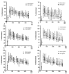
Results: After venous ligation, there was a 25% to 45% decrease in the degree of enhancement compared with preligation values on the ligated side. The decrease in cumulative enhancement (integrated over the entire time course) was statistically significant. The occlusion technique was verified by confirming a statistically significant decrease in blood flow determined by the microsphere method.
Conclusions: Dynamic Gd-DTPA-enhanced fast MRI technique can detect acute changes in bone marrow perfusion due to venous occlusion. This technique may have applications in the early detection of nontraumatic AVN.
Figures


Similar articles
-
Bone marrow perfusion evaluated with gadolinium-enhanced dynamic fast MR imaging in a dog model.Radiology. 1991 May;179(2):535-9. doi: 10.1148/radiology.179.2.2014306. Radiology. 1991. PMID: 2014306
-
Functional assessment of canine kidneys after acute vascular occlusion on Gd-DTPA-enhanced dynamic echo-planar MR imaging.Invest Radiol. 2001 Nov;36(11):659-76. doi: 10.1097/00004424-200111000-00006. Invest Radiol. 2001. PMID: 11606844
-
Value of a dynamic MR scan in predicting vascular ingrowth from free vascularized scapular transplant used for treatment of avascular femoral head necrosis.Microsurgery. 1995;16(10):673-8. doi: 10.1002/micr.1920161004. Microsurgery. 1995. PMID: 8676730
-
Detection of acute avascular necrosis of the femoral head in dogs: dynamic contrast-enhanced MR imaging vs spin-echo and STIR sequences.AJR Am J Roentgenol. 1992 Dec;159(6):1255-61. doi: 10.2214/ajr.159.6.1442396. AJR Am J Roentgenol. 1992. PMID: 1442396
-
Contrast-enhanced fat saturation magnetic resonance imaging for studying the pathophysiology of osteonecrosis of the hips.Skeletal Radiol. 1992;21(6):375-9. doi: 10.1007/BF00241816. Skeletal Radiol. 1992. PMID: 1523433
Cited by
-
Osteonecrosis of the Femoral Head.J Am Acad Orthop Surg Glob Res Rev. 2022 May 1;6(5):e21.00176. doi: 10.5435/JAAOSGlobal-D-21-00176. J Am Acad Orthop Surg Glob Res Rev. 2022. PMID: 35511598 Free PMC article.
-
Bone marrow blood supply in gadolinium-enhanced magnetic resonance imaging.Skeletal Radiol. 1994 Aug;23(6):455-7. doi: 10.1007/BF00204608. Skeletal Radiol. 1994. PMID: 7992112
-
Femoral head-neck junction reconstruction, after iatrogenic bone resection.J Hip Preserv Surg. 2015 Jul;2(2):190-3. doi: 10.1093/jhps/hnv016. Epub 2015 Feb 3. J Hip Preserv Surg. 2015. PMID: 27011838 Free PMC article.
-
Assessment of bone perfusion with contrast-enhanced magnetic resonance imaging.Orthop Clin North Am. 2009 Apr;40(2):249-57. doi: 10.1016/j.ocl.2008.12.003. Orthop Clin North Am. 2009. PMID: 19358910 Free PMC article.
-
[Atraumatic and aseptic osteonecrosis of large joints].Radiologe. 2019 Jul;59(7):647-662. doi: 10.1007/s00117-019-0560-3. Radiologe. 2019. PMID: 31250028 German.
References
-
- Mitchell DG. Using MR imaging to probe the pathophysiology of osteonecrosis. Radiology. 1989;171:25–26. - PubMed
-
- Turner DA, Templeton AC, Selzer PM, Rosenberg AG, Petasnick JP. Femoral capital osteonecrosis: MR findings of diffuse marrow abnormalities without focal lesions. Radiology. 1989;171:135–140. - PubMed
-
- Hungerford DS. The Hip, Proceedings of the 7th Opens Scientific Meeting of the Hip Society. St. Louis, MO: Mosby; 1979. Bone marrow pressure, venography, and core decompression in ischemic necrosis of the femoral head; pp. 218–237.
-
- Cova M, Kang YS, Tsukamoto H, Jones LC, McVeigh E, Neff BL, Herold CJ, Scott WW, Jr, Hungerford DS, Zerhouni EA. Bone marrow perfusion evaluated with Gadolinium-enhanced dynamic fast MR imaging in a dog model. Radiology. 1991;179:535–539. - PubMed
-
- Hungerford DS, Lennox DW. The importance of increased intraosseous pressure in the development of osteonecrosis of the femoral head: Implication for treatment. Orthop Clin North Am. 1985;16:635–654. - PubMed
MeSH terms
Substances
Grants and funding
LinkOut - more resources
Full Text Sources
Medical
