. 2005 Aug;132(16):3777-86.
doi: 10.1242/dev.01935. Epub 2005 Jul 14.

Cerebral hypoplasia and craniofacial defects in mice lacking heparan sulfate Ndst1 gene function

Affiliations

Cerebral hypoplasia and craniofacial defects in mice lacking heparan sulfate Ndst1 gene function

Kay Grobe et al. Development. 2005 Aug.

Abstract

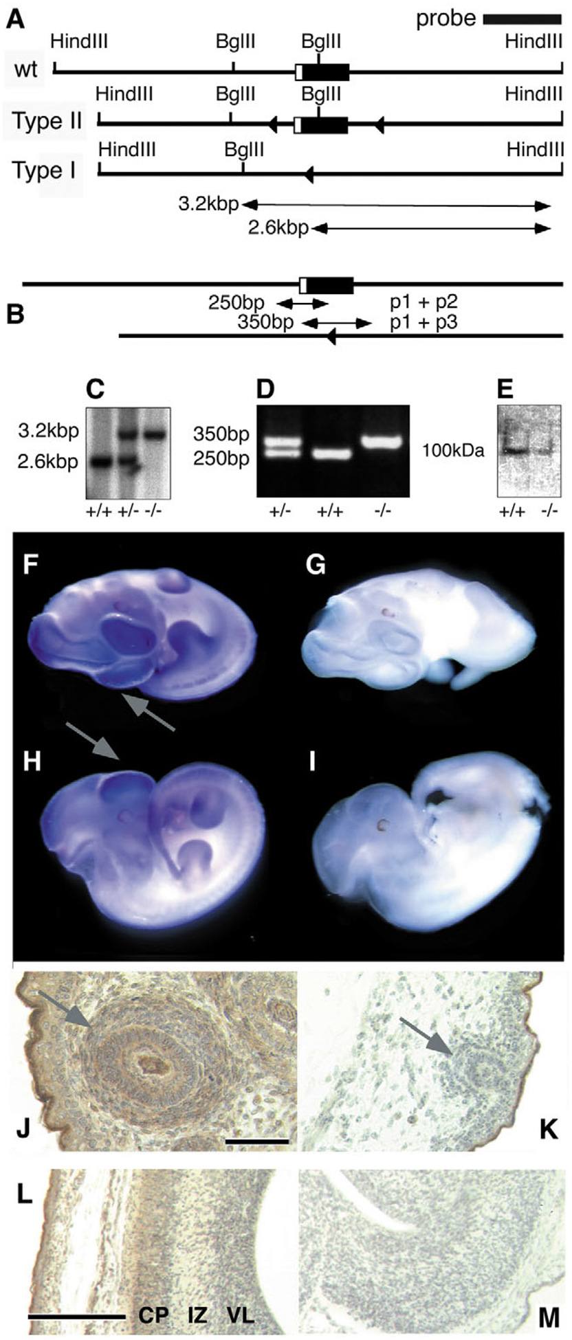
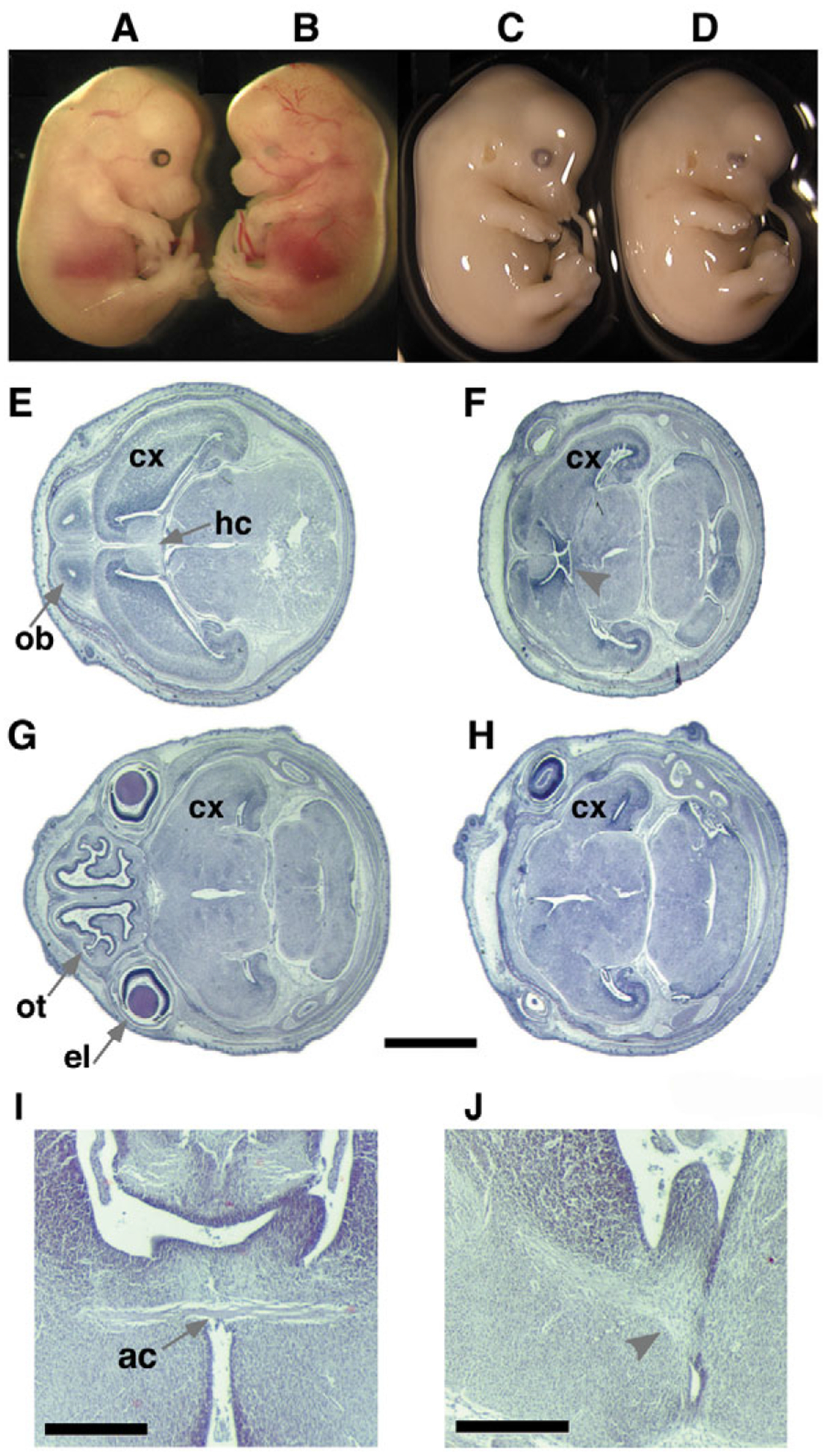
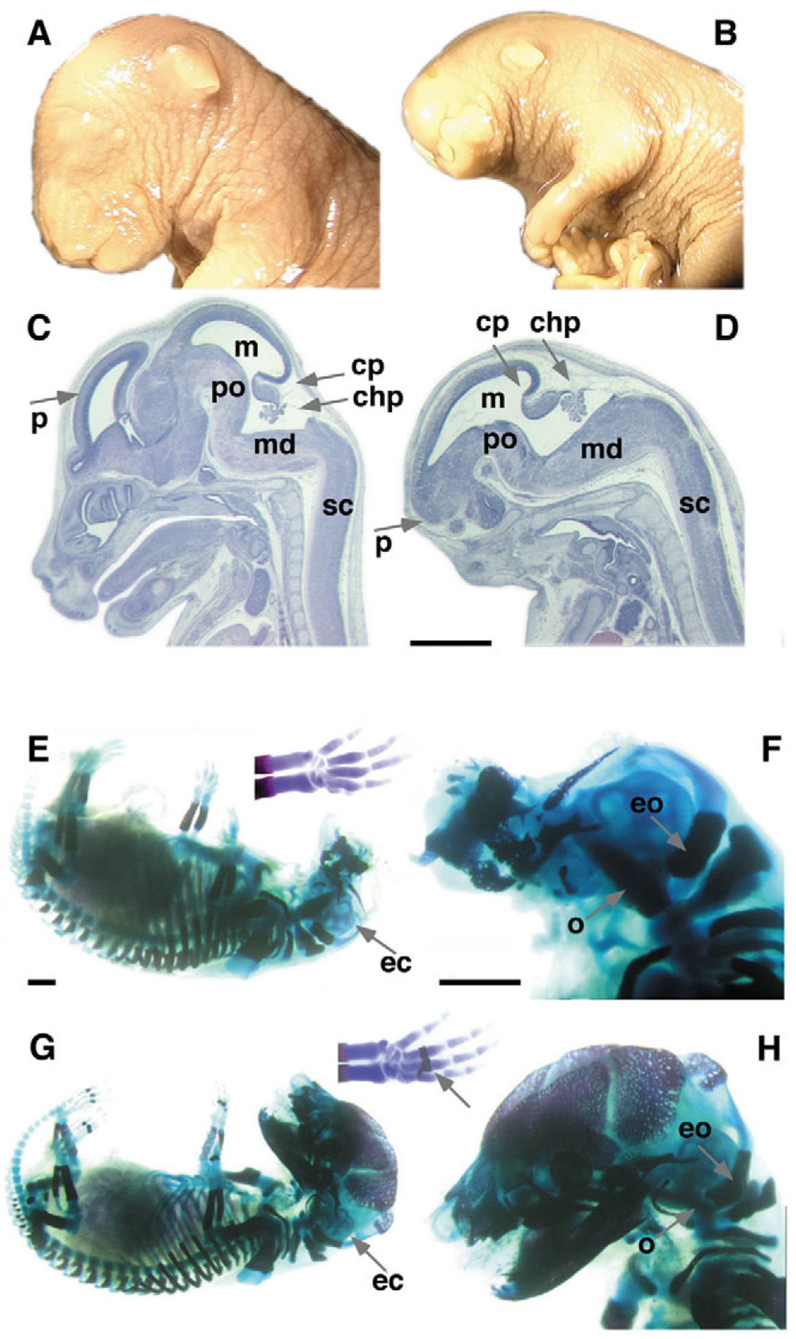
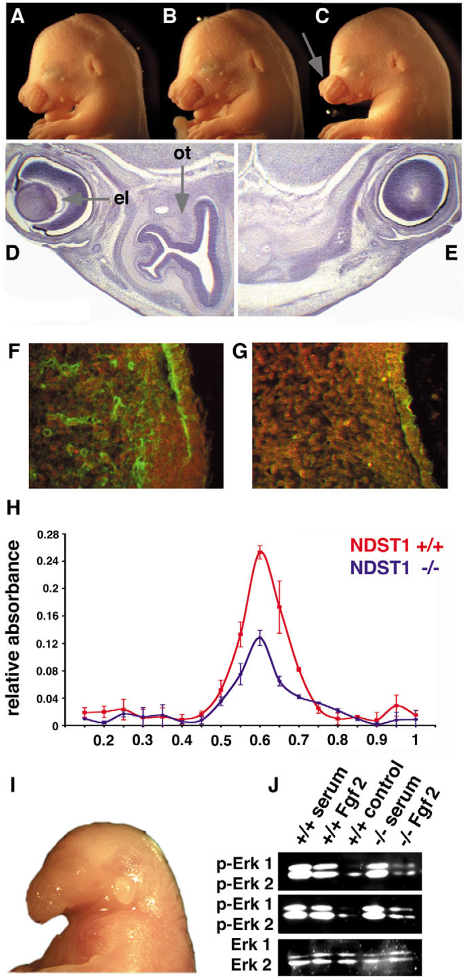
Mutant mice bearing a targeted disruption of the heparan sulfate (HS) modifying enzyme GlcNAc N-deacetylase/N-sulfotransferase 1 (Ndst1) exhibit severe developmental defects of the forebrain and forebrain-derived structures, including cerebral hypoplasia, lack of olfactory bulbs, eye defects and axon guidance errors. Neural crest-derived facial structures are also severely affected. We show that properly synthesized heparan sulfate is required for the normal development of the brain and face, and that Ndst1 is a modifier of heparan sulfate-dependent growth factor/morphogen signalling in those tissues. Among the multiple heparan sulfate-binding factors potentially affected in Ndst1 mutant embryos, the facial phenotypes are consistent with impaired sonic hedgehog (Shh) and fibroblast growth factor (Fgf) interaction with mutant heparan sulfate. Most importantly, the data suggest the possibility that defects in heparan sulfate synthesis could give rise to or contribute to a number of developmental brain and facial defects in humans.


Figures

Fig. 1.
Fig. 1.
Disruption of the Ndst1 gene by targeted recombination. (A) Maps of the wild-type Ndst1 locus, the Type II ‘floxed’ allele and a Type I deletion allele, obtained after breeding of Type II mice with ZP3-Cre mice. Lox-sites located in intron sequences are shown as triangles. (B) PCR analysis. Deletion of exon 2 in Ndst1−/− mice yields a 350 bp product, whereas wild-type mice produce a 250 bp amplification product. Heterozygous mice yield both amplification products. (C) Southern blot analysis of DNA. DNA digestion using restriction endonucleases HindIII and BglII yields a 2.6 kbp (wild type) and a 3.2 kbp (Ndst1−/−) band. (D) PCR analysis of the same samples as in (C). (E) Western blotting of total embryo extract after SDS-PAGE. Samples were detected by an affinity-purified rabbit-anti-mouse Ndst1 antiserum (Grobe and Esko, 2002). The weak signal seen in the knockout lane is due to cross-reactivity with Ndst2–4, all of which are expressed in the developing embryo (Aikawa et al., 2001). (F-I) Detection of Ndst1 expression in the developing embryo. (F,H) Whole-mount in situ hybridization in E11.5 wild-type embryo showed strongest expression (blue stain) in the forebrain and frontonasal/maxillary processes (arrows). (G,I) Ndst1−/− embryo probed with an antisense riboprobe showed no reactivity. (J-M) Immunohistochemical detection of Ndst1 expression in E16.5 embryos using the affinity-purified rabbit-anti-mouse Ndst1 antiserum (Grobe and Esko, 2002). Ndst1 protein is strongly expressed (brown) in frontonasal mesenchyme (J) and telencephalic walls (L). In the developing face, hair follicles show the highest expression levels (arrow). (K,M) The hair follicles and telencephalic walls are not stained in Ndst1−/− tissues. CP, cortical plate; IZ, intermediate zone; VL, ventricular layer. (M) There is a lack of stratification in the Ndst1 mutant cortex. Scale bar: 10 μm in J; 100 μm in L.
Fig. 2.
Fig. 2.
Phenotypes of mildly affected Ndst1 mutant E13.5 embryos. (A,C) Wild-type littermate controls. (B,D) Mutant embryos show lack of eyes and reduced frontonasal process (B) and (D) lack of eye lens. (E,G) Wild-type littermate controls. (F,H) Mutant embryos with mild defects in external features show severe defects in the frontonasal process and forebrain (E16.5, horizontal sections). Lack of olfactory tracts (ot), olfactory bulbs (ob), the eye lens (el) and the hippocampal commissure (hc, arrowhead) is evident. The cortex (cx) is reduced in size and stratification and the cleavage along transverse, sagittal and horizontal axes is defective. Scale bar: 1 mm. (I,J) Coronal sections of a E18.5 wild-type (I) and Ndst1−/− (J) brain show the lack of the anterior commissure (ac) in the mutant embryo (arrowhead). Axons disperse instead of crossing the midline. Scale bar: 500 μm.
Fig. 3.
Fig. 3.
Anatomy and histology of strongly affected E18.5 Ndst1 mutant embryos. (A,C) Wild-type embryo, (B,D) Ndst1 mutant. Gross examination revealed severe frontonasal dysmorphism, including the lack of eyes but normal outer ear. (D) Hypoplastic prosencephalon (p). The midbrain (m), pons (po), medulla (md), the cerebellar primordium (cp), choroid plexus (chp) and spinal cord (sc) appear normal (sagittal sections). (E-H) Bone (red) and cartilage (blue) stain of E18.5 embryos. (E,F) Severely affected Ndst1 mutant embryos show lack of all neural crest cell-derived bones of the viscerocranium and neurocranium. Some lack or delay of ossification occurs, especially in the vertebrae and (shortened) limb digits (compare with wild type, arrow in G), which may be fused (E). The ear capsule (ec) is intact in these embryos (E,F), as are the occipital (o) and exoccipital (eo) bones, which are derived from paraxial mesoderm. (G,H) Wild-type littermate controls. Scale bars: 1 mm.
Fig. 4.
Fig. 4.
Ndst1; Shh interact genetically and biochemically. Ptch1 protein expression is reduced in the developing face of Ndst1−/− embryos and Fgf signalling is also affected. (A-C) Craniofacial morphology in E15.5 embryos. (A) Ndst+/− and (B) Shh+/− littermate controls. (C) Some Ndst1; Shh compound heterozygous mice showed hypoplastic maxillary processes (arrow). (D,E) Haematoxylin/Eosin staining of horizontal sections of embryos in A,C, respectively. The olfactory epithelium (ot) is absent in the Ndst1, Shh compound heterozygous mouse, and development of the eye lens (el) is also affected, both resembling features found in the Ndst1−/− mutant embryos (Fig. 2F,H). (F) Wild-type embryo, (G) Ndst1−/− embryo. (F,G) Reduced expression of the Shh receptor Ptch1 in the frontonasal process of Ndst1−/− mutant E15.5 embryos, as assessed by immunohistochemical staining. Three wt and mutant embryos were analyzed, representative sections are shown. Green fluorescence indicates Ptch1 expression. (H) More AP-Shh binds to E14.5 wild-type GAGs (red line) than to GAGs isolated from mutant embryos (blue line). Equal amounts of GAGs isolated from these embryos were coupled with Affi-Gel, loaded with recombinant, soluble alkaline phosphatase fused to Shh (AP-Shh) followed by elution with increasing NaCl concentrations ranging from 0–1 M in 50 mM increments. (I,J) Impaired Fgf signalling in Ndst1 mutant embryos. (I) Twenty-five percent of all Ndst1−/− embryos showed developmental defects of the first branchial arch, including agnathia strongly reminiscent of neural- and neural-crest specific Fgf8 mutant mice (Trumpp et al., 1999). One representative E18.5 Ndst1 mutant embryo is shown. (J) Fgf-dependent Erk1/2 phosphorylation is significantly reduced in Ndst1 mutant mesenchymal cells isolated from two mutant embryos (top and middle). Cultured cells derived from facial mesenchyme of two E14.5 wild-type and two mutant littermates were stimulated with 10% serum in DMEM or 10 ng/ml Fgf2 in DMEM for 5 minutes before analysis. Phosphorylation of Erk1 and Erk2 (p-Erk1 and p-Erk2) were observed in the wild type after addition of serum or Fgf2; however, Fgf2 addition alone failed to stimulate Erk1/2 phosphorylation in the Ndst1 mutant cells. Control refers to no stimulation. (Bottom) Detection of total Erk1 and Erk2 with anti-Erk antibodies.
Fig. 5.
Fig. 5.
Enhanced apoptosis, decreased proliferation and impaired glia development in Ndst1−/− embryos. (A,B) Apoptosis is significantly enhanced in the forebrains of mutant embryos. Skin (arrowhead) serves as a positive control. (A) TUNEL staining reveals very high levels of cell death (blue nuclei) in the neopallial cortex (arrow) of E15.5 Ndst1−/− embryos, a region expressing Ndst1 at high levels (Fig. 1L). (B) Wild-type littermate controls. (C) Cells residing in the ependymal and ventricular layers of the diencephalon in Ndst1−/− embryos also undergo apoptosis. (D) Wild-type littermate control. v, ventricle. Horizontal sections. (E,F) Proliferation (brown stain) in a E17.5 mutant (F) embryo compared with the wild type (E) lateral telencephalic wall. In mutant mice, the proliferative rate is reduced in the lateral telencephalic wall of the brain (F), and dividing cells are more clustered within the VZ if compared with the wild type. Horizontal sections. (G,H) Reduced Gfap (red) staining indicating lower numbers of glia cells in mutant E18.5 forebrain (H). The wild-type brain (G) shows presence of those cells, which provide a scaffold for lateral neural cell migration from the ventricular zone. Scale bars: 50 μm. Horizontal sections. (I) Quantitation of apoptotic cells of the neopallial cortex (1, inset) and the ependymal layer (2) of the dorsal part of the third ventricle of three mutant and wild-type embryos. (J) Quantitation of BrdU positive nuclei in the posterior (3), anterior (4), lateral (5) and medial (6) telencephalic ventricular zones of three mutant and wild-type E17.5 embryos. Horizontal sections. A moderate reduction in proliferation could only be observed in the lateral area of Ndst1−/− embryos. Coronal sections also revealed similar proliferative rates in the medial and parietal cortex of wild-type and mutant embryos (not shown). Analysis of three E15.5 mutant and wild-type embryos showed no differences in proliferation (not shown).

References

    1. Abzhanov A and Tabin CJ (2004). Shh and Fgf8 act synergistically to drive cartilage outgrowth during cranial development. Dev. Biol 273, 134–148. - PubMed
    1. Ahlgren SC and Bronner-Fraser M (1999). Inhibition of sonic hedgehog signaling in vivo results in craniofacial neural crest cell death. Curr. Biol 9, 1304–1314. - PubMed
    1. Aikawa J, Grobe K, Tsujimoto M and Esko JD (2001). Multiple Isozymes of Heparan Sulfate/Heparin GlcNAc N-Deacetylase/N-Sulfotransferase: Structure and Activity of the Fourth Member, NDST4. J. Biol. Chem 276, 5876–5882. - PubMed
    1. Allen BL and Rapraeger AC (2003). Spatial and temporal expression of heparan sulfate in mouse development regulates FGF and FGF receptor assembly. J. Cell Biol 163, 637–648. - PMC - PubMed
    1. Bennett KL, Bradshaw J, Youngman T, Rodgers J, Greenfield B, Aruffo A and Linsley PS (1997). Deleted in colorectal carcinoma (DCC) binds heparin via its fifth fibronectin type III domain. J. Biol. Chem 272, 26940–26946. - PubMed

Publication types

MeSH terms