. 2005 Aug 19;97(4):354-62.
doi: 10.1161/01.RES.0000177669.29525.78. Epub 2005 Jul 14.

Mitochondria-derived reactive oxygen species dilate cerebral arteries by activating Ca2+ sparks

Affiliations

Mitochondria-derived reactive oxygen species dilate cerebral arteries by activating Ca2+ sparks

Qi Xi et al. Circ Res. .

Abstract

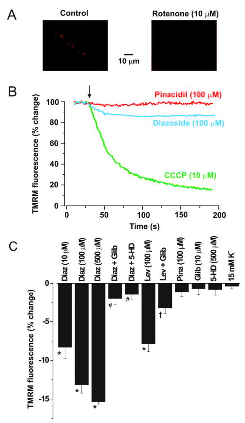
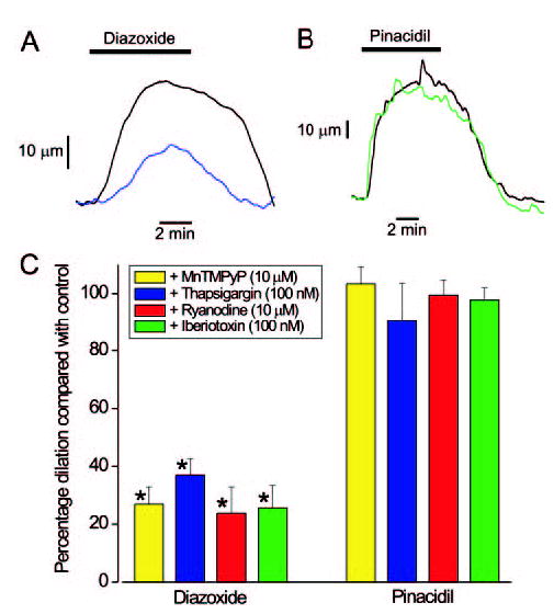
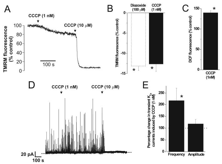
Mitochondria regulate intracellular calcium (Ca2+) signals in smooth muscle cells, but mechanisms mediating these effects, and the functional relevance, are poorly understood. Similarly, antihypertensive ATP-sensitive potassium (KATP) channel openers (KCOs) activate plasma membrane KATP channels and depolarize mitochondria in several cell types, but the contribution of each of these mechanisms to vasodilation is unclear. Here, we show that cerebral artery smooth muscle cell mitochondria are most effectively depolarized by diazoxide (-15%, tetramethylrhodamine [TMRM]), less so by levcromakalim, and not depolarized by pinacidil. KCO-induced mitochondrial depolarization increased the generation of mitochondria-derived reactive oxygen species (ROS) that stimulated Ca2+ sparks and large-conductance Ca2+-activated potassium (KCa) channels, leading to transient KCa current activation. KCO-induced mitochondrial depolarization and transient KCa current activation were attenuated by 5-HD and glibenclamide, KATP channel blockers. MnTMPyP, an antioxidant, and Ca2+ spark and KCa channel blockers reduced diazoxide-induced vasodilations by >60%, but did not alter dilations induced by pinacidil, which did not elevate ROS. Data suggest diazoxide drives ROS generation by inducing a small mitochondrial depolarization, because nanomolar CCCP, a protonophore, similarly depolarized mitochondria, elevated ROS, and activated transient KCa currents. In contrast, micromolar CCCP, or rotenone, an electron transport chain blocker, induced a large mitochondrial depolarization (-84%, TMRM), reduced ROS, and inhibited transient KCa currents. In summary, data demonstrate that mitochondria-derived ROS dilate cerebral arteries by activating Ca2+ sparks, that some antihypertensive KCOs dilate by stimulating this pathway, and that small and large mitochondrial depolarizations lead to differential regulation of ROS and Ca2+ sparks.


Figures

Figure 1
Figure 1
Regulation of cerebral artery smooth muscle cell mitochondrial potential. A, Confocal image illustrating TMRM fluorescence localization and reduction by rotenone. B, Original traces with arrow indicating time of drug application. C, Average changes in TMRM fluorescence with diazoxide (10 μmol/L, n = 9;100 μmol/L, n = 5; 500 μmol/L, n = 5), diazoxide (100 μmol/L) + glibenclamide (1 μmol/L, n = 5), diazoxide (100 μmol/L) + 5-HD (500 μmol/L, n = 6), levcromakalim (100 μmol/L, n = 4), levcromakalim (100 μmol/L) + glibenclamide (1 μmol/L, n = 6), pinacidil (100 μmol/L, n = 5), glibenclamide (10 μmol/L, n = 6), 5-HD (500 μmol/L, n = 4), and 15 mmol/L K+ (n = 5). *, #, and † illustrate P < 0.05 when compared with control, diazoxide, (100 μmol/L), or levcromakalim, respectively.
Figure 2
Figure 2
Regulation of transient KCa currents by KATP channel modulators in cells voltage-clamped at −40 mV. A, Diazoxide activates transient KCa currents, and addition of rotenone blocks transient KCa currents. B, Diazoxide-induced transient KCa current activation is attenuated by 5-HD. C through D, Average effects on transient KCa current frequency and amplitude of diazoxide (10 μmol/L, n = 7; 100 μmol/L, n = 23), diazoxide (100 μmol/L) + glibenclamide (10 μmol/L, n = 7), diazoxide (100 μmol/L) + 5-HD (500 μmol/L, n = 5), diazoxide (100 μmol/L) applied in the presence of diltiazem (50 μmol/L, n = 4), rotenone (10 μmol/L) applied in the presence of diazoxide (100 μmol/L, n = 5), levcromakalim (100 μmol/L, n = 4), pinacidil (100 μmol/L, n = 9), glibenclamide (10 μmol/L, n = 5), and 5-HD (500 μmol/L, n = 4). * and # illustrate P < 0.05 when compared with control or diazoxide (100 μmol/L), respectively.
Figure 3
Figure 3
Diazoxide (100 μmol/L) elevates Ca2+ spark frequency and enhances coupling of sparks to KCa channels. A, Simultaneous measurements of Ca2+ sparks (lower traces) and transient KCa currents (upper traces) at −40 mV. Images illustrating time course of the Ca2+ spark labeled as † in A are shown below traces. In this cell in control, Ca2+ sparks were observed primarily at one location during 15 seconds of acquisition, but one spark occurred at a second position. In diazoxide, all sparks occurred at the primary discharge site. B, Diazoxide increased mean Ca2+ spark frequency (from 0.58 ± 0.15 to 0.76 ± 0.09 Hz, n = 12 cells), but did not change amplitude (control, 1.96 ± 0.09, n = 120; diazoxide, 1.99 ± 0.11, n = 193, P > 0.05). C, Amplitude correlation of Ca2+ sparks and evoked transient KCa currents in control (black squares) and diazoxide (red circles) with first-order polynomial linear fit and 95% confidence bands to each data-set (slope ± SE: control, 31 ± 2, r = 0.08; diazoxide, 43 ± 2, r = 0.25, P < 0.0001 for each). Diazoxide increased effective coupling of Ca2+ sparks to KCa channels (P = 0.034). *P < 0.05 when compared with control.
Figure 4
Figure 4
Regulation of ROS in smooth muscle cells of intact arteries. A, DCF fluorescence in smooth muscle cells of the same intact artery segment in control and diazoxide (100 μmol/L). B, Average changes in DCF fluorescence with diazoxide (100 μmol/L, n = 9), pinacidil (100 μmol/L, n = 5), rotenone (10 μmol/L, n = 6), CCCP (1 μmol/L, n = 6), or vehicle (DMSO, time control, n = 5). Catalase (2000 U/mL, n = 5) attenuated, and MnT-MPyP (10 μmol/L, n = 6) or rotenone (10 μmol/L, n = 6) essentially abolished, diazoxide (100 μmol/L)-induced DCF fluorescence elevations. C, Confocal images of DHE fluorescence in cerebral artery sections in control and after diazoxide (100 μmol/L). D, Diazoxide elevated mean DHE fluorescence in smooth muscle cells (n = 5). * and # indicate P < 0.05 when compared with control or diazoxide, respectively. NS, nonsignificant.
Figure 5
Figure 5
Diazoxide-induced transient KCa current activation is abolished by antioxidants. A, Inclusion of SOD and catalase (300 U/mL of each, conventional whole-cell) in the pipette solution prevented diazoxide-induced transient KCa current activation (−40 mV). B, Frequency histogram illustrating effect of diazoxide on transient KCa currents when using active (black bars, data taken from cell illustrated in A), or boiled (white bars) SOD + catalase. C, Mean change in transient KCa currents in cells dialyzed with active (black bars, n = 4), or boiled (white bars, n = 6), SOD + catalase. In SOD + catalase, the apparent reduction in transient KCa currents with diazoxide occurs because of rundown. Frequency rundown was similar in control (0.016 Hz/min) and diazoxide (0.017 Hz/min). *P < 0.05.
Figure 6
Figure 6
Diazoxide dilates pressurized cerebral arteries because of an elevation in ROS and RyR and KCa channel activation. A, Diazoxide (100 μmol/L) induces a reversible dilation in a pressurized (60 mm Hg) cerebral artery (black line). In the same cerebral artery in the continued presence of thapsigargin (100 nmol/L), diazoxide induces a smaller dilation (blue line). B, Pinacidil (100 μmol/L)-induced dilations in the same artery were similar in control (black line) and in the continued presence of iberiotoxin (100 nmol/L, green line). C, Mean effects of MnT-MPyP (yellow), thapsigargin (blue), ryanodine (red), or iberiotoxin (green) on dilations induced by diazoxide (100 μmol/L, n = 7, 5, 10, and 7, respectively), or pinacidil (100 μmol/L, n = 7, 5, 5 and 7, respectively). Thapsigargin, ryanodine, and iberiotoxin reduced mean arterial diameter between 10 and 16 μm, and MnTMPyP did not alter diameter (see online data supplement). *P < 0.05 when compared with control dilations induced by the same KATP channel opener in the same arteries.
Figure 7
Figure 7
Differential regulation of transient KCa currents by small and large mitochondrial depolarizations. A, Original recording illustrating TMRM intensity changes in the same cell with 1 nmol/L and 10 μmol/L CCCP. B, Mean changes in TMRM intensity induced by 1 nmol/L CCCP (n = 5) and 100 μmol/L diazoxide (reproduced from Figure 1C for comparison). C, 1 nM CCCP elevates DCF fluorescence (n = 6). D, 1 nmol/L CCCP activates transient KCa currents, whereas 10 μmol/L CCCP blocks these events in a voltage-clamped (−40 mV) cell. E, Mean change in transient KCa currents induced by 1 nmol/L CCCP (n = 8). *P < 0.05 when compared with control.

Comment in

References

    1. Jaggar JH, Porter VA, Lederer WJ, Nelson MT. Calcium sparks in smooth muscle. Am J Physiol. 2000;278:C235–C256. - PubMed
    1. Perez GJ, Bonev AD, Nelson MT. Micromolar Ca2+ from sparks activates Ca2+-sensitive K+ channels in rat cerebral artery smooth muscle. Am J Physiol. 2001;281:C1769–C1775. - PubMed
    1. Jaggar JH, Leffler CW, Cheranov SY, Tcheranova DES, Cheng X. Carbon monoxide dilates cerebral arterioles by enhancing the coupling of Ca2+ sparks to Ca2+-activated K+ channels. Circ Res. 2002;91:610–617. - PubMed
    1. Cheranov SY, Jaggar JH. Mitochondrial modulation of Ca2+ sparks and transient KCa currents in smooth muscle cells of rat cerebral arteries. J Physiol. 2004;556:755–771. - PMC - PubMed
    1. Wang YX, Zheng YM, Abdullaev I, Kotlikoff MI. Metabolic inhibition with cyanide induces calcium release in pulmonary artery myocytes and Xenopus oocytes. Am J Physiol. 2003;284:C378–C388. - PubMed

Publication types

MeSH terms