. 2005 Sep;30(1):108-17.
doi: 10.1016/j.mcn.2005.06.004.

Purified mouse dopamine neurons thrive and function after transplantation into brain but require novel glial factors for survival in culture

Affiliations

Purified mouse dopamine neurons thrive and function after transplantation into brain but require novel glial factors for survival in culture

A E Donaldson et al. Mol Cell Neurosci. 2005 Sep.

Erratum in

  • Mol Cell Neurosci. 2005 Dec;30(4):559

Corrected and republished in

Abstract

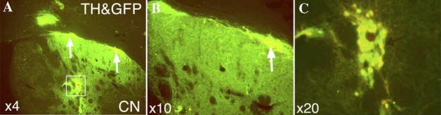
Cell replacement therapy in Parkinson's disease depends on a reliable source of purified dopamine (DA) neurons (PDN) and the identification of factors relevant to their survival. Our goal was to genetically tag and purify by flow cytometry embryonic midbrain DA neurons from a transgenic mouse line carrying 11 kb of human tyrosine hydroxylase promoter driving expression of the enhanced green fluorescent protein (GFP) for studies in vivo and in vitro. A 99% purification of GFP(+) cells was achieved. When transplanted into 6-hydroxydopamine-treated rat striatum, PDN survived, became well-integrated and produced recovery from amphetamine-induced motor behaviors. However, when grown in culture, PDN died within days of plating. No known growth factors prevented PDN death as did incubation with novel factors in glia/glial-conditioned media. We conclude that GFP-tagged DA neurons can be purified to homogeneity and can survive and function when grown with glial factors in vitro or after transplantation in vivo.


Figures

Fig. 1
Fig. 1
Dissection, FACS analysis and recovery of PDN. (A) Shown is the ventral midbrain regions microdissected from the brains of TH-GFP mice prior to transplantation. (B) Following dissociation of midbrain regions in panel A, GFP-positive cells (green) were plated in culture and counterstained for the general nuclear marker, Bis-benzimide (blue). Note that, despite the uniformly green appearance of microdissected tissue in panel A, the majority of cells with blue nuclei do not stain green for GFP in panel B. Because of the high level of GFP fluorescence, blue-green nuclei are not readily distinguished in the green GFP+ cells. (C) FACS analysis from a typical experiment of midbrain cells from TH-GFP mice. Shown in the left panel is the pool of healthy cells (circumscribed) which were passed through a Beckman Coulter Elite ESP sorter equipped with a 15 mW argon laser and a 100 μm nozzle tip (excitation wavelength of 488 nM). Shown in the right panel is the FACS analysis demonstrating several peaks in fluorescence, extending up to 4 logs in signal intensity.
Fig. 2
Fig. 2
Localization of cellular signals in PDN at various times in vitro. Cells were examined for GFP (A), the general nuclear marker Bis-benzimide (B) and images merged (C) 30 min after plating. Note the overlap of green and blue markers within nearly all cells, indicating that only GFP+ cells had been collected by FACS. Arrows indicate the few Bis+ cells which are GFP (97.3% ± 2% overlap; n = 4 platings). One day later, sister cultures of GFP+ cells were fixed and stained for TH and images merged (D) (98.7% ± 1% overlap; n = 4 platings). Note that all GFP+ cells stained positively for TH (yellow cells with green GFP+ nuclei and red TH+ processes); exceptions indicated by arrows. In addition, cells also stained for DA phenotypic markers AADC (E) and Nurr 1 (F).
Fig. 3
Fig. 3
The co-localization of endogenous GFP fluorescence and TH immunocytochemistry in PDN 12 weeks after their transplantation into the striatum of 6-OHDA lesioned rats. (A) Low power view through one brain section of the graft showing PDN perikarya (rectangle, arrows) and the area of terminal plexus in the striatum. (B) Higher power views of the re-innervated striatum and PDN (arrow) in panel A. (C) Higher power view of the PDN shown in rectangle in panel A. Total survival of PDN in all brain sections through the graft = 5201 ± 71.
Fig. 4
Fig. 4
Analysis of rotational behavior after transplantation of PDN into the 6-OHDA lesioned striatum. The ipsilateral rotational score after amphetamine challenge was tested at 3 and 6 weeks after 6-OHDA injection for all rats (n = 21). The rotation score for control rats (6-OHDA, no PDN; n = 12) remained at high levels (10.6 ± 3.4 turns/min) for the next 12 weeks. In contrast, in rats where PDN were transplanted into the central striatum (n = 9), there was a significant decrease in the number of rotations (3.9 ± 1.7 turns/min; **P < 0.01).
Fig. 5
Fig. 5
Time course of survival in cultures of PDN or unpurified (mixed) VM cells grown on various media. GFP+ (green) PDN cells (A–C, E–G) or VM cells (D, H) were stained with ethidium (red) from the Live/Dead Cell-mediated cytotoxicity kit in order to identify dying cells. Cultures were grown either on DM (A–D) or SM (E–H) for 30 min (A), 1 day (B, E), 3 days (C, F) or 5 days (D, H). Note that all cultures were re-fed fresh media at 2 –3 days.
Fig. 6
Fig. 6
Survival of PDN or VM cells on glial factors. PDN (A, B) or mixed VM cells (C, D) were grown on strial glial CM (A, C) or on DM containing GDNF (100 ng/ml) (B, D). Note that the survival of GFP+ PDN was supported by glial CM but not GDNF. In contrast, TH+ VM cells, at the densities used, exhibited no marked change in survival over control with the addition of GCM or GDNF.
Fig. 7
Fig. 7
PCR-based analysis of c-Ret in PDN. (A) Gel electrophoresis of amplification products. Lane 1: no template control (NC), lane 2: positive control of β-actin (PC), lane 3: adult striatum (CN), lane 4: adult substantia nigra (SN), and lane 5: purified dopamine neurons (PDN). (B) Table comparing c-ret PCR products in PDN and other DA tissues by quantitative analysis (as described in Experimental methods and table legend).

References

    1. Akerud P, Alberch J, Eketjall S, Wagner J, Arenas E. Differential effects of glial cell line-derived neurotrophic factor and neurturin on developing and adult substantia nigra dopaminergic neurons. J. Neurochem. 1999;73:70–78. - PubMed
    1. Barker RA, Dunnett SB, Faissner A, Fawcett JW. The time course of loss of dopaminergic neurons and the gliotic reaction surrounding grafts of embryonic mesencephalon to the striatum. Exp. Neurol. 1996;141:79–93. - PubMed
    1. Bilang-Bleuel A, Revah F, Colin P, Locquet I, Robert JJ, Mallet J, Horellou P. Intrastriatal injection of an adenoviral vector expressing glial-cell-line-derived neurotrophic factor prevents dopaminergic neuron degeneration and behavioral impairment in a rat model of Parkinson disease. Proc. Natl. Acad. Sci. U. S. A. 1997;94:8818–8823. - PMC - PubMed
    1. Bjorklund LM, Sanchez-Pernaute R, Chung S, Anderson T, Chen IYC, McNaught KSP, Brownell AL, Jenkins BG, Wahlestedt C, Kim KS, Isacson O. Embryonic stem cells develop into functional dopaminergic neurons after transplantation in a Parkinsonian rat model. Proc. Natl. Acad. Sci. U. S. A. 2002;99:2344–2349. - PMC - PubMed
    1. Brundin P, Karlsson J, Emgard M, Schierle GS, Hansson O, Petersen A, Castilho RF. Improving the survival of grafted dopaminergic neurons: a review over current approaches. Cell. Transplant. 2000;9:179–195. - PubMed

Publication types

MeSH terms