Comparative Study
. 2005 Jul 26;102(30):10551-6.
doi: 10.1073/pnas.0501497102. Epub 2005 Jul 15.

An archaeal antioxidant: characterization of a Dps-like protein from Sulfolobus solfataricus

Affiliations
Comparative Study

An archaeal antioxidant: characterization of a Dps-like protein from Sulfolobus solfataricus

Blake Wiedenheft et al. Proc Natl Acad Sci U S A. .

Abstract

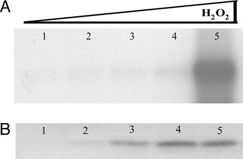
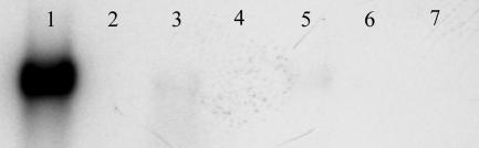
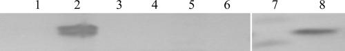
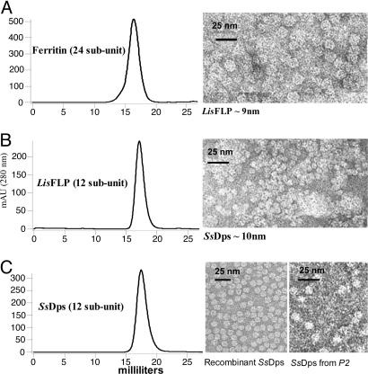
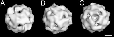
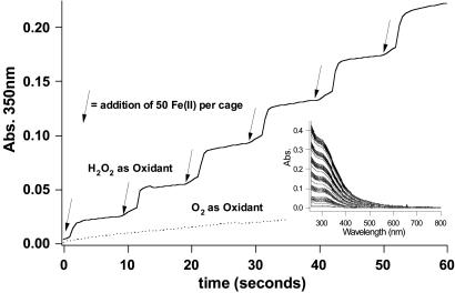
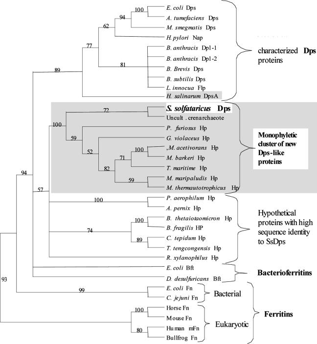
Evolution of an oxygenic atmosphere required primordial life to accommodate the toxicity associated with reactive oxygen species. We have characterized an archaeal antioxidant from the hyperthermophilic acidophile Sulfolobus solfataricus. The amino acid sequence of this approximately 22-kDa protein shares little sequence similarity with proteins with known function. However, the protein shares high sequence similarity with hypothetical proteins in other archaeal and bacterial genomes. Nine of these hypothetical proteins form a monophyletic cluster within the broad superfamily of ferritin-like diiron-carboxylate proteins. Higher order structural predictions and image reconstructions indicate that the S. solfataricus protein is structurally related to a class of DNA-binding protein from starved cells (Dps). The recombinant protein self assembles into a hollow dodecameric protein cage having tetrahedral symmetry (SsDps). The outer shell diameter is approximately 10 nm, and the interior diameter is approximately 5 nm. Dps proteins have been shown to protect nucleic acids by physically shielding DNA against oxidative damage and by consuming constituents involved in Fenton chemistry. In vitro, the assembled archaeal protein efficiently uses H2O2 to oxidize Fe(II) to Fe(III) and stores the oxide as a mineral core on the interior surface of the protein cage. The ssdps gene is up-regulated in S. solfataricus cultures grown in iron-depleted media and upon H2O2 stress, but is not induced by other stresses. SsDps-mediated reduction of hydrogen peroxide and possible DNA-binding capabilities of this archaeal Dps protein are mechanisms by which S. solfataricus mitigates oxidative damage.


Figures

Fig. 1.
Fig. 1.
SsDps expression in response to oxidative stress. (A) Northern blot analysis of RNA extracted from exponential growth phase (OD650 ≈0.5) S. solfataricus cells. Lane 1, standard growth conditions (0 μM H2O2); lane 2, 5 μM H2O2; lane 3, 10 μM H2O2; lane 4, 20 μM H2O2; lane 5, 30 μM H2O2. Approximately 1.5 μg of total RNA was loaded in each lane. (B) Western blot analysis of total protein extract from logarithmic (OD650 ≈0.5) S. solfataricus cells. Loads are normalized according to number of cells, and lane assignments are as above. Densitometry and loading controls are available in Fig. 8.
Fig. 2.
Fig. 2.
Ssdps expression is specific to oxidative stress. Shown is Northern blot analysis of RNA extracted from S. sulfataticus cultures. Lane 1, cells cultured in media supplemented with 30 μMH2O2; lane 2, late log phase cells; lane 3, 300 KJ UV, which accounts for RNA degradation as shown in the load control; lane 4, sucrose as the sole carbon source; lane 5, SSV RH (Sulfolobus spindle-shaped virus, Ragged Hills)-infected; lane 6, 11 h heat shock at 90°C; lane 7, 11 h cold shock at 60°C. Approximately 0.75 μg of total RNA was loaded in each lane. Loading controls are available in Fig. 9.
Fig. 3.
Fig. 3.
SsDps expression in response to iron. Western blot analysis of total protein extract from exponential growth phase (OD650 ≈0.5) S. solfataricus cells. Loads were normalized according to number of cells. Lane 1, standard growth conditions; lane 2, iron-extracted media; lane 3, iron-extracted media supplemented with 1.25 μM Fe2SO4; lane 4, iron-extracted media supplemented with 1.25 mM Fe2SO4; lane 5, iron-extracted media supplemented with 5.0 mM Fe2SO4; lane 6, iron-extracted media supplemented with 5.0 mM Fe2Cl3; lane 7, molecular weight standard; lane 8, 2 ng of purified SsDps.
Fig. 4.
Fig. 4.
Size exclusion liquid chromatography elution profiles and TEM images of corresponding peaks. Retention times according to size exclusion liquid chromatography are shown for the 24-subunit horse spleen ferritin (A), the 12-subunit ferritin-like protein (Flp) from L. innocua (B), and the 12-subunit Dps-like protein from S. solfataricus (C). The L. innocua FLP and the Dps-like protein from S. solfataricus have retention times consistent with a 12-subunit, 260-kDa protein, whereas the 24-subunit ferritin from horse spleen elutes earlier. Transmission electron microscopy of each peak reveals intact cage diameters of ≈13 nm (A), ≈9 nm (B), and ≈10 nm (C), commensurate with each protein's retention time.
Fig. 5.
Fig. 5.
3D image reconstruction of the assembled SsDps cage. Surface-shaded views of reconstructed negative-stained images displayed along the twofold (2F) axis (A), and along the two nonequivalent environments at each end of the threefold (3F) axis (B and C). (Scale bar: 2.5 nm).
Fig. 6.
Fig. 6.
SsDps-catalyzed mineralization of iron. SsDps efficiently uses H2O2 to oxidize iron in a stepwise progression (1H2O2:2Fe) (solid line). In contrast, O2 serves as a relatively poor oxidant of iron in this reaction (dotted line). (Inset) Iron confined within the SsDps cage remains soluble as shown in the full-spectrum.
Fig. 7.
Fig. 7.
Phylogenetic analysis of the ferritin-like diiron-carboxylate superfamily. The Dps protein from S. solfataricus is more closely related to a group of hypothetical proteins than it is to other characterized members of the ferritin-like diiron-carboxylate superfamily. H. salinarum DpsA (shaded) and S. solfataricus Dps are currently the only examples of archaeal Dps proteins; all other characterized Dps proteins are bacterial. Database gene identification numbers are as follows: E. coli Dps (Escherichia coli) gi:16128780, A. tumefaciens Dps (Agrobacterium tumefaciens) gi:15889746, M. smegmatis Dps (Mycobacterium smegmatis) gi:17887432, H. pylori Nap (Helicobacter pylori) gi:15611298, B. anthracis Dpl-1 (Bacillus anthracis) gi: 21730371, B. anthracis Dpl-2 (Bacillus anthracis) gi: 21730378, B. Brevis Dps (Bacillus brevis) gi:31615600, B. subtilis Dps (Bacillus subtilis) gi:16080351, L. innocua Flp (Listeria innocua) gi:16800011, H. salinarum DpsA (Halobacterium salinarum sp. NRC-1) gi:15791220, S. solfataricus Dps (Sulfolobus solfataricus) gi:15898865, Uncult. crenarchaeote (uncultured crenarchaeote non-heme iron-containing protein) gi:14548145, P. furiosus Hp (Pyrococcus furiosus) gi:18977565, G. violaceus Hp (Gloeobacter violaceus) gi:37523861, M. acetivorans Hp (Methanosarcina acetivorans) gi:20091704, M. barkeri Hp (Methanosarcina barkeri) gi:48837814, T. maritima Hp (Thermotoga maritima) gi: 15644274, M. maripaludis Hp (Methanococcus maripaludis) gi:45358735, M. thermautotrophicus Hp (Methanothermobacter thermautotrophicus) gi:7482214, P. aerophilum Hp (Pyrobaculum aerophilum) gi:18313528, A. pernix Hp (Aeropyrum pernix) gi:14601419, B. thetaiotaomicron Hp (Bacteroides thetaiotaomicron) gi:29349231, B. fragilis HP (Bacteroides fragilis) gi:53714735, C. tepidum Hp (Chlorobium tepidum) gi:21674150, T. tengcongensis Hp (Thermoanaerobacter tengcongensis) gi:20808605, R. xylanophilus Hp (Rubrobacter xylanophilus) gi:46106146, E. coli Bft (Escherichia coli) gi:16131215, D. desulfuricans Bft (Desulfovibrio desulfuricans) gi:14326006, E. coli Fn (Escherichia coli) gi:16129855, C. jejuni Fn (Campylobacter jejuni) gi:15791972, Horse Fn (Equus caballus L-chain ferritin) gi:406209, Mouse Fn (Mus musculus L-chain ferritin) gi:55154579, Human mFn (Homo sapiens mitochondrial ferritin) gi:29126241, and Bullfrog Fn (Rana catesbeiana) gi:85895. Protein abbreviations are as follows: Hp, hypothetical protein; Dps, DNA-binding protein from nutrient-starved cells; Bft, bacterioferritin; Fn, ferritin; mFn, mitochondrial ferritin; Dpl, Dps-like protein; Flp, ferritin-like protein; and Nap, neutrophil-activating protein. Numbers at branching nodes are bootsrap values from 10,000 resamplings.

References

    1. Touati, D., Jacques, M., Tardat, B., Bouchard, L. & Despied, S. (1995) J. Bacteriol. 177, 2305-2314. - PMC - PubMed
    1. Chen, L., James, L. P. & Helmann, J. D. (1993) J. Bacteriol. 175, 5428-5437. - PMC - PubMed
    1. Chen, L. & Helmann, J. D. (1995) Mol. Microbiol. 18, 295-300. - PubMed
    1. Jenney, F. E., Jr., Verhagen, M. F. J. M., Cui, X. Y. & Adams, M. W. W. (1999) Science 286, 306-309. - PubMed
    1. Imlay, J. A. (2003) Annu. Rev. Microbiol. 57, 395-418. - PubMed

Publication types