. 2005 Aug;25(15):6303-13.
doi: 10.1128/MCB.25.15.6303-6313.2005.

Binding of hnRNP L to the pre-mRNA processing enhancer of the herpes simplex virus thymidine kinase gene enhances both polyadenylation and nucleocytoplasmic export of intronless mRNAs

Affiliations

Binding of hnRNP L to the pre-mRNA processing enhancer of the herpes simplex virus thymidine kinase gene enhances both polyadenylation and nucleocytoplasmic export of intronless mRNAs

Shouhong Guang et al. Mol Cell Biol. 2005 Aug.

Abstract

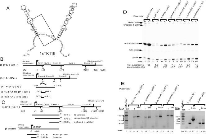
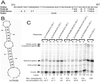
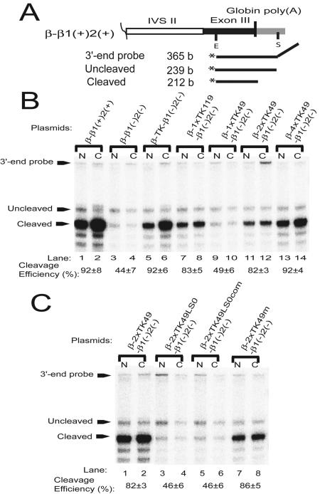
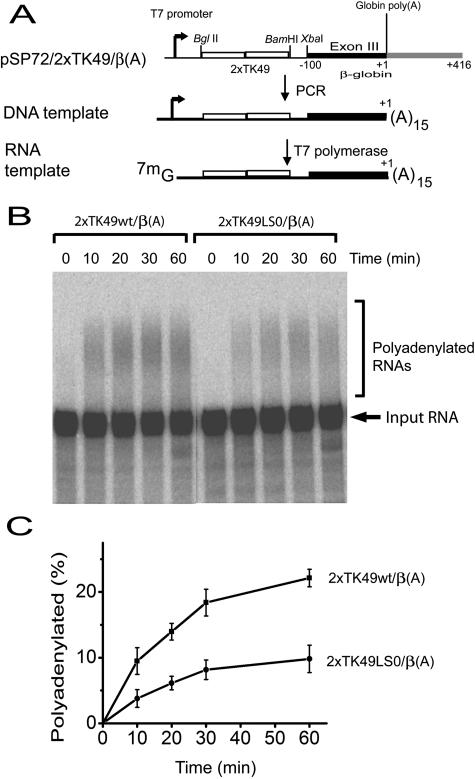
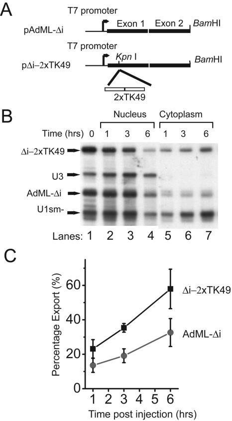
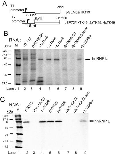
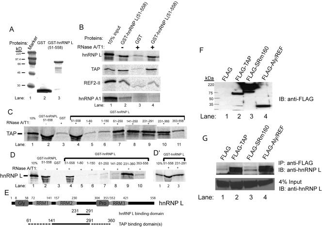
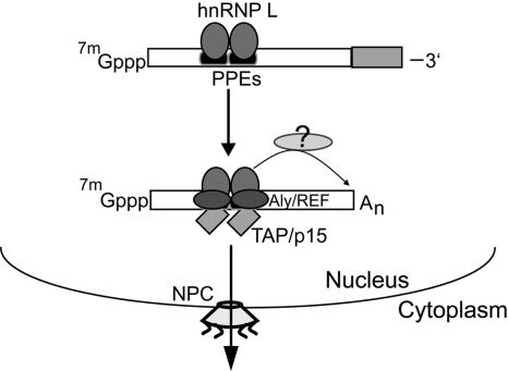
Liu and Mertz (Genes Dev. 9:1766-1780, 1995) previously identified a 119-nt pre-mRNA processing enhancer (PPE) element within the herpes simplex virus type 1 thymidine kinase gene that enables intron-independent gene expression in higher eukaryotes by binding heterogeneous nuclear ribonucleoprotein L (hnRNP L). Here, we identify a 49-nt subelement within this PPE that enhanced stability, polyadenylation, and cytoplasmic accumulation of transcripts synthesized in CV-1 cells from an intronless variant of the human beta-globin gene when present in two or more tandem copies. This 2xTK49 PPE also enhanced (i) the efficiency of polyadenylation of intronless beta-globin RNA in a cell-free polyadenylation system and (ii) the kinetics of nucleocytoplasmic export of an intronless variant of adenovirus major late leader region RNA in Xenopus oocytes. This 2xTK49 PPE bound only hnRNP L. Analysis of 2xTK49 PPE mutants showed a strong positive correlation existed between binding hnRNP L and enhancement of intronless beta-globin gene expression. hnRNP L was found to associate with both the mRNA export factor TAP and the exon-exon junction complex protein Aly/REF. Thus, we conclude that hnRNP L plays roles in enhancing stability, polyadenylation, and nucleocytoplasmic export; it does so, at least in part, by directly recruiting to intronless PPE-containing RNAs cofactors normally recruited to intron-containing RNAs.


Figures

FIG. 1.
FIG. 1.
A 49-nt sequence from the HSV-TK gene can enhance cytoplasmic accumulation of intronless β-globin-like transcripts when present in two or more tandem copies. (A) Computer-predicted secondary structure of 1×TK119 PPE. Secondary structure was determined using the mFOLD program (37, 52). The boxed region includes bases previously determined by cluster mutagenesis to be important for PPE function (32). (B) Structures of plasmids containing HSV-TK's full-length coding region, 1×TK119, and 1×TK49 inserted into the NcoI site in exon I of a cDNA variant of the human β-globin gene. All numbers are given relative to the site of transcription initiation from each gene. Black rectangles indicate exons. Open rectangles indicate introns. Gray rectangles indicate sequences 3′ of the site of cleavage for polyadenylation. Rectangles are not drawn to scale. N, NcoI; B, BamHI; E, EcoRI. (C) Structure of the human β-globin and human β-actin probes used for S1 nuclease mapping analysis. Probes were amplified by PCR and end-labeled with 32P indicated by the * as described in Materials and Methods. The sizes of the DNA fragments resulting from protection by hybridization with the corresponding RNAs are indicated. S, SspI. (D) Autoradiogram of quantitative S1 nuclease mapping analysis of the human β-globin-like RNAs accumulated in the nucleus and cytoplasm of CV-1PD cells transfected with the indicated plasmids. CV-1PD cells were cotransfected in parallel with 2 μg of each of the indicated plasmids together with 1 μg of the SV40 T antigen-encoding plasmid pRSV-Tori. Nuclear (N) and cytoplasmic (C) RNAs were harvested 48 h later and analyzed by concurrent S1 nuclease mapping with the 5′-end-labeled β-globin and β-actin probes shown in panel C. The amount of β-globin-like RNA accumulated in the cytoplasm was internally normalized to the amount of cellular β-actin RNA present in the same sample. The data were calculated as percentages relative to the amount of β-globin RNA accumulated in the cytoplasm of cells transfected in parallel with pβ-β1(+)2(+) RNA after internal normalization as well to the relative amounts of DpnI-resistant globin-encoding plasmid DNA present in these cells. The numbers below the lanes are means ± SEMs of data obtained from three independent experiments similar to the one shown here. (E) β-Globin-like RNAs accumulated in the cytoplasm are unspliced. Portions of the cytoplasmic RNA samples from the experiment shown in panel A were reverse transcribed and then amplified by PCR with the primers described in Materials and Methods (+RT, lanes 3, 6, 9, 12, 16, and 19). As controls, PCR amplification reactions were performed on the RNA samples without prior reverse transcription (−RT, lanes 2, 5, 8, 11, 15, and 18) and on the plasmid DNAs used in the transfections (DNA, lanes 4, 7, 10, 13, 17, and 20). Shown here is a photograph of an ethidium bromide-stained, 1% agarose gel in which the PCR products were electrophoresed. Lanes 1 and 14 contain size markers.
FIG. 2.
FIG. 2.
Effects of mutations in 2×TK49 on enhancement of intron-independent expression of human β-globin gene. (A) Sequences of TK49 PPE and its cluster base substitution mutants. (B) Computer-predicted secondary structure of 1×TK49. (C) Autoradiogram of quantitative S1 nuclease mapping analysis of β-globin-like RNAs accumulated in the nucleus (N) and cytoplasm (C) by 48 h after cotransfection of CV-1PD cells with the indicated plasmids and pRSV-Tori. The S1 nuclease mapping probes were the 5′-end-labeled ones shown in Fig. 1C. The samples were analyzed as described in the legend to Fig. 1D. The numbers shown here are means ± standard errors of the mean (SEMs) of data obtained from three independent experiments similar to the one shown here.
FIG. 3.
FIG. 3.
PPEs also enhance 3′ end processing of intronless β-globin-like RNAs in situ. (A) Structure of β-globin 3′ end probe used in S1 mapping analysis. Probes were amplified by PCR and 3′-end-labeled with 32P. The sizes of the DNA fragments resulting from protection by hybridization with the corresponding RNAs are indicated. (B and C) Autoradiograms of quantitative S1 nuclease mapping analysis of the 3′ ends of the β-globin-like RNAs accumulated in the nucleus and cytoplasm of CV-1PD cells. Portions of the RNA samples from the experiment shown in Fig. 1 and 2 were mapped with the 3′-end-labeled probe shown in panel A. The numbers below the lanes are means ± SEMs of the cleaved RNA (N+C) divided by the cleaved plus uncleaved RNA (N+C) times 100% of data obtained from three independent experiments similar to the one shown here.
FIG. 4.
FIG. 4.
Presence of 2×TK49 enhances polyadenylation in a cell-free system. (A) Structures of the plasmids employed in synthesis of the RNAs used in the cell-free polyadenylation assay. Plasmids pSP72/2×TK49wt/β(A) and pSP72/2×TK49LS0/β(A) were PCR amplified to generate double-stranded linear DNAs containing a T7 promoter, wild-type or LS0 mutant 2×TK49 PPE sequence, the 3′ half of β-globin's exon III, and 15 thymidine nucleotides. Radiolabeled, 5′-m7G-capped RNAs were synthesized from these linear DNA templates with T7 RNA polymerase. The arrow indicates the site of transcription initiation. The open rectangles indicate the TK sequences inserted between the BglII and BamHI sites in pSP72. The black rectangle indicates the β-globin exon III sequences relative to β-globin's natural cleavage site for polyadenylation. The gray rectangle indicates the β-globin sequences downstream of its cleavage site for polyadenylation. (B) Autoradiogram showing enhancement of polyadenylation of PPE-containing RNA. The 5′-m7G-capped, 32P-labeled RNA templates prepared as outlined in panel A were incubated in parallel with HeLa cell nuclear extract prepared as described in Materials and Methods in the presence of 1 mM ATP at 30°C for the indicated times. The resulting RNAs were phenol extracted, ethanol precipitated, and sized by electrophoresis in an 8 M urea-6% polyacrylamide gel. (C) Data averaged from four experiments similar to the one shown here indicating the means ± SEMs of the percentage of polyadenylated RNA relative to total input RNA as a function of time incubated at 30°C.
FIG. 5.
FIG. 5.
2×TK49 PPE enhances kinetics and efficiency of nucleocytoplasmic export of intronless RNA. (A) Structures of plasmids employed in synthesis of RNAs used in Xenopus oocyte export experiment. The 2×TK49 PPE (open rectangles) was inserted into pAdML-Δi, a plasmid containing a cDNA version of the first two exons of the adenovirus major late leader region (solid rectangles) present downstream of a T7 promoter. Template DNAs were linearized with BamHI prior to transcription with T7 RNA polymerase in the presence of [32P]UTP and the cap analogue m7G(5′)ppp(5′)G. (B) Autoradiogram of RNA present in the nuclei versus cytoplasm of Xenopus oocytes at the indicated times after nuclear coinjection of a mixture of the indicated 32P-labeled RNAs. The oocytes were manually dissected at the indicated times postinjection as described previously (42). The RNAs were purified and sized by electrophoresis in an 8 M urea-6% polyacrylamide gel. (C) Summary of data (means ± SEMs) obtained from three pools of oocytes analyzed in parallel as described in panel B. Percentage export was calculated as the amount of the indicated RNA in the cytoplasm relative to the total amount of that RNA in the nucleus plus cytoplasm at the indicated time.
FIG. 6.
FIG. 6.
2×TK49 is bound only by hnRNP L. (A) Structures of the plasmids used to synthesize the RNA probes employed in UV-cross-linking analysis. Symbols are the same as described in the legend to Fig. 4A. (B) Autoradiogram of SDS-PAGE analysis of the proteins that UV-cross-linked with the indicated variants of the HSV-TK PPE. 5′-m7G-capped, 32P-labeled RNAs were synthesized in parallel with T7 RNA polymerase, incubated with 20 μg of HeLa cell nuclear extract, exposed to UV, and incubated with RNases A and T1. Afterward, the proteins were sized by SDS-12% PAGE. Lane 1 contains 14C-labeled Rainbow markers (Amersham Pharmacia). (C) Autoradiogram of the UV-cross-linked proteins from panel B sized by SDS-12% PAGE after digestion with RNases A and T1 and immunoprecipitation with the hnRNP L-specific monoclonal antibody 4D11.
FIG. 7.
FIG. 7.
hnRNP L binds itself, TAP, and Aly/REF. (A) Coomassie brilliant blue staining of an SDS-PAGE analysis of GST and GST-hnRNP L fusion protein expressed in E. coli and purified by glutathione Sepharose-4B. (B) Autoradiogram of GST pull-down analysis. [35S]methionine-labeled, nearly full-length hnRNP L, TAP, REF2-II, and hnRNP A1 were synthesized with a rabbit reticulocyte lysate system. Lysates were pretreated with RNases A and T1 (+) or not pretreated (−) before addition of the indicated GST fusion protein. The proteins that bound were sized by SDS-12% PAGE and exposed to a PhosphorImager. (C and D) Domains of hnRNP L involved in binding TAP and itself. GST pull-down assays were performed with equimolar amounts of each of the indicated deletion variants of hnRNP L synthesized in E. coli and 35S-labeled TAP (C) or full-length hnRNP L (D). (E) Schematic showing known domains of hnRNP L. The numbers indicate positions of amino acid residues. RRM, RNA recognition motif; Gly, glycine-rich region; Pro, proline-rich region. Below the schematic is indicated the tentative mapping of the hnRNP L- and TAP-binding domains as determined from the data presented here. (F) FLAG-tagged proteins present in human 293T cells 48 h after transient transfection with expression plasmids encoding the indicated FLAG-tagged proteins. Whole-cell extracts were electrophoresed in an SDS-12% polyacrylamide gel and probed by immunoblotting with the anti-FLAG M2 antibody. (G) Immunoblot showing coimmunoprecipitation of endogenous hnRNP L with FLAG-tagged TAP and Aly/REF. The whole-cell extracts from panel F were incubated with RNase A followed by immunoprecipitation of the FLAG-tagged protein-containing complexes with anti-FLAG M2 affinity gel, electrophoresis in an SDS-12% polyacrylamide gel, and immunoblotting with the anti-hnRNP L monoclonal antibody 4D11. The bottom panel represents 4% of the amount of hnRNP L present in each whole-cell extract prior to immunoprecipitation.
FIG. 8.
FIG. 8.
Proposed role of hnRNP L in mediating nucleocytoplasmic export of PPE-containing intronless mRNAs. hnRNP L functions as an adaptor molecular, binding both PPEs and the mRNA export factors TAP and Aly/REF, bringing multiple TAP-p15 complexes to RNAs containing multiple PPEs to efficiently mediate their export. Likely, hnRNP L also associates with other as-yet-unidentified factors, recruiting the 3′ end cleavage/polyadenylation machinery to the RNA as well.

Similar articles

Cited by

References

    1. Ausubel, F. M., R. Brent, R. E. Kingston, D. D. Moore, J. G. Seidman, J. A. Smith, and K. Struhl. 1987. Preparation of nuclear and cytoplasmic extracts from mammalian cells, p. 12.1.1-12.1.9. In V. B. Chanda (ed.), Current protocols in molecular biology. John Wiley & Sons, Inc., New York, N.Y.
    1. Bray, M., S. Prasad, J. W. Dubay, E. Hunter, K.-T. Jeang, D. Rekosh, and M.-L. Hammarskjöld. 1994. A small element from the Mason-Pfizer monkey virus genome makes human immunodeficiency virus type 1 expression and replication Rev-independent. Proc. Natl. Acad. Sci. USA 91:1256-1260. - PMC - PubMed
    1. Buchman, A. R., and P. Berg. 1988. Comparison of intron-dependent and intron-independent gene expression. Mol. Cell. Biol. 8:4395-4405. - PMC - PubMed
    1. Chen, I.-H. B., K. S. Sciabica, and R. M. Sandri-Goldin. 2002. ICP27 interacts with the RNA export factor Aly/REF to direct herpes simplex virus type 1 intronless mRNAs to the TAP export pathway. J. Virol. 76:12877-12889. - PMC - PubMed
    1. Collis, P., M. Antoniou, and F. Grosveld. 1990. Definition of the minimal requirements within the human β-globin gene and the dominant control region for high level expression. EMBO J. 9:233-240. - PMC - PubMed

Publication types